Journal List > Korean J Neurotrauma > v.14(1) > 1058870

Kim, Suh, Kang, Lee, Lee, Lee, and Kang: Predictable Values of Decompressive Craniectomy in Patients with Acute Subdural Hematoma: Comparison between Decompressive Craniectomy after Craniotomy Group and Craniotomy Only Group

Abstract

Objective

Patients with traumatic acute subdural hematoma (ASDH) often require surgical treatment. Among patients who primarily underwent craniotomy for the removal of hematoma, some consequently developed aggressive intracranial hypertension and brain edema, and required secondary decompressive craniectomy (DC). To avoid reoperation, we investigated factors which predict the requirement of DC by comparing groups of ASDH patients who did and did not require DC after craniotomy.

Methods

The 129 patients with ASDH who underwent craniotomy from September 2007 to September 2017 were reviewed. Among these patients, 19 patients who needed additional DC (group A) and 105 patients who underwent primary craniotomy only without reoperation (group B) were evaluated. A total of 17 preoperative and intraoperative factors were analyzed and compared statistically. Univariate and multivariate analyses were used to compare these factors.

Results

Five factors showed significant differences between the two groups. They were the length of midline shifting to maximal subdural hematoma thickness ratio (magnetization transfer [MT] ratio) greater than 1 (p<0.001), coexistence of intraventricular hemorrhage (IVH) (p<0.001), traumatic intracerebral hemorrhage (TICH) (p=0.001), intraoperative findings showing intracranial hypertension combined with brain edema (p<0.001), and bleeding tendency (p=0.02). An average value of 2.74±1.52 was obtained for these factors for group A, which was significantly different from that for group B (p<0.001).

Conclusion

An MT ratio >1, IVH, and TICH on preoperative brain computed tomography images, intraoperative signs of intracranial hypertension, brain edema, and bleeding tendency were identified as factors indicating that DC would be required. The necessity for preemptive DC must be carefully considered in patients with such risk factors.

Introduction

Acute subdural hematoma (ASDH) is one of the leading causes of death in trauma patients and present in 12% to 29% among the patients with severe traumatic brain injury.31230) Surgical treatment of ASDH depends on patient neurologic status which can be evaluated by pupil response, size, Glasgow Coma Scale (GCS) score, and brain computed tomography (CT) scan. Bullock et al.3) recommended surgical treatment in patients with ASDH with the hematoma thickness greater than 10 mm or midline shifting greater than 5 mm on brain CT scan. Patients not in the above criteria with GCS score less than 9, decrease in GCS scores by 2 from the initial status, or abnormal pupil change should be considered for surgical intervention.3)
The general surgical procedure for ASDH is craniotomy with removal of hematoma and decompressive craniectomy (DC) if necessary. The decision of the surgical technique depends on individual surgeon's experience in preoperative neurologic status, preoperative CT findings, and intraoperative findings. In some studies, more craniotomies than decompressive craniectomies were performed as a surgical treatment of choice for ASDH.1424) Even though DC has its own complications and requires a subsequent cranioplasty, preemptive DC can provide more effective control of intracranial pressure (ICP) and aggressive brain edema. Some patients who underwent craniotomy for removal of hematoma suffered refractory intracranial hypertension and brain edema, as a result required reoperation with DC. To avoid reoperation, we investigated predictable values that could indicate DC as a surgical treatment of choice by comparing groups that did and did not require reoperation using DC after craniotomy in ASDH patients.

Materials and Methods

We retrospectively reviewed 155 cases of ASDH surgically managed with craniotomy in our hospital from September 2007 to September 2017. The 26 exclusions were made in patients younger than 16 years old, who underwent bilateral craniotomy, patients with non-traumatic ASDH, and ASDH in the posterior fossa. After exclusion there were 129 patients, and they were sorted into two groups; 24 patients in DC after craniotomy group, 105 patients in only craniotomy group. Among 24 cases in group A, 5 patients who underwent DC due to epidural hematoma, rebleeding of subdural hematoma (SDH) after primary craniotomy were excluded. Finally, 19 patients who needed additional reoperation using DC to control intracranial hypertension and brain edema (group A), and 105 patients who performed primary craniotomy without reoperation (group B) were analyzed.
We hypothesized that patients with underlying conditions inducing bleeding tendency, more severe brain injury, and diffuse intracerebral combined lesions with ASDH would tend to develop more aggressive intracranial hypertension and brain edema. Patients' age, gender, liver cirrhosis, chronic kidney disease, history of medication inducing bleeding tendency, and history of alcohol ingestion were collected through retrospective review of the medical records. The data of neurologic status at the time of the admission (pupil response, GCS scores) were also collected. Preoperative brain CT findings were also reviewed and the length of maximal SDH thickness and midline shifting were measured. We also calculated the length of midline shifting to maximal SDH thickness ratio (magnetization transfer [MT] ratio) to depict if there is extra mass effect other than ASDH or not. Combined lesions such as traumatic subarachnoid hemorrhage (TSAH), intraventricular hemorrhage (IVH), and traumatic intracerebral hemorrhage (TICH) were also evaluated. TICH is defined as a well-defined appearance hematoma with a diameter greater than 10 mm.5)
Craniotomy was performed based on indications suggested by Bullock et al.3) All patient's operative records were reviewed to estimate existence of intracranial hypertension, brain edema sign (macroscopic brain bulging, abrupt hematoma drainage after durotomy, grossly invisible brain pulsation), and sign of bleeding tendency (existence of multiple contusion, diffuse oozing without bleeding focus). We defined the prior as operative findings 1, and the later as operative findings 2. Total bleeding amount was also reviewed. DC after craniotomy was performed when patients showed worsening GCS scores or pupil response with worsening midline shifting more than 5 mm in follow-up brain CT.
Data were analyzed using SPSS software for personal computers (SPSS version 21; IBM Corp., Armonk, NY, USA). Continuous variables were analyzed by using unpaired Student's t-test and Categorical variables were analyzed by using Pearson χ2 test. Variables with statistical significance were selected as predictable values and underwent multivariate logistic regression analysis. A p-value of less than 0.05 was considered as statistically significance.

Results

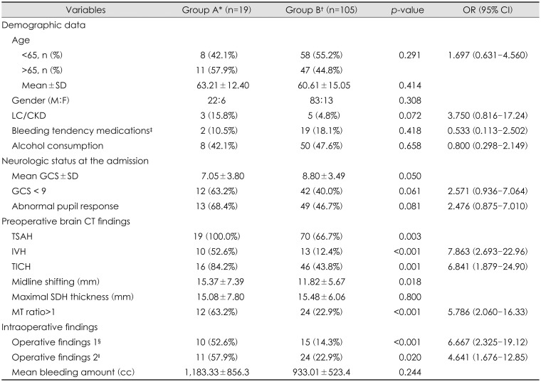
Of total 124 ASDH patients treated with craniotomy, 19 (15.3%) underwent reoperation using DC. Comparison of total 17 variables was performed between group A and group B (Table 1). Mean age was higher in group A (63.21±12.4, p=0.414) and it also had higher proportion (57.9%) of elders (age, >65; odds ratio [OR], 1.697; 95% confidence interval [CI], 0.631–4.560; p=0.291). There was higher proportion of men in both group (p=0.308). Demographic data showed no significant difference between two groups. Past medical history with bleeding tendency such as liver cirrhosis, chronic kidney disease, antiplatelet/anticoagulant drugs, and alcohol consumption history (greater than two drinks per day for male, greater than one drink per day for female) was also analyzed, but we could not find significant differences.
Patient's neurologic status at the admission was defined by GCS score and pupil response. Average GCS score was lower in group A (7.05±3.80, p=0.050). We divided patients into two groups by GCS score of 9 as it had been considered as severely injured state in other articles.330) Group A showed higher proportion of GCS less than 9 (3–8) (63.2%; OR, 2.571; p=0.061). Abnormal pupil response defined as pupil size other than normal size (3 mm) or light reflex. Group A showed higher proportions of abnormal pupil response (68.4%; OR, 2.476; 95% CI, 0.875–7.010; p=0.081). Neurologic status was worse in group A but had no significant differences.
Group A's preoperative brain CT findings were compared to those of group B, which showed significantly higher proportions of combined lesions like TSAH (100%, 0.003), IVH (52.6%; OR, 7.863; 95% CI, 2.698–22.96; p<0.001), and TICH (84.2%; OR, 6.841; 95% CI, 1.879–24.90; p=0.001). All of group A had TSAH, so it was considered improper to get OR. Midline shifting length was significantly longer in group A than that of group B (15.37±7.39 mm; p=0.018), but maximal SDH thickness was longer in group B than the other (15.48±6.06 mm; p=0.800). It demonstrated higher proportions of MT ratio >1 in group A, and there was a significant difference (63.2%; OR, 5.786; 95% CI, 2.060–16.33; p<0.001).
Intraoperative finding was divided into operative findings 1 and 2. Both operative findings showed significantly higher proportions in group A (operative findings 1, 52.6%; OR, 6.667; 95% CI, 2.325-19.12; p<0.001; operative findings 2, 57.9%; OR, 4.641; 95% CI, 1.676–12.85; p=0.020). Mean bleeding was higher in group A (1,183.33±856.3 cc; p=0.244), but did not show any significant difference.
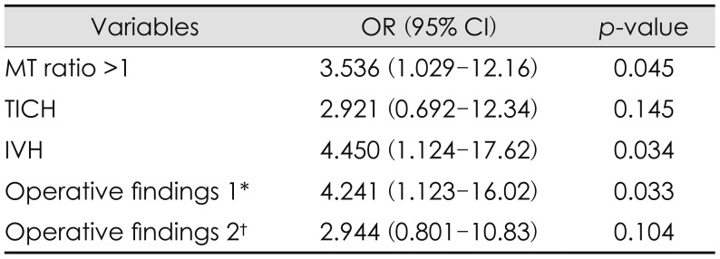
Among 17 candidate variables, six values (IVH, TICH, midline shifting length, MT ratio, operative findings 1, and 2 have significant relation to the need for DC. Midline shifting length was modified as midline shifting >15 mm to clarify the criteria of predictable value. It showed higher proportions in group A (52.6%) than group B (30.5%) but did not have statistical significance (OR, 2.535; 95% CI, 0.940–6.834; p=0.060); therefore excluded. Finally we got 5 predictable values, MT ratio >1, TICH, IVH, operative findings 1 and 2. Average number of predictable values in group A was 2.74 (p<0.001) and proportion of predictable values >2 was 52.6% while only 7.6% in group B (OR, 13.47; 95% CI, 4.251–42.70; p<0.001) (Table 2). These predictable values were analyzed through multivariate logistic regression (Table 3). MT ratio >1, IVH, and operative findings 1 showed higher ORs with statistical significance than TICH and operative findings 2.

Discussion

Performing DC or craniotomy in ASDH patients has remained an unresolved topic. Several comparative studies between these two operative techniques were conducted. Some preferred craniotomy because DC had shown unfavorable outcome and was considered more invasive with possible severe complications such as rebleeding of contusions, developing of contralateral mass lesion, and external cerebral herniation.6152731) On the other hand, other studies preferred DC in selective patients because it would supply more flexible ICP control and provide extra space for edematous brain tissue that was prepared for the refractory intracranial hypertension and further brain edema.8161928) Phan et al.20) meta-analyzed six comparative studies between DC and craniotomy groups, and depicted that DC was associated with worse postoperative outcome. It could be interpreted that these studies were biased to select patients with greater severity who ultimately underwent DC. Therefore, merely comparing these two groups has limitations to provide clues to make a decision on operational technique in ASDH patient. So, we investigated the patients who needed further DC among the patients who had undergone craniotomy.
In consideration of performing craniotomy in patients with ASDH, postoperative progression of brain edema, refractory intracranial hypertension, and additional bleeding risks should be estimated. If there is higher possibility to have these features, preemptive DC would be better alternatives than craniotomy. High initial intraoperative ICP monitoring, age, early hypotension, and combined lesions were identified as independent risk factors of reoperations using DC by Zhao et al.33) In addition, we identified preoperative brain CT finding of IVH, MT ratio >1, TICH, operative findings 1, 2 as predictable values.
Group A showed older average age and higher proportions of age >65 but did not show statistical significance. In the study of Zhao et al.33) which has a larger sample size in DC after craniotomy group (n=41), it was decided that age is also an independent risk factor for salvage DC. Potts et al.21) also suggested that age was a predictive value for unfavorable outcome of patients who underwent DC. Cerebral atrophy is more common in elders which creates extra cranial space, therefore possibly more endurable to brain edema and consequent intracranial hypertension. However, Oertel et al.18) depicted the association of elder's fragile microvascular structure and tendency of developing progressive hemorrhagic injury after head trauma. Degenerated blood brain barrier is more permeable to posttraumatic inflammatory factors and vulnerable to osmotic stress, consequently leading to vasogenic and cytotoxic edema.910) Even though age was not identified as a predictable value in our study, it still could be considered as a possible predictor.
Huang et al.11) used Rotterdam CT score as a prognosticator in DC patients, and patients with higher Rotterdam CT score had worse outcome. Since IVH is part of this scoring system, our results of higher IVH occurrence in group A correlated with postoperative change of these patients. MT ratio and TICH provide information on mass effect other than SDH in ASDH patients. Even though both midline shifting length, and SDH thickness themselves did not show correlation, when it was combined as MT ratio, it showed higher proportion of MT ratio >1 in group A. We got this idea from the fact that group A had more frequent intracranial combined lesion which may give extra mass effect in addition to ASDH, consequently inducing brain edema and intracranial hypertension. Other studies also showed significant correlation of TICH, IVH with poorer prognosis of acute traumatic SDH patients.411121728) Therefore MT ratio which can describe the existence of additional mass effect was measured as a predictable value.
Although craniotomy is sufficient to reduce the elevated ICP by removal of SDH, remaining mass effects by other types of hematoma cannot be solved solely by craniotomy because ICP reduction effect is limited due to bone closure. This will result in sustained increased ICP which cause brain ischemia and compromised brain oxygen, which leads to additional brain edema.2933) Operative findings 1 is the witnessing of this phenomenon. The operative findings 2 is related to bleeding tendency. Although successful bleeding control was done intraoperatively, TICH can be reformed by the negative pressure of cavitated space or solely due to the hemorrhagic progression tendency of TICH.5) Enlarged hematoma also gives additional mass effect after craniotomy, ultimately increasing the ICP. Therefore, even though it is subsided enough to close the bone flap, if operative findings 1, 2 were combined with predictable values from preoperative CT, it should be carefully considered to leave the bone flap without replacement.
Surgeons should be cautious when selecting DC as the primary operation. In some cases decompression creates excessive pressure gradient across the injured capillary and induces subsequent edema aggravation. This was proved by Cooper et al.7) in their animal models. This was also associated with developing of contralateral mass lesion.25) Besides drastic pressure change by removal of bone flap impairs autoregulatory capacity which induces hyperemia or oligoemia.2226) They are related to early stage complications of DC like blossoming of contusion, or ischemic damages.2) Due to these complications, DC was reserved as a second tier therapy after all possible medical therapeutic modalities were applied, so indiscreet use of preemptive DC should be avoided.3032) Even though preemptive DC has its own limitation and drawback, it would be preferred in selective cases. Bor-Seng-Shu et al.1 demonstrated that ICP is effectively decreased and cerebral perfusion pressure is increased significantly after DC. This consequently supplies more oxygen to the damaged tissue which helps to normalize abnormal metabolic parameters, finally promoting the recovery and preventing secondary ischemic insults.23) The predictable values we identified were related to the postoperative progression of brain edema, refractory intracranial hypertension, and additional bleeding risks. In patients with these values preemptive DC can be carefully considered, so they will make it possible to avoid unnecessary reoperation by using DC.
This study has several limitations. This is a retrospective analysis in a single medical center, so selection bias may have occurred. Although we performed both univariate, and multivariate analysis to reinforce the confidentiality of predictable values, due to small sample size, further studies with larger sample size are required such as Randomised Evaluation of Surgery with Craniectomy for patients Undergoing Evacuation of ASDH (RESCUE-ASDH) trial.13) Operative findings 1, and 2 were defined as predictable values but have subjective components in itself, so additional efforts are needed to uniform the descriptive terms of operative findings to objectificate these values. Despite these limitations, predictable values that we found can be referred to when making a decision as to whether to perform DC or craniotomy in ASDH patients.

Conclusion

MT ratio, IVH, TICH in brain CT image, and intraoperative signs of intracranial hypertension, brain edema (operative findings 1), and bleeding tendency (operative findings 2) can be considered as predictable values for re-operation using salvage DC after craniotomy. Despite the complications that may occur after DC, Preemptive DC can be considered a reasonable option to prepare for postoperative refractory intracranial hypertension and brain edema in patients with these values.

Notes

The authors have no financial conflicts of interest.

References

1. Bor-Seng-Shu E, Figueiredo EG, Amorim RL, Teixeira MJ, Valbuza JS, de Oliveira MM, et al. Decompressive craniectomy: a meta-analysis of influences on intracranial pressure and cerebral perfusion pressure in the treatment of traumatic brain injury. J Neurosurg. 2012; 117:589–596. PMID: 22794321.
crossref
2. Bor-Seng-Shu E, Paiva WS, Figueiredo EG, Fujimoto Y, de Andrade AF, Fonoff ET, et al. Posttraumatic refractory intracranial hypertension and brain herniation syndrome: cerebral hemodynamic assessment before decompressive craniectomy. Biomed Res Int. 2013; 2013:750809. PMID: 24377095.
crossref
3. Bullock MR, Chesnut R, Ghajar J, Gordon D, Hartl R, Newell DW, et al. Surgical management of acute subdural hematomas. Neurosurgery. 2006; 58:S16–S24. PMID: 16710968.
crossref
4. Caroli M, Locatelli M, Campanella R, Balbi S, Martinelli F, Arienta C. Multiple intracranial lesions in head injury: clinical considerations, prognostic factors, management, and results in 95 patients. Surg Neurol. 2001; 56:82–88. PMID: 11580939.
5. Cepeda S, Gómez PA, Castaño-Leon AM, Munarriz PM, Paredes I, Lagares A. Contrecoup traumatic intracerebral hemorrhage: A geometric study of the impact site and association with hemorrhagic progression. J Neurotrauma. 2016; 33:1034–1046. PMID: 26391755.
crossref
6. Chen SH, Chen Y, Fang WK, Huang DW, Huang KC, Tseng SH. Comparison of craniotomy and decompressive craniectomy in severely head-injured patients with acute subdural hematoma. J Trauma. 2011; 71:1632–1636. PMID: 22027888.
crossref
7. Cooper PR, Hagler H, Clark WK, Barnett P. Enhancement of experimental cerebral edema after decompressive craniectomy: implications for the management of severe head injuries. Neurosurgery. 1979; 4:296–300. PMID: 450227.
8. Coplin WM, Cullen NK, Policherla PN, Vinas FC, Wilseck JM, Zafonte RD, et al. Safety and feasibility of craniectomy with duraplasty as the initial surgical intervention for severe traumatic brain injury. J Trauma. 2001; 50:1050–1059. PMID: 11426120.
crossref
9. Donkin JJ, Vink R. Mechanisms of cerebral edema in traumatic brain injury: therapeutic developments. Curr Opin Neurol. 2010; 23:293–299. PMID: 20168229.
crossref
10. Hopp S, Nolte MW, Stetter C, Kleinschnitz C, Sirén AL, Albert-Weissenberger C. Alleviation of secondary brain injury, posttraumatic inflammation, and brain edema formation by inhibition of factor XIIa. J Neuroinflammation. 2017; 14:39. PMID: 28219400.
crossref
11. Huang YH, Deng YH, Lee TC, Chen WF. Rotterdam computed tomography score as a prognosticator in head-injured patients undergoing decompressive craniectomy. Neurosurgery. 2012; 71:80–85. PMID: 22382208.
crossref
12. Koç RK, Akdemir H, Oktem IS, Meral M, Menku A. Acute subdural hematoma: outcome and outcome prediction. Neurosurg Rev. 1997; 20:239–244. PMID: 9457718.
13. Kolias AG, Adams H, Timofeev I, Czosnyka M, Corteen EA, Pickard JD, et al. Decompressive craniectomy following traumatic brain injury: developing the evidence base. Br J Neurosurg. 2016; 30:246–250. PMID: 26972805.
crossref
14. Kolias AG, Scotton WJ, Belli A, King AT, Brennan PM, Bulters DO, et al. Surgical management of acute subdural haematomas: current practice patterns in the United Kingdom and the Republic of Ireland. Br J Neurosurg. 2013; 27:330–333. PMID: 23530712.
crossref
15. Kwon YS, Yang KH, Lee YH. Craniotomy or decompressive craniectomy for acute subdural hematomas: surgical selection and clinical outcome. Korean J Neurotrauma. 2016; 12:22–27. PMID: 27182498.
crossref
16. Li LM, Kolias AG, Guilfoyle MR, Timofeev I, Corteen EA, Pickard JD, et al. Outcome following evacuation of acute subdural haematomas: a comparison of craniotomy with decompressive craniectomy. Acta Neurochir (Wien). 2012; 154:1555–1561. PMID: 22752713.
crossref
17. Marshall LF, Marshall SB, Klauber MR, van Berkum Clark M, Eisenberg HM, Jane JA, et al. A new classification of head injury based on computerized tomography. J Neurosug. 1991; 75:S14–S20.
crossref
18. Oertel M, Kelly DF, McArthur D, Boscardin WJ, Glenn TC, Lee JH, et al. Progressive hemorrhage after head trauma: predictors and consequences of the evolving injury. J Neurosurg. 2002; 96:109–116. PMID: 11794591.
crossref
19. Paci GM, Sise MJ, Sise CB, Sack DI, Shackford SR, Kureshi SA, et al. Preemptive craniectomy with craniotomy: what role in the management of severe traumatic brain injury? J Trauma. 2009; 67:531–536. PMID: 19741396.
crossref
20. Phan K, Moore JM, Griessenauer C, Dmytriw AA, Scherman DB, Sheik-Ali S, et al. Craniotomy versus decompressive craniectomy for acute subdural hematoma: Systematic review and meta-analysis. World Neurosurg. 2017; 101:677–685.e672. PMID: 28315797.
crossref
21. Potts MB, Chi JH, Meeker M, Holland MC, Claude HJ 3rd, Manley GT. Predictive values of age and the Glasgow Coma Scale in traumatic brain injury patients treated with decompressive craniectomy. Acta Neurochir Suppl. 2008; 102:109–112. PMID: 19388299.
crossref
22. Rangel-Castilla L, Gasco J, Nauta HJ, Okonkwo DO, Robertson CS. Cerebral pressure autoregulation in traumatic brain injury. Neurosurg Focus. 2008; 25:E7.
crossref
23. Robertson CS, Valadka AB, Hannay HJ, Contant CF, Gopinath SP, Cormio M, et al. Prevention of secondary ischemic insults after severe head injury. Crit Care Med. 1999; 27:2086–2095. PMID: 10548187.
crossref
24. Rush B, Rousseau J, Sekhon MS, Griesdale DE. Craniotomy versus craniectomy for acute traumatic subdural hematoma in the United States: A national retrospective cohort analysis. World Neurosurg. 2016; 88:25–31. PMID: 26748175.
crossref
25. Stiver SI. Complications of decompressive craniectomy for traumatic brain injury. Neurosurg Focus. 2009; 26:E7.
crossref
26. Ter Minassian A, Dube L, Guilleux AM, Wehrmann N, Ursino M, Beydon L. Changes in intracranial pressure and cerebral autoregulation in patients with severe traumatic brain injury. Crit Care Med. 2002; 30:1616–1622. PMID: 12130988.
crossref
27. Tsermoulas G, Shah O, Wijesinghe HE, Silva AH, Ramalingam SK, Belli A. Surgery for acute subdural hematoma: Replace or remove the bone flap? World Neurosurg. 2016; 88:569–575. PMID: 26523763.
crossref
28. Ucar T, Akyuz M, Kazan S, Tuncer R. Role of decompressive surgery in the management of severe head injuries: prognostic factors and patient selection. J Neurotrauma. 2005; 22:1311–1318. PMID: 16305319.
crossref
29. Weiner GM, Lacey MR, Mackenzie L, Shah DP, Frangos SG, Grady MS, et al. Decompressive craniectomy for elevated intracranial pressure and its effect on the cumulative ischemic burden and therapeutic intensity levels after severe traumatic brain injury. Neurosurgery. 2010; 66:1111–1118. PMID: 20386136.
crossref
30. Wen L, Wang H, Wang F, Gong JB, Li G, Huang X, et al. A prospective study of early versus late craniectomy after traumatic brain injury. Brain Inj. 2011; 25:1318–1324. PMID: 21902550.
crossref
31. Woertgen C, Rothoerl RD, Schebesch KM, Albert R. Comparison of craniotomy and craniectomy in patients with acute subdural haematoma. J Clin Neurosci. 2006; 13:718–721. PMID: 16904897.
crossref
32. Wong GK, Hung YW, Chong C, Yeung J, Chi-Ping Ng S, Rainer T, et al. Assessing the neurological outcome of traumatic acute subdural hematoma patients with and without primary decompressive craniectomies. Acta Neurochir Suppl. 2010; 106:235–237. PMID: 19812956.
crossref
33. Zhao HX, Liao Y, Xu D, Wang QP, Gan Q, You C, et al. The value of intraoperative intracranial pressure monitoring for predicting re-operation using salvage decompressive craniectomy after craniotomy in patients with traumatic mass lesions. BMC Surg. 2015; 15:111. PMID: 26467905.
crossref
TABLE 1

General characteristics of study population and odd ratios for each variable

kjn-14-14-i001

*Patients reoperated with decompressive craniectomy after craniotomy, patients only operated with primary craniotomy, antiplatelets, anticoagulants, §macroscopic brain bulging, abrupt hematoma drainage after durotomy, or grossly invisible brain pulsation, existence of multiple contusion, or diffuse oozing without bleeding focus. OR: odds ratio, CI: confidence interval, SD: standard deviation, M: male, F: female, LC: liver cirrhosis, CKD: chronic kidney disease, GCS: Glasgow Coma Scale, CT: computed tomography, TSAH: traumatic subarachnoid hemorrhage, IVH: intraventricular hemorrhage, TICH: traumatic intracerebral hemorrhage, SDH: subdural hematoma, MT ratio: magnetization transfer ratio

TABLE 2

Predictable values for reoperation

kjn-14-14-i002

*Patients reoperated with decompressive craniectomy after craniotomy, patients only operated with primary craniotomy. OR: odds ratio, CI: confidence interval

TABLE 3

Odd ratio of predictable values using multiple logistic regression analysis

kjn-14-14-i003

*Macroscopic brain bulging, abrupt hematoma drainage after durotomy, or grossly invisible brain pulsation, existence of multiple contusion, or diffuse oozing without bleeding focus. OR: odds ratio, CI: confidence interval, MT ratio: magnetization transfer ratio, TICH: traumatic intracerebral hemorrhage, IVH: intraventricular hemorrhage

TOOLS
Similar articles