Journal List > Korean J Neurotrauma > v.13(2) > 1058868

So and Yun: The Combined Use of Cardiac Output and Intracranial Pressure Monitoring to Maintain Optimal Cerebral Perfusion Pressure and Minimize Complications for Severe Traumatic Brain Injury

Abstract

Objective

To show the effect of dual monitoring including cardiac output (CO) and intracranial pressure (ICP) monitoring for severe traumatic brain injury (TBI) patiens. We hypothesized that meticulous treatment using dual monitoring is effective to sustain maintain minimal intensive care unit (ICU) complications and maintain optimal ICP and cerebral perfusion pressure (CPP) for severe TBI patiens.

Methods

We included severe TBI, below Glasgow Coma Scale (GCS) 8 and head abbreviation injury scale (AIS) >4 and performed decompressive craniectomy at trauma ICU of our hospital. We collected the demographic data, head AIS, injury severity score (ISS), initial GCS, ICU stay, sedation duration, fluid therapy related complications, Glasgow Outcome Scale (GOS) at 3 months and variable parameters of ICP and CO monitor.

Results

Thirty patients with severe TBI were initially selected. Thirteen patients were excluded because 10 patients had fixed pupillary reflexes and 3 patients had uncontrolled ICP due to severe brain edema. Overall 17 patients had head AIS 5 except 2 patients and 10 patients (58.8%) had multiple traumas as mean ISS 29.1. Overall complication rate of the patients was 64.7%. Among the parameters of CO monitoring, high stroke volume variation is associated with fluid therapy related complications (p=0.043) and low cardiac contractibility is associated with these complications (p=0.009) statistically.

Conclusion

Combined use of CO and ICP monitors in severe TBI patients who could be necessary to decompressive craniectomy and postoperative sedation is good alternative methods to maintain an adequate ICP and CPP and reduce fluid therapy related complications during postoperative ICU care.

Introduction

Importance of post-operative management of intracranial pressure (ICP) and cerebral perfusion pressure (CPP) in severe traumatic brain injury (TBI) patients cannot be emphasized enough since it has direct influence over patient treatment outcome.15) In order to reach the appropriate CPP and ICP, meticulous control of mean arterial pressure (MAP), is key, however, this is usually a difficult process since severe TBI patients usually have combined multiple injuries rather than single isolated head injuries. The complications arising from fluid therapy to control the ICP and MAP, such as volume depletion, pneumonia, pulmonary edema due to volume overload and congestive heart failure (CHF) secondary acute renal failure (ARF) may result in high patient's morbidity and mortality. Up to now, most effective monitoring methods for MAP regulation are continuous arterial-line monitoring through an arterial catheter and central venous pressure (CVP) through a central catheter.9)
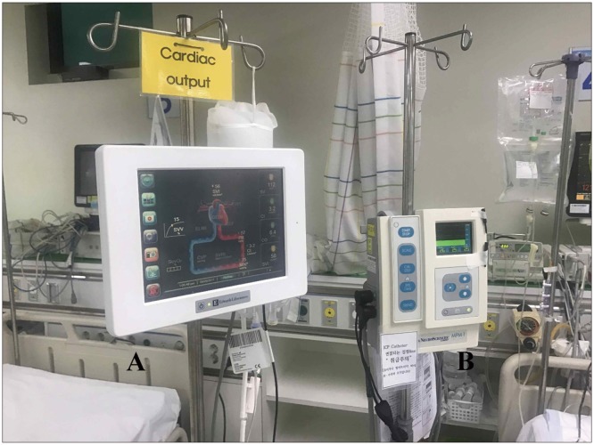
Recently, a new cardiac output (CO) device which accurately measures CO and MAP is being widely used for intensive care unit (ICU) critical care.1118) Dynamic parameters of fluid responsiveness in CO monitoring device-stroke volume variation (SVV) and stroke volume index (SVI)-can be obtained from analysis of arterial waveform.12) Differences in systolic pressure and pulse pressure are generated by changes in stroke volume during respiratory cycle. The amount of variation (SVV) correlates well with volume responsiveness.17) Also, SVI is known to be represented as the measure of cardiac performance.12) Therefore, we can estimate the patient's volume status and cardiac function by using these two parameters. In this study, we aimed to maintain optimal ICP and reduce fluid therapy related complications in severe TBI patients who received CPP, MAP control by using ICP monitoring and arterial line based CO monitoring simultaneously.

Materials and Methods

Patient population and clinical data

A total of 883 head trauma patients who were admitted the regional trauma center at our hospital between January 2015 and December 2016 were reviewed. We defined severe TBI as patients with initial Glasgow Coma Scale (GCS) 3–8 and head abbreviation injury scale (AIS) 4–5 (including subdural 4–5 or epidural 5 or intracerebral hematoma 4–5) and requiring decompressive craniectomy. Among these 883 patients, we included the cases of 30 patients with severe TBI who underwent decompressive craniectomy with ICP monitor and CO monitor insertion simultaneously. From the selected 30 patients, we excluded 13 more patients who had already fixed state of both pupillary reflex and uncontrolled ICP due to postoperative severe brain edema and who had past medical disease such as pneumonia or pulmonary edema or renal failure or heart failure or cancer disease etc. Therefore, we excluded overall 13 patients with 10 fixed states of both pupillary reflex and 3 uncontrolled ICP due to severe brain edema or herniation. Clinical information was obtained through a retrospective review of the medical record in these consecutive patients. We collected the demographic data, head AIS, injury severity score (ISS), initial GCS, ICU stay, sedation duration, fluid therapy related complications during sedation period, Glasgow Outcome Scale (GOS) at 3 months and variable parameters of ICP and CO monitor. This retrospective study was approved by our Institutional Review Board.

Procedures of ICP monitoring and cardiovascular monitoring

We used CO monitor as FloTrac™ (Vigileo™; Edwards Lifesciences, Irvine, CA, USA) which worked as arterial pressure based system (Figure 1A). Furthermore, we used ICP monitor as Integra Camino® (Camino Laboratories, San Diego, CA, USA) system which were inserted by subdural catheter (Figure 1B.) during intraoperative procedure (Figure 2). All patients were treated with analgesic based sedation (propofol or dexmedetomidine or remifentanil) for postoperative critical care in neuro ICU during 5 to 7 days postoperatively. We used only dual monitor during sedation period but, ICP monitor did not last over 1 week due to infection. We evaluated the clinical data of each patient with continuous CO and ICP monitor parameters such as ICP, MAP, CPP, SVV and SVI data for 3 to 7 days postoperatively and fluid therapy related complications during ICU care and GOS at 3 months. All parameter were measured every hour and calculated as mean value. Also, we determined that a fluid therapy related complications were diagnosed only by clinical physician's confirms or formal consultations during the period that dual monitors were applied. According to previous guideline, uncontrolled ICP was defined as mean value of 22 mmHg more or above 22 mmHg over 5 times per day.6) Also, uncontrolled CPP was defined as mean value of 50 mmHg less.6)

Treatment threshold and protocol

We treated our patient treatment goal for optimizing ICP as below 22 mmHg and optimizing CPP (CPP target) as 60 and 70, according to the guideline of brain trauma foundation 4th edition.6) We defined volume deficit when SVV was above 13% and normal volume state when SVV was ranging from 3% to 13%. Also the cardiac contractibility was defined with SVI where 40 and 50 to be normal range, below 40 as low contractibility, and above 50 as high contractibility.12) In case of volume deficit, we tried to a fluid challenge with either 500 mL of crystalloid or 250 mL of colloid solution. In case of the low cardiac contractibility, we tried to maintain proper MAP or CPP by injecting an inotropic or vasopressor agent. In case of the high contractibility and normal volume status, we tried to adjust volume status of the patients by injecting diuretics.12) After hemodynamic stabilization, we reassessed that the end organ perfusion parameters as following: urine output greater than 0.5 mL/kg per hour, clearance of arterial lactate, central venous oxygen saturation greater than 70%, and A-V CO2 less than 6 are good indicators of effective systemic resuscitation.10)

Statistical analysis

The correlation of each factor with the occurrence of fluid therapy related complications was analyzed statistically by univariate regression analysis using the χ2 test or Fisher's exact test (SPSS 20.0 version; SPSS Inc., Chicago, IL, USA). Both group with and without complication were subdivided according to the volume deficit (SVV>13%) and ICP and CPP and the existence of multiple trauma to calculate the odds ratio using multivariate analysis. A p-value of less than 0.05 were considered statistically significant.

Results

Patients' characteristics

The demographic characteristics of the 17 patients with severe TBI are summarized in Table 1. All seventeen patients underwent ICP and CO monitoring. Most of patients were male (16/17) and mean age is 56.5 years old ranging from 28 to 83. All patients had head AIS 5 except 2 patients and 10 patients (58.8%) had multiple traumas as mean ISS 29.1. Initial GCS was ranging from 3 to 10 as mean 6.8 and postoperative GCS (after 1 week) is ranging from 4 to 13 as mean 8.6. The mean ICU stay day of these patients was 21.1 days ranging from 5 to 41. The mean duration of postoperative sedation was 6.5 days ranging from 3 to 10. The 3 months GOS was score 1 (death) in 4 (23.5%) patients, score 2 (severe disability) in 1 (5.9%) patients, score 3 (moderate disability) in 8 (47.1%) patients and score 4 (mild disability) in 4 (23.5%) patients. All mortality cases were associated with severe brain edema or brain herniation due to progressive hemorrhage or secondary edema, which were not related with fluid therapy associated complications or its sequelae.

Clinical data of dual monitors' parameter and complications

We evaluated continuous recordings of ICP and CO monitoring during postoperative sedation period but, we only recorded the data of ICP and MAP in Table 2 for up to postoperative 3 days because other data did not show any statistical significance. The mean duration of continuous recordings in ICP and CO monitor was 5.8 days including ranging from 5 to 7. The mean value of initial ICP was 18.4 mmHg with ranging from 11 to 35 and the average ICP for the total duration of monitoring was 21.8 mmHg with ranging from 11 to 55. The mean value of initial SVV was 9.4% with ranging from 7 to 16. The mean SVV for the total recording period was 10.9% with ranging from 5 to 18 (Table 2). The mean value of initial MAP was 89.2 mmHg with ranging from 76 to 102 and the mean MAP for whole recording period was 87.3 mmHg with ranging from 76 to 102. The rate of CPP target was 70.6% (12/17) and the rate of volume deficit (SVV>13%) was 35.3% (6/17). However, the rate of low cardiac contractibility (SVI<40) were 29.4% (5/17) and the rate of high or normal cardiac contractibility (SVI>50 or 40–50) was 70.6% (12/17: high 3, normal 9). Overall complication rate of the patients was 64.7% (11/17) including 21 events with 8 patients of pulmonary edema, 6 patients of pneumonia, 1 patient of acute respiratory distress syndrome (ARDS), 1 patient of prerenal ARF and 5 patients of CHF (Table 2). To determine the factors associated with complication, we divided two groups as complication group and non-complication group. These complications are restricted as fluid or volume therapy related one such as pneumonia, pulmonary edema, ARDS, prerenal ARF and CHF in ICU. Among the parameters of CO monitoring system, high SVV (volume deficit) is associated with fluid therapy related complications (p=0.043) and low cardiac contractibility (abnormal cardiac function) is associated with these complications (p=0.009) statistically (Table 3). We found that the control of ICP and CPP in complication group (uncontrolled ICP: 6/7, uncontrolled CPP: 3/4) was difficult and resulted in an increase for mortality (4/5) although the following is not statistically significant. Only one patient was diagnosed as a septic shock due to pneumonia or pulmonary edema.

Discussion

The brain trauma foundation has established guideline for ICP monitoring in severe TBI.619) These studies demonstrated that superior survival was observed in severe TBI patients with ICP monitoring. However, other studies revealed that use of the ICP monitoring in isolated severe TBI had no survival benefit and was associated with occurrences of more complications which increased utilization of hospital resources.12) There are three methods for lowering ICP via vasoconstriction without reducing cerebral blood flow (CBF). First, decreasing blood viscosity in patients with intact viscosity autoregulation results in decreased ICP without significant changes in CBF. The sustained ICP lowering effects of hypertonic saline and mannitol likely result in part from the impact on blood viscosity.41416) Second, in patients with intact pressure autoregulation, increasing CPP will trigger vasoconstriction which decreases CBV and ICP; CBF remains constant in these circumstances.15) Third, sedation (e.g., propofol, pentobarbiturate infusion) reduces cerebral metabolic rate of oxygen, which not only protects cells from secondary injuries and insults, but also reduces ICP through metabolic autoregulatory pathways. If the patients undergo sedation therapy, sudden drop of blood pressure can often occurs, lowering MAP which results in imbalance of CPP causing uncontrolled brain edema or vicious cycle. Therefore, many guidelines suggested that comatose patients with severe TBI should be mechanically ventilated and monitored with an invasive arterial line, a central venous catheter, and an ICP pressure probe.9)
Usually severe TBI patients have combined multiple traumatic injuries which makes treatment very complicated. In order to maintain adequate CBF in severe TBI patients accurate monitoring of ICP and CPP is crucial. An ICP monitor can provide objective and exact values in order to maintain the CBF in such patients. However, the first goal of severe TBI is normalization of brain tissue oxygenation and CPP. If patients suffered multiple traumas which could result in major blood losses such as hemoperitoneum, hemothorax or major fracture of extremities, maintaining an optimum MAP and CPP is very challenging because MAP value is reversely proportionate to ICP value.19) In this situation, volume status and cardiac contractibility in patients are important indicators for optimizing systemic treatment such as fluid resuscitation or injection inotropics or diuretics. Also with the introduction of advanced CO monitoring, it is possible to continuously monitor MAP and CO which enables physicians to deduce the change in CPP. Therefore, the goal of advanced neuromonitoring in patients with severe brain injury should be to allow early detection of fluid therapy related complications such as pneumonia, pulmonary edema, ARDS, ARF, CHF and electrolyte imbalance etc.
Numerous supportive methods have been introduced in order to optimize treatment in TBI patients6910) In the past, CVP or body weights were the only methods to notify volume status of the patient indirectly.9) Recently, various methods have been introduced to determine volume status with safe and accuracy.9101112) These recent device are more accurate and easier to manipulate for physicians to evaluate patients' volume status and cardiac function. One recently developed CO monitoring system (Vigileo™; Edwards Life sciences) is also based on arterial pulse contour analysis. A special blood flow sensor (FloTrac), which is connected to an arterial line (radial, brachial, axillary or femoral arteries) is needed with no external calibration is necessary.111318) This device calculates CO on a continuous basis (every 20s) by multiplying pulse rate by calculated stroke volume which analyzed the impact of vascular tone on pressure and adjustment for actual vascular tone based on waveform analysis.5) SVV is the ability of the heart to increase stroke volume in response to an increase in preload. It is estimated dynamic parameters volume challenge maneuver, respiratory variation.1012) Based on this, a mathematical model was developed. Several studies have been performed concerning the accuracy of Vigileo CO monitoring that include a variety of patients with different software versions of the device.1718) Other limitations of this device include lack of validation in a pediatric population, total dependence on arterial line waveform for accurate calculations, the presence of dysrhythmias or improper dampening from kinking, shock state and significant valvular disease that can prevent accurate results.20)
Low SVV is good indicator of fluid resuscitation for TBI patients under sedation and a ventilator. After fluid resuscitation, systemic hemodynamic resuscitation should always precede brain targeted interventions.10) Assessment of fluid responsiveness may be possible by following global end diastolic volume index (GEDVI), SVV, and pulse pressure variation. CVP is a poor indicator for fluid responsiveness or intravascular volume status. SVV greater than 10% to 14% and GEDVI less the 600 mL/m2 generally indicate that the patients will respond to a fluid challenge with either 500 mL of crystalloid or 250 mL of colloid solution.10)
The sudden development of hypoxemic respiratory failure following a catastrophic central nervous system event, which cannot be attributed to other causes of ARDS, is the only universally agreed upon characteristic of neurogenic pulmonary edema (NPE).78) In patients with TBI, the incidence of NPE has been estimated to be up to 20%.3) It appears that the clinical manifestations of this surge may vary depending on the individual circumstance. Although this article does not include the diagnosis and early detection of ARDS or NPE, it would be considered an important issue in our following paper.
Although our study is based upon retrospective data, there are no reports to evaluate for fluid therapy related complications in severe TBI patients with CO and ICP monitor at the same time. The mortality of the patients due to complication during ICU care rather than one due to failure of control ICP and progressive hemorrhage can be very unfortunate event for neurosurgeons. Therefore, as we have seen in our study, the dual monitor setups would be a very useful alternative treatment to maintain optimal CPP, ICP and minimal fluid therapy related complications.
There are some limitations in this study. First, it is a retrospective study design with a small population. Second, the mean value of parameter in CO and ICP monitor is not accurate one due to a flow recording continuously and subdural type ICP monitor is less accurate than intraparenchymal or intraventricular type. Third, it is unreliable due to there are many heterogeneous factors in investigating one related to complications. Last, it is difficult to distinguish between NPE and fluid related pulmonary edema. Therefore, further studies will be required to ascertain the long term follow up results and recruit more cases of TBI patients using dual monitor and compare with control group for more accurate effect of combined monitor.

Conclusion

Advanced hemodynamic monitoring is a cornerstone of the management of critically ill or severe neurologic patients particularly who need sedation therapy In severe TBI patients who undergo sedation therapy after decompressive craniectomy to maintain an adequate CBF, combined use of CO monitor and ICP monitor is a good method to reduce fluid therapy related complications.

Notes

The authors have no financial conflicts of interest.

References

1. Aiolfi A, Benjamin E, Khor D, Inaba K, Lam L, Demetriades D. Brain trauma foundation guidelines for intracranial pressure monitoring: Compliance and effect on outcome. World J Surg. 2017; 41:1543–1549. PMID: 28188356.
crossref
2. Piccinini A, Lewis M, Benjamin E, Aiolfi A, Inaba K, Demetriades D. Intracranial pressure monitoring in severe traumatic brain injuries: a closer look at level 1 trauma centers in the United States. Injury. 2017; 48:1944–1950. PMID: 28495204.
crossref
3. Bratton SL, Davis RL. Acute lung injury in isolated traumatic brain injury. Neurosurgery. 1997; 40:707–712. PMID: 9092843.
crossref
4. Burke AM, Quest DO, Chien S, Cerri C. The effects of mannitol on blood viscosity. J Neurosurg. 1981; 55:550–553. PMID: 6792325.
crossref
5. Button D, Weibel L, Reuthebuch O, Genoni M, Zollinger A, Hofer CK. Clinical evaluation of the FloTrac/Vigileo system and two established continuous cardiac output monitoring devices in patients undergoing cardiac surgery. Br J Anaesth. 2007; 99:329–336. PMID: 17631509.
6. Carney N, Totten AM, O'Reilly C, Ullman JS, Hawryluk GW, Bell MJ, et al. Guidelines for the Management of Severe Traumatic Guidelines for the management of severe traumatic brain injury, fourth edition. Neurosurgery. 2017; 80:6–15. PMID: 27654000.
7. Colice GL. Neurogenic pulmonary edema. Clin Chest Med. 1985; 6:473–489. PMID: 3907948.
crossref
8. Davison DL, Terek M, Chawla LS. Neurogenic pulmonary edema. Crit Care. 2012; 16:212. PMID: 22429697.
crossref
9. Kipnis E, Ramsingh D, Bhargava M, Dincer E, Cannesson M, Broccard A, et al. Monitoring in the intensive care. Crit Care Res Pract. 2012; 2012:473507. PMID: 22970356.
crossref
10. Lee K. The neuroICU book. New York, NY: McGraw-Hill;2012.
11. Mayer J, Boldt J, Wolf MW, Lang J, Suttner S. Cardiac output derived from arterial pressure waveform analysis in patients undergoing cardiac surgery: validity of a second generation device. Anesth Analg. 2008; 106:867–872. PMID: 18292432.
12. McGee WT. A simple physiologic algorithm for managing hemodynamics using stroke volume and stroke volume variation: physiologic optimization program. J Intensive Care Med. 2009; 24:352–360. PMID: 19736180.
crossref
13. Mehta Y, Chand RK, Sawhney R, Bhise M, Singh A, Trehan N. Cardiac output monitoring: comparison of a new arterial pressure waveform analysis to the bolus thermodilution technique in patients undergoing off-pump coronary artery bypass surgery. J Cardiothorac Vasc Anesth. 2008; 22:394–399. PMID: 18503927.
crossref
14. Muizelaar JP, Lutz HA 3rd, Becker DP. Effect of mannitol on ICP and CBF and correlation with pressure autoregulation in severely head-injured patients. J Neurosurg. 1984; 61:700–706. PMID: 6432972.
crossref
15. Muizelaar JP, Ward JD, Marmarou A, Newlon PG, Wachi A. Cerebral blood flow and metabolism in severely head-injured children. Part 2: Autoregulation. J Neurosurg. 1989; 71:72–76. PMID: 2738644.
16. Muizelaar JP, Wei EP, Kontos HA, Becker DP. Mannitol causes compensatory cerebral vasoconstriction and vasodilation in response to blood viscosity changes. J Neurosurg. 1983; 59:822–828. PMID: 6413661.
crossref
17. Prasser C, Bele S, Keyl C, Schweiger S, Trabold B, Amann M, et al. Evaluation of a new arterial pressure-based cardiac output device requiring no external calibration. BMC Anesthesiol. 2007; 7:9. PMID: 17996086.
crossref
18. Scheeren TW, Wiesenack C, Compton FD, Zukunft B, Hoffmann C, Zidek W, et al. Performance of a minimally invasive cardiac output monitoring system (Flotrac/Vigileo). Br J Anaesth. 2008; 101:279–280. PMID: 18614598.
crossref
19. Talving P, Karamanos E, Teixeira PG, Skiada D, Lam L, Belzberg H, et al. compliance with Brain Trauma Foundation guidelines and effect on outcomes: a prospective study. J Neurosurg. 2013; 119:1248–1254. PMID: 23971954.
20. Yeo LL, Seow SC, Loh JP, Phua J. Comparison of cardiac output measurement by arterial waveform analysis and pulmonary artery catheter in mitral stenosis. Ann Acad Med Singapore. 2010; 39:655–657. PMID: 20838709.
FIGURE 1

Hemodynamic monitoring system (FloTrac™/Vigileo™; Edwards Lifesciences, Irvine, CA, USA) (A). Intracranial pressure monitoring system Camino® (Camino Laboratories, San Diego, CA, USA) in our trauma intensive care unit (B).

kjn-13-96-g001
FIGURE 2

Intraoperative intracranial pressure monitoring monitor by subdural probe (white arrow) insertion.

kjn-13-96-g002
TABLE 1

Clinical characteristics and outcome of 17 patients with severe traumatic brain injury treated with cardiovascular and intracranial pressure monitoring system

kjn-13-96-i001

M: male, F: female, Dx: diagnosis, T-ICH: traumatic intracerebral hematoma, T-SDH: traumatic subdural hematoma, T-SAH: traumatic subarachnoid hemorrhage, MRF: multiple rib fracture, FBF: facial bone fracture, PBF: pelvic bone fracture, Cbr: cerebral, EDH: epidural hematoma, EF: extremity fracture, AIS: abbreviation injury scale, ISS: Injury Severity Score, GCS: Glasgow Coma Scale, ICU: intensive care unit, GOS: Glasgow Outcome Scale

TABLE 2

Clinical data (ICP, MAP, CPP, SVV, SVI) of ICP and cardiac output monitoring system during postoperative 3 days

kjn-13-96-i002

*CPP target: 60–70 mmHg. ICP: intracranial pressure, MAP: mean arterial pressure, CPP: cerebral perfusion pressure, SVV: stroke volume variation, nor: normal, SVI: stroke volume index, Cx: complication, GOS: Glasgow Outcome Scale, Pn: pneumonia, Pul: pulmonary, CHF: congestive heart failure, ARF: acute renal failure, ARDS: acute respiratory destress syndrome

TABLE 3

Statistical analysis for factors associated with postoperative fluid therapy related complications*

kjn-13-96-i003

*Fluid therapy related complication: pneumonia, pulmonary edema, acute renal failure, congestive heart failure acute respiratory stress syndrome. SVV: stroke volume variation, SVI: stroke volume index, ICP: intracranial pressure, CPP: cerebral perfusion pressure

TOOLS
Similar articles