Abstract
Objective
The LiquoGuard® system is a new ventricular-type monitoring device that facilitates intracranial pressure (ICP)-controlled or volume-controlled drainage of cerebrospinal fluid (CSF). The purpose of this study is to report the authors' experience with the LiquoGuard® ICP monitoring system, as well as the clinical safety, usefulness, and limitations of this device in the management of patients with traumatic brain injury (TBI).
Methods
Intraventricular ICP monitoring was performed on 10 patients with TBI using the LiquoGuard® monitoring system. ICP measurements, volume of drained CSF, and clinical outcomes were analyzed and discussed.
Results
ICP monitoring was performed on 10 patients for a mean duration of 6.9 days. With a mean 82,718 records per patient, the mean initial ICP was 16.4 mm Hg and the average ICP across the total duration of monitoring was 15.5 mm Hg. The mean volume of drained CSF was 29.2 cc/day, with no CSF drained in 4 patients. Seven of 10 patients showed 1 or 2 episodes of abnormal ICP measurements. No patient exhibited complications associated with ICP monitoring.
Conclusion
The LiquoGuard® system is a versatile tool in the management of TBI patients. Its use is both reliable and feasible for ICP monitoring and therapeutic drainage of CSF. However, episodes of abnormal ICP measurements were frequently observed in patients with slit ventricles, and further study may be needed to overcome this issue.
Traumatic brain injury (TBI) is one of the major causes of death and disability where limited improvement in outcome is achieved despite the advances of medical and surgical care.8131620) The Brain Trauma Foundation (BTF) established guidelines [American Association of Neurological Surgeons (AANS), ver. 2007]7) on the use of intracranial pressure (ICP) monitoring in TBI, but its use has not been popular among neurosurgeons for various clinical reasons.16) Many neurosurgeons find insertion of ICP monitoring devices unfamiliar and cumbersome, and procedures of ICP monitoring may put the hemodynamically unstable patient at additional risk.30) Despite the debate on the usefulness of ICP monitoring, however, a future demand for evidence-based medical care and surgical management of TBI creates a need for such monitoring.
Various types of ICP monitoring devices are available for clinical use. Commonly used intraparenchymal ICP monitors are simpler in terms of insertion, but less accuracy and difficulty in baseline adjustment are known drawbacks of this type.26) Although insertion of intraventricular ICP monitors is technically more difficult, ICP measurements are more accurate and therapeutic drainage of cerebrospinal fluid (CSF) to lower elevated ICP is possible.16) Known drawbacks of conventional ventricular ICP monitoring devices include overdrainage or underdrainage of CSF which may reduce the reliability of measurements and risk patient safety.131) The LiquoGuard® system (Möller Medical GmbH, Fulda, Germany) is a new ventricular-type monitoring device that facilitates ICP-controlled or volume-controlled drainage of CSF. The aim of this study is to report the authors' experience with the LiquoGuard® ICP monitoring system, as well as the clinical safety, usefulness, and limitation of this device in the management of patients with TBI.
The analysis was performed in 10 patients admitted with severe TBI at the International St. Mary's Hospital, Catholic Kwandong University, from February 2014 to June 2015. Patients with severe TBI were defined as: Glasgow Coma Scale (GCS) scores of 3 to 8 and an abnormal computed tomography (CT) scan revealing hematoma, contusion, edema, or compressed basal cisterns.6) All patients were managed according to the established guidelines of the BTF (AANS, ver. 2007).7)
After a brief physical and neurologic examination, laboratory tests and radiographic imaging were performed for all patients on arrival at the emergency department. A brain CT scan was performed if the patient was hemodynamically stable. If the CT scan revealed the need for emergency decompressive surgery, the patient underwent surgery and was excluded from this study. In patients without the need for emergency surgery, a repeated CT scan was taken 4 hours after the initial scan. ICP monitoring was done in patients with probable intracranial hypertension on repeated CT scan who had no need for decompressive surgery.
Placement of an external ventricular drainage (EVD) catheter was done in the operating room under neuronavigation guidance (Medtronic Navigation, Inc., Minneapolis, MN, USA). The navigation system was routinely used since entricles in patients with intracranial hypertension are usually compressed, which makes proper placement of an EVD catheter difficult for reliable ICP monitoring. After induction of general anesthesia, a routine skin incision and a burr hole were made at Kocher's point for EVD placement. Any type of EVD catheter can be used with the LiquoGuard® ICP monitoring system. After the catheterization, the ICP monitoring device was connected postoperatively in the neurointensive care unit. Simultaneous ICP monitoring and drainage of CSF were done with the parameters of ICP pressure set to 15 to 20 mm Hg and CSF flow set to 0 to 10 cc/min. The duration of ICP monitoring and CSF drainage was determined by the ICP measurements obtained and the patient's clinical condition. The duration of monitoring did not exceed 2 weeks to prevent possible complications.
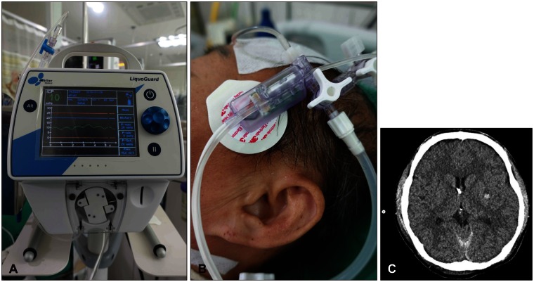
The LiquoGuard® is a ventricular-type ICP monitoring system with an external transducer. An EVD catheter is connected to an external sensor where simultaneous pressure sensing and CSF drainage are done. This pressure sensor is attached to the patient's head at the level of foramen of Monro, and then to the main console and drainage bag (Figure 1). The uniqueness in the design of the LiquoGuard® lies in the pressure sensor and peristaltic pump on the console, which permits pressure-regulated or flow-controlled drainage of CSF in a closed system. An air filter is not included in the device, which reduces the risk of infection and the possibility of system error. Additionally, the LiquoGuard® has 2 pressure transducers and 2 microcontrollers on its sensor that make self-detection possible for sensor failures, inconsistencies in measured values, and baseline drifts. The alarm system helps detect abrupt changes in measurements that may be due to intracranial pathology or occlusion of the catheter. Recordings of ICP can be reviewed on the main console, and data may be transferred via digital storage devices.
Data were analyzed using the Statistical Package for Social Sciences (SPSS) software for personal computers (SPSS ver. 21; IBM Corp., Armonk, NY, USA). Correlation analyses were used for continuous variables. A probability value of less than 0.05 was considered statistically significant.
Ten patients underwent ICP monitoring with the LiquoGuard® monitoring system. Five (50%) patients were male, and 5 (50%) were female. The mean age was 46.4 years with a range of 18 to 68 years. The diagnoses of patients were as listed in Table 1. The average GCS on admission was 6.4 (range, 4-12). On the patients' initial CT scans, the average midline shift was 1.4 cm.
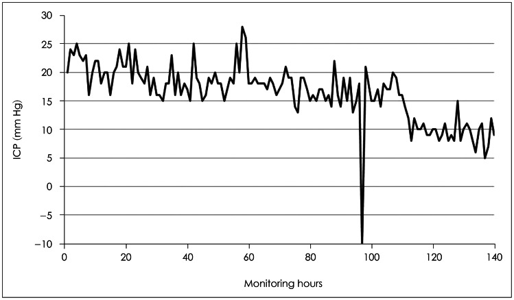
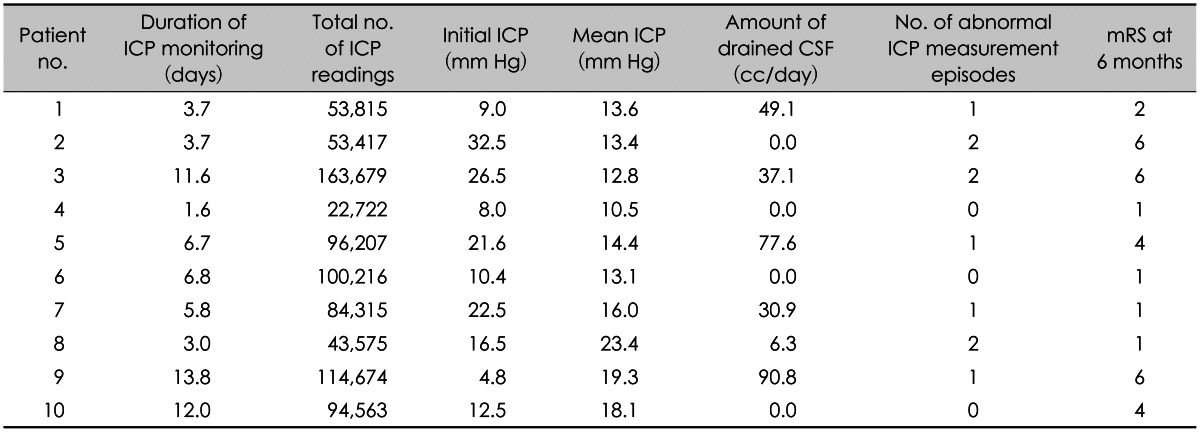
The mean duration of ICP monitoring was 6.9 days (range, 1.6-13.8 days) (Table 2). ICP readings were recorded every 6 seconds with a mean 82,718 records per patient (range, 22,722-163,679). Mean initial ICP was 16.4 mm Hg (range, 4.8-32.5 mm Hg) and the average ICP across the total duration of monitoring was 15.5 mm Hg (range, 10.5-23.4 mm Hg). Two patients underwent decompressive surgery after cessation of ICP monitoring due to intolerable intracranial hypertension. The mean amount of drained CSF was 29.2 cc/day (range, 0-90.8 cc/day). Among the 10 patients, no CSF was drained in 4 patients. During the whole duration of monitoring, 7 (70%) patients showed 1 or 2 episodes of abnormal ICP measurements (Figure 2). The 6 months modified Rankin Scale (mRS) was good (mRS of ≤3) in 5 (50%) patients and poor (mRS ≥4) in the other 5 (50%) patients. Correlational analyses of the duration of ICP monitoring, initial ICP, mean ICP, amount of drained CSF, and 6 months mRS revealed that only the duration of ICP monitoring was significantly related to the 6 months mRS outcome (p=0.05). No patient showed complications associated with ICP monitoring in our series.
Besides the direct injury to the brain, intracranial hypertension often presents as the major clinical problem following TBI.24) Pathologically elevated ICP reduces cerebral perfusion pressure and causes widespread secondary damage by cerebral ischemia in the vital brain structures.23) Recognition of ICP in TBI patients is crucial regarding choice of therapy, and ICP-dependent treatment strategies are reported to benefit patient outcomes.910)
In 2007, the BTF recommended ICP monitoring for all severe TBI patients with a GCS between 3 to 8 and an abnormal CT scan. Although some authors have questioned the risk associated with ICP monitoring, numerous reports describe its benefit in producing favorable patient outcomes, supporting more widespread use of this technique.34591011) Moreover, ICP monitoring in the future will be essential for the practice of evidence-based medicine and will help improve neurotrauma critical care.
There are various types of U. S. Food and Drug Administration-approved ICP monitoring devices available for use. They can be classified according to the location of the sensor probe, i.e., intraparenchymal, ventricular, subdural, and epidural.33) Of these, intraparenchymal devices are the most frequently used and studied as they are simple to use and their measurements directly reflect the pressure in the cerebral cortex.152526) However, intraparenchymal devices also carry the known limitations of zero drift and mechanical device failure.22225262732) Zero drift occurs due to the inability of the device to be recalibrated in relation to atmospheric pressure and was reported to occur in 50 to 60% of cases, with drift greater than 5 mm Hg in about 20% of cases.21427) This device is also prone to mechanical failure because the fiber-optic transducers and cables are susceptible to excessive kinking or bending.25) The subdural and epidural monitors are similar to the intraparenchymal type and may be useful in specific circumstances, but their measurements are known to be less reliable than the intraparenchymal type due to their placement locations.628)
Intraventricular ICP monitors are the most accurate and provide the most reliable ICP measurements.61217) However, a fluid-filled system is necessary for accurate ICP measurement, and limitations exist when the patient's ventricles are compressed or slit, which is frequently observed in patients with TBI.283033) In addition, correct intraventricular placement of the catheter is difficult in these cases. Despite the limitations of intraventricular monitoring, it is the only monitoring technique that allows simultaneous drainage of CSF for lowering ICP.61728)
Traditional intraventricular monitors use tip-sensors at the end of the ventricular catheter for measurement of ICP. The ventricular catheter and sensor are connected to a monitor for ICP measurement and a chamber, which is leveled to a reference point, for drainage of CSF. The disadvantage of this system lies in the hydraulic drainage of CSF, in which overdrainage or underdrainage is possible.131) Overdrainage or underdrainage frequently occurs when a patient is being transferred for radiologic study or when a patient is coughing. Frequent leveling of the CSF chamber also is needed when a patient's ICP changes since overdrainage can lead to ventricle collapse and inaccurate ICP measurements.
The LiquoGuard® system is designed to overcome the disadvantages of conventional intraventricular monitors. Firstly, the use of a fluid pump controls the drainage of CSF in preset pressure zones, thereby minimizing the risk of underdrainage or overdrainage. Thus, neurosurgeons and nursing personnel can reduce the time and effort spent leveling the drip chamber, and drainage of CSF is within the expected amount. Secondly, a fluid-filled closed system reduces CSF contact with air, which lessens the possible risk of infection. Thirdly, the LiquoGuard® system uses 2 pressure transducers, which allows for a double-check of unrecognized sensor malfunctions. The main console has an alarm system that can be set for various monitoring circumstances, e.g., an abrupt change in pressure, a sensor malfunction, or CSF drainage status, which make early detection of catheter occlusion or intracranial changes possible. Lastly, common EVD catheters can be connected for monitoring, which enable the use of a neuronavigation system for easy insertion. Further, cost savings is achieved compared with single-use tip-sensors.
To the best our knowledge, there are only a few reports on the use of the LiquoGuard® system and only one in the field of neurosurgery.192129) Linsler et al.21) compared the results of simultaneous ICP monitoring with the LiquoGuard® system and tip-sensor intraventricular monitors in 15 patients with TBI and subarachnoid hemorrhage. They reported that the LiquoGuard® system provided adequate ICP measurements and had various advantages over tip-sensor monitors but found episodes of unreliable measurements in 2 patients due to slit ventricles. In our study, we also experienced episodes of unreliable measurements in 7 of 10 (70%) patients. In the 3 patients who did not show abnormal ICP measurements, CSF was not drained as the ICP readings were below the level of drainage pressure. We think that slit ventricles, sensor malfunctions, or inadequate manipulation of the device may underlie the high incidence of abnormal measurement episodes in the current study. In all cases, the ICP measurement returned to reasonable values within minutes to hours.
Due to the small number of patients studied, the clinical outcome of the patients did not correlate with initial ICP, mean ICP, or amount of drained CSF. Only the duration of ICP monitoring was associated with clinical outcome, which may be due to a prolonged need for monitoring in cases of poor clinical status.
Our group has previously reported on the clinical usefulness of ventricular-type monitoring in comparison with intraparenchymal-type monitoring.18) In the course of that study, we found that leveling of the CSF chamber for adequate CSF drainage was time-consuming and difficult. The use of the LiquoGuard® system reduced the drawbacks of conventional intraventricular-type monitoring, without any associated complications.
This report is only a preliminary examination of the use of the LiquoGuard® system in 10 TBI patients. A detailed study of a larger patient population may be needed to reveal the true clinical advantage of LiquoGuard® over conventional ICP monitoring devices.
From our preliminary study, the LiquoGuard® system is a versatile tool in the management of TBI patients in that its use is both reliable and feasible for ICP monitoring and therapeutic drainage of CSF. However, episodes of abnormal ICP measurements were frequently observed in patients with slit ventricles, and further study of this device may be needed under these circumstances.
References
1. Açikbaş SC, Akyüz M, Kazan S, Tuncer R. Complications of closed continuous lumbar drainage of cerebrospinal fluid. Acta Neurochir (Wien). 2002; 144:475–480. PMID: 12111503.
2. Al-Tamimi YZ, Helmy A, Bavetta S, Price SJ. Assessment of zero drift in the Codman intracranial pressure monitor: a study from 2 neurointensive care units. Neurosurgery. 2009; 64:94–98. discussion 98-99PMID: 19145157.
3. Alali AS, Gomez D, Sathya C, Burd RS, Mainprize TG, Moulton R, et al. Intracranial pressure monitoring among children with severe traumatic brain injury. J Neurosurg Pediatr. 2015; 1–10. PMID: 26273741.
4. Becker DP, Miller JD, Ward JD, Greenberg RP, Young HF, Sakalas R. The outcome from severe head injury with early diagnosis and intensive management. J Neurosurg. 1977; 47:491–502. PMID: 903803.
5. Bowers SA, Marshall LF. Outcome in 200 consecutive cases of severe head injury treated in San Diego County: a prospective analysis. Neurosurgery. 1980; 6:237–242. PMID: 7383286.
6. Bratton SL, Chestnut RM, Ghajar J, McConnell Hammond FF, Harris OA, Hartl R, et al. Guidelines for the management of severe traumatic brain injury. VII. Intracranial pressure monitoring technology. J Neurotrauma. 2007; 24(Suppl 1):S45–S54. PMID: 17511545.
7. Bratton SL, Chestnut RM, Ghajar J, McConnell Hammond FF, Harris OA, Hartl R, et al. Guidelines for the management of severe traumatic brain injury. VIII. Intracranial pressure thresholds. J Neurotrauma. 2007; 24(Suppl 1):S55–S58. PMID: 17511546.
8. Brazinova A, Mauritz W, Leitgeb J, Wilbacher I, Majdan M, Janciak I, et al. Outcomes of patients with severe traumatic brain injury who have Glasgow Coma Scale scores of 3 or 4 and are over 65 years old. J Neurotrauma. 2010; 27:1549–1555. PMID: 20597653.
9. Cremer OL, van Dijk GW, van Wensen E, Brekelmans GJ, Moons KG, Leenen LP, et al. Effect of intracranial pressure monitoring and targeted intensive care on functional outcome after severe head injury. Crit Care Med. 2005; 33:2207–2213. PMID: 16215372.
10. Dang Q, Simon J, Catino J, Puente I, Habib F, Zucker L, et al. More fateful than fruitful? Intracranial pressure monitoring in elderly patients with traumatic brain injury is associated with worse outcomes. J Surg Res. 2015; 198:482–488. PMID: 25972315.
11. Dawes AJ, Sacks GD, Cryer HG, Gruen JP, Preston C, Gorospe D, et al. Intracranial pressure monitoring and inpatient mortality in severe traumatic brain injury: a propensity score-matched analysis. J Trauma Acute Care Surg. 2015; 78:492–501. discussion 501-502PMID: 25710418.
12. Exo J, Kochanek PM, Adelson PD, Greene S, Clark RS, Bayir H, et al. Intracranial pressure-monitoring systems in children with traumatic brain injury: combining therapeutic and diagnostic tools. Pediatr Crit Care Med. 2011; 12:560–565. PMID: 20625341.
13. Fakhry SM, Trask AL, Waller MA, Watts DD. IRTC Neurotrauma Task Force. Management of brain-injured patients by an evidencebased medicine protocol improves outcomes and decreases hospital charges. J Trauma. 2004; 56:492–499. discussion 499-500PMID: 15128118.
14. Fernandes HM, Bingham K, Chambers IR, Mendelow AD. Clinical evaluation of the Codman microsensor intracranial pressure monitoring system. Acta Neurochir Suppl. 1998; 71:44–46. PMID: 9779140.
15. Gelabert-González M, Ginesta-Galan V, Sernamito-García R, Allut AG, Bandin-Diéguez J, Rumbo RM. The Camino intracranial pressure device in clinical practice. Assessment in a 1000 cases. Acta Neurochir (Wien). 2006; 148:435–441. PMID: 16374566.
16. Haddad SH, Arabi YM. Critical care management of severe traumatic brain injury in adults. Scand J Trauma Resusc Emerg Med. 2012; 20:12. PMID: 22304785.
17. Kasotakis G, Michailidou M, Bramos A, Chang Y, Velmahos G, Alam H, et al. Intraparenchymal vs extracranial ventricular drain intracranial pressure monitors in traumatic brain injury: less is more? J Am Coll Surg. 2012; 214:950–957. PMID: 22541986.
18. Lee CS, Lim YC, Kim SH, Cho JM. Comparison of ventricular type and parenchymal type intracranial pressure (ICP) monitoring for the severe traumatic brain injury patients. Korean J Neurotrauma. 2012; 8:128–133.
19. Leopardi M, Tshomba Y, Kahlberg A, Baccellieri D, Melissano G, Chiesa R. Automated lumbar drainage for the control of the cerebrospinal fluid pressure during surgery for thoracoabdominal aortic aneurysms. Ann Vasc Surg. 2015; 29:1050.
20. Li LM, Timofeev I, Czosnyka M, Hutchinson PJ. Review article: the surgical approach to the management of increased intracranial pressure after traumatic brain injury. Anesth Analg. 2010; 111:736–748. PMID: 20686006.
21. Linsler S, Schmidtke M, Steudel WI, Kiefer M, Oertel J. Automated intracranial pressure-controlled cerebrospinal fluid external drainage with LiquoGuard. Acta Neurochir (Wien). 2013; 155:1589–1594. discussion 1594-1595PMID: 23188469.
22. Martínez-Mañas RM, Santamarta D, de Campos JM, Ferrer E. Camino intracranial pressure monitor: prospective study of accuracy and complications. J Neurol Neurosurg Psychiatry. 2000; 69:82–86. PMID: 10864608.
23. Miller JD, Butterworth JF, Gudeman SK, Faulkner JE, Choi SC, Selhorst JB, et al. Further experience in the management of severe head injury. J Neurosurg. 1981; 54:289–299. PMID: 7463128.
24. Miller MT, Pasquale M, Kurek S, White J, Martin P, Bannon K, et al. Initial head computed tomographic scan characteristics have a linear relationship with initial intracranial pressure after trauma. J Trauma. 2004; 56:967–972. discussion 972-973PMID: 15179234.
25. Münch E, Weigel R, Schmiedek P, Schürer L. The Camino intracranial pressure device in clinical practice: reliability, handling characteristics and complications. Acta Neurochir (Wien). 1998; 140:1113–1119. discussion 1119-1120PMID: 9870055.
26. Piper I, Barnes A, Smith D, Dunn L. The Camino intracranial pressure sensor: is it optimal technology? An internal audit with a review of current intracranial pressure monitoring technologies. Neurosurgery. 2001; 49:1158–1164. discussion 1164-1165PMID: 11846910.
27. Poca MA, Sahuquillo J, Arribas M, Báguena M, Amorós S, Rubio E. Fiberoptic intraparenchymal brain pressure monitoring with the Camino V420 monitor: reflections on our experience in 163 severely head-injured patients. J Neurotrauma. 2002; 19:439–448. PMID: 11990350.
28. Raboel PH, Bartek J Jr, Andresen M, Bellander BM, Romner B. Intracranial pressure monitoring: invasive versus non-invasive methods-a review. Crit Care Res Pract. 2012; 2012:950393. PMID: 22720148.
29. Riambau V, Capoccia L, Mestres G, Matute P. Spinal cord protection and related complications in endovascular management of B dissection: LSA revascularization and CSF drainage. Ann Cardiothorac Surg. 2014; 3:336–338. PMID: 24967177.
30. Shafi S, Diaz-Arrastia R, Madden C, Gentilello L. Intracranial pressure monitoring in brain-injured patients is associated with worsening of survival. J Trauma. 2008; 64:335–340. PMID: 18301195.
31. Staykov D, Speck V, Volbers B, Wagner I, Saake M, Doerfler A, et al. Early recognition of lumbar overdrainage by lumboventricular pressure gradient. Neurosurgery. 2011; 68:1187–1191. discussion 1191PMID: 21273925.
32. Talving P, Karamanos E, Teixeira PG, Skiada D, Lam L, Belzberg H, et al. Intracranial pressure monitoring in severe head injury: compliance with Brain Trauma Foundation guidelines and effect on outcomes: a prospective study. J Neurosurg. 2013; 119:1248–1254. PMID: 23971954.
33. Vender J, Waller J, Dhandapani K, McDonnell D. An evaluation and comparison of intraventricular, intraparenchymal, and fluidcoupled techniques for intracranial pressure monitoring in patients with severe traumatic brain injury. J Clin Monit Comput. 2011; 25:231–236. PMID: 21938526.
TABLE 1
CT: computed tomography, GCS: Glasgow Coma Scale, T-SAH: traumatic subarachnoid hemorrhage, FCCD: fracture compound comminuted depressed, T-SDH: traumatic subdural hematoma, T-IVH: traumatic intraventricular hemorrhage, EDH: epidural hematoma, DAI: diffuse axonal injury, ICH: intracerebral hemorrhage