

 PDF
PDF ePub
ePub Citation
 Print
Print


A 36-year-old man with abdominal pain presented with a 9-month history of low back pain after traffic accident. He was a chronic daily alcohol drinker, but had any other medical problem. He was diagnosed as a herniation of nucleus pulposus previously and had been treated by acupuncture and pain killer. In spite of treatment, wax and wane pattern low back pain continued, and left ankle pain newly developed. Abdominal pain, which was dominant on the right side, also appeared at 5 months after the accident. Abdominal pain occurred abruptly and disappeared by rest, and it was not associated with meal. We could not find abnormal finding on esophagogastroduodenoscopy, total colonoscopy, abdominal ultrasonography, and laboratory examination. Erythrocyte sedimentation rate (2 mm/hr) and C-reactive protein (0.113 mg/dL) level were not elevated.
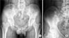
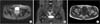
Back pain began when he was younger than 40 years old, aggravated at night, and was not improved with rest. Initially, we thought that he might have inflammatory back pain, and ankle pain was one of extra-axial manifestations of spondyloarthropathy. Simple radiography (Fig. 1) showed suspicious cam type femoroacetabular impingement at right. Sacroiliac joint magnetic resonance imaging (Fig. 2) showed avascular necrosis (AVN) of the femoral head, Association Research Circulation Osseous (ARCO) stage II [1]. The T1-weighted image revealed a large osteonecrotic lesion. Low-signal-intensity bands or lines within the femoral head were seen surrounding the area that corresponded to ischemic bone on T1- and T2-weighted images. The appearance on T2-weighted image is known as the "double-line sign" and was considered highly specific for AVN of the femoral head [2].
Finally, he was diagnosed as AVN of the femoral head. Abdominal pain was considered as a referred pain. Referred abdominal pain results from the integration of nervous impulses arising in the two main divisions of the nervous system [3]. Clinical suspicion should be raised for not only spondyloarthropathy but also AVN of the femoral head in cases of low back pain with abdominal pain.
 XML Download