

 PDF
PDF ePub
ePub Citation
 Print
Print


INTRODUCTION
Dental caries is one of the major oral diseases, which threatened the oral health of people. For example, dental caries can lead to a toothache, which can be stressful12). The usage of fluoride is one of the most effective methods for caries prevention3). Fluoride can promote remineralization reducing tooth mineral solubility by exchanging for hydroxyl groups and reducing carbonate content4).
Fluoride varnish is easy and simple method relatively comparing other professionally applied topical fluoride products such as rinses, gels, foams, and drops5678). The fluoride varnish can release fluoride over a longer period of 3 months contacting tooth surface as a thin film9). Beltran-Aguilar et al.10) suggested that the fluoride varnish was safe and easy to apply and could set in contact with intraoral moisture. Benson et al.11) reported that fluoride varnish application with six-month interval could decrease the prevalence of white spot about 70%. The Center for Disease Control (CDC)12) and American Dental Association (ADA)13) also reported that the application of fluoride varnish was more effective on high caries risk children than other. Twetman and Peterson14) examined 1,022 children, 4-5 years of age using WHO criteria and followed for two years. They concluded that the fluoride varnish had a cariostatic effect and caries prediction was influenced by the overall fluoride exposure. Therefore, fluoride varnish can be an excellent prevention of dental caries, especially in the high caries risk group.
Recently, calcium phosphate based delivery system using tricalcium phosphate (TCP) was introduced by 3M manufacturer15). This system was developed in that remineralization ability of fluoride is limited by the availability of calcium and phosphate ions16). Elkassas and Arafa17) concluded that fluoride varnish containing TCP showed the highest remineralization ability comparing to other fluoride varnish.
Most research about an in-vitro test of fluoride varnish has investigated the amount of fluoride release1819). Evidently, the amount of fluoride release means remineralization because the amount of fluoride release is directly proportional to the amount of remineralization20). However, some research suggested that remineralization did not depend on the amount of fluoride release21). The manual of fluoride varnish containing TCP by manufacturer wrote even only the release of fluoride by time22). Conclusively, the information about remineralization ability of fluoride varnish is scarce. Therefore, the aim of this study was to measure the amount of remineralization of fluoride varnish containing TCP and without TCP by time.
Materials and Methods
1. Specimen preparation
This in-vitro study used extracted bovine lateral incisors without dental caries. Enamel specimens of 5 mm diameter were created by drilling into the labial surface of bovine incisors. Specimens then were embedded in epoxy resin using the mounting mold. Specimens were flattened and polished with silicon carbide paper (600-2,000 grid) under cooling water. Vickers hardness number (VHN) was measured using a Vickers microhardness tester (SHIMADZU, JP/HMV-2, Japan), and VHN was 319.51±13.83 in control groups, 318.90±11.11 in non-TCP groups, and 319.17±11.70 in TCP groups. They were randomly allocated to 18 groups over time (Table 1).
2. Early carious lesion
Artificial lesions were created in the enamel specimens by immersion into a solution of 0.1 M lactic acid and 0.2% carbopol during 48 hours at 37℃. After immersion, the specimens were washed out with distilled water for 10 seconds and dried by compressive air for 3 seconds. All specimens were stored under the 100% humidity until the experiments.
3. Amount of mineral loss before experiment (ΔFbefore)
The amount of mineral loss (ΔFbefore) was measured by quantitative light-induced fluorescence (QLF, Inspektor Research System; QLF Pro™, Amsterdam, Netherlands). This QLF was equipped with a xenon micro-discharge arc lamp as the light source and an optical filter system producing blue light with a maximum wavelength of 370 nm. To avoid interference of the fluorescence images, such as bubbles of artificial saliva or water, specimens were thoroughly dried before the images were taken in the dark room.
4. Fluoride varnish application and artificial saliva immersion
Test fluoride varnishes (10 mg) were applied to the enamel surface of the specimen and dried four minutes. There was no application of fluoride varnish in the control groups. All the specimens were placed in the artificial saliva to mimic oral environment. Artificial saliva was prepared by mixing 0.021 M Na2HPO4/NaH2PO4 (Sigma-Aldrich, Switzerland), a pH 7.0 solution containing 36 mM NaCl (Sigma-Aldrich, USA), 0.96 mM CaCl2 (Acros Organics, USA), and sodium carboxymethylcellulose 1.0% (Sigma-Aldrich, USA)23). Twelve specimens were randomly allocated to each group by application time (0.5, 1, 3, 6, 12, and 24 hours) of fluoride varnish.
5. Amount of mineral loss after experiment (ΔFafter)
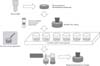
Specimens were taken out in the artificial saliva, and the fluoride varnishes on the surfaces of specimens were removed using No.15 scalpel blade (Hu-Friedy, Chicago, Illinois). The remnants of fluoride varnish were eliminated by powered toothbrush (Oral-B advance power, Braun, Cincinnati, Ohio, USA) using 300-gram weight (Weight, Arirang Science, Korea) for 30 seconds (Fig. 1)24). Then, specimens were washed out with distilled water, and dried using compressive air for three seconds. ΔFafter was re-measured by QLF as the same method of ΔFbefore. The overall procedure of this in-vitro test was shown in the Fig. 1.
6. Statistical analysis
ΔFbefore and ΔFafter were analyzed by paired t-tests for continuous variables. The differences between ΔFbefore and ΔFafter by the group were analyzed using one-way ANOVA followed multiple comparisons Tukey's post hoc test. This study could not use repeated measure ANOVA because the time of this study means different groups by application time of fluoride varnish before removal of varnish film.
All analyses were performed using SPSS software version 19.0 (SPSS. Chicago. IL. USA). Significance was determined at α=0.05 in all the tests.
RESULTS
ΔF significantly decreased at 3, 6, and 24 hours in control group, whereas ΔF in TCP group significantly increased at 6, 12, and 24 hours between before and after fluoride varnish application. ΔF in the non-TCP group also significantly increased at 12, and 24 hours between before and after fluoride varnish application. On the other hand, the amount of mineral loss increased in the control group (Table 2).
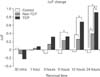
The difference between ΔFbefore and ΔFafter (ΔΔF) by time showed increasing tendency. There was a significant difference between TCP group and control group after 6 hours. The non-TCP group showed a significant difference after 24 hours comparing control group. The TCP group showed significant differences comparing both non-TCP and control group, after 12 hours (Fig. 2).
DISCUSSION
It is critical information to dental clinicians when is the optimal resuming time to brush teeth after application of fluoride varnish. However, there were few reports about fluoride release by time after application of fluoride varnish. Ritwik et al.18) studied the amount of fluoride release from four commercial fluoride varnishes. In their study, three fluoride varnishes among them showed a plateau of fluoride release after four hours of the application. Carvalho et al.19) also showed that the highest fluoride release occurred during the first eight hours after fluoride varnish application.
However, Elkassas and Arafa17) suggested that short-term gradual fluoride release could not guarantee a directly proportional relationship to net remineralization ability because the remineralization was very complicated process concerned with calcium, phosphate and other additives of commercial fluoride varnish. They investigated the remineralization effect of five commercial fluoride varnishes by surface microhardness and surface roughness and concluded that fluoride varnish containing TCP showed the highest remineralization tendency with the greatest resistance to acid challenge.
However, their study did not give information on short-term remineralization time by the hour. Moreover, Lippert and Juthani20) compared the fluoride dose-response of different caries lesions, and suggested that hardness test could not measure mineral content, which could be an indicator about remineralization. Therefore, the amount of remineralization was measured by QLF, not only by the hardness test.
This study evaluated the remineralization ability of fluoride varnish over time using QLF. QLF is an illumination system which can detect initial lesion and monitor progress and remineralization of dental caries. The blue light from 50-W xenon arc lamp, which has a peak intensity 370 nm wavelength, is guided to the tooth. Then, dental fluorescence emitted, and that color is predominantly green. Decalcified area showed increased fluorescence, and is seen darker than normal tooth structure. QLF showed the amount of mineral loss as the amount of fluorescence quantitatively25). Using this QLF system, quantification of the mineral loss or remineralization can be measured26). QLF can observe non-cavity dental caries and initial dental caries lesion, and show the excellent ability of early detection of dental caries2728).
In this study, TCP groups showed a significant increase of remineralization at 6, 12, and 24 hours comparing to control group. A significant difference was shown between TCP and other groups (control and non-TCP) at 12 and 24 hours. Non-TCP groups showed a significant increase of remineralization at 12 and 24 hours compared to control groups only. There were significant differences between ΔFbefore and ΔFafter witnin TCP groups at 6, 12, and 24 hours. Within non-TCP groups, significant differences were shown at 12 and 24 hours between ΔFbefore and ΔFafter. Whereas, the control group did not show any significant difference at any time within groups. This increasing remineralization ability of fluoride varnish over time corresponds to the result by Elkassas and Arafa17).
This study showed the remineralization ability of the fluoride varnish containing tricalcium phosphate by time using QLF. This study was unique in that the remineralization effect of fluoride varnish was investigated by short-term hour using QLF. Manual of fluoride varnish has instructed for the dentist to their patient to resume to brushing teeth after six hours. The recommendation of the application period of fluoride varnish is minimally twice per year, or three-month interval especially in the high caries risk group by CDC12) or ADA13). That guideline was a sketchy knowledge based fluoride releasing time of fluoride varnish. However, results of our study suggested the optimal resuming time to brushing could be longer than six hours. In conclusion, this study suggests that the longer the resuming time to brushing teeth after application of fluoride varnish was, the better the remineralization effect was.
Evidently, the suggestions could not apply clinically now, because this study was an only in-vitro test. Moreover, this in-vitro study used not human enamel teeth but bovine enamel teeth. Bovine enamel teeth were more easily demineralized than human enamel teeth20). Therefore, further studies need to adopt a clinical trial design and in-vitro study using human enamel teeth.


 XML Download