

 PDF
PDF ePub
ePub Citation
 Print
Print


Abstract
Objectives
The aim of this study was to evaluate applicability of the Qraycam device for detecting caries and filling body during tooth examinations.
Methods
Fifty-two subjects aged 25 to 34 years were recruited for tooth examination. Two examiners (an epidemiologic expert and a non-expert) performed visual tooth examination using only dental operating light, dental mirror, and air-drying without a dental explorer. Pictures or videos of every tooth surface were obtained under visual ray and 405 nm blue ray, respectively, by using Qraycam. The two examiners then evaluated these videos or images more than 7 days after visual examination. Agreement between the two examiners and area under the receiver operating characteristics (ROC) curve (AUC) were evaluated based on the gold standard derived from visual and Qraycam examinations.
Results
The results of the visual, visible ray image, and 405 nm blue ray image examinations showed very good kappa agreement with the gold standard for both examiners. The 405 nm blue ray image examination showed higher kappa agreement than visible ray image examination, and was similar to visual examination. Accuracy of detecting caries was enhanced by using 405 nm blue ray images from Qray-cam. Accuracy of detecting filling body on 405 nm blue ray image examination was almost same as that by visual examination.
Go to : 
References
1. World Health Organization. Oral health surveys, Basic methods. 5th ed. Geneva: World Health Organization Press;2013. p. 35–56.
2. Lee CK, Kim JS, Yoo SH. Comparative study on the rate of dental enamel demineralization using a QLF. J Korean Acad Pediatr Dent. 2004; 31:506–515.
3. Lee HS, Hyun HK, Jang KT. Remineralization effect of interproximal caries adjacent to glass ionomer restorations: In vitro study using QLF. J Korean Acad Pediatr Dent. 2011; 38:244–249.

4. Gmür R1, Giertsen E, van der Veen MH, de Josselin de Jong E, ten Cate JM, Guggenheim B. In vitro quantitative light-induced fluorescence to measure changes in enamel mineralization. Clin Oral Investig. 2006; 10:187–195.
5. Stookey GK. Quantitative light fluorescence: a technology for early monitoring of the caries process. Dent Clin North Am. 2005; 49:753–770.
6. Pretty IA, Pender N, Edgar WM, Higham SM. The in vitro detection of early enamel de- and re-mineralization adjacent to bonded orthodontic cleats using quantitative light-induced fluorescence. Eur J Orthod. 2003; 25:217–223.
7. Ando M, Hall AF, Eckert GJ, Schemehorn BR, Analoui M, Stookey GK. Relative ability of laser fluorescence techniques to quantitate early mineral loss in vitro. Caries Res. 1997; 31:125–131.
8. Gomez J, Tellez M, Pretty IA, Ellwood RP, Ismail AI. Non-cavitated carious lesions detection methods: a systematic review. Community Dent Oral Epidemiol. 2013; 41:54–66.
9. Kim BI. QLF Concept and Clinical Implementation. The Journal of Korean Dental Association. 2011; 49:443–450.
10. Pretty IA. Caries detection and diagnosis: novel technologies. J Dent. 2006; 34:727–739.
11. Feng Y, Yin W, Hu D, Zhang YP, Ellwood RP, Pretty IA. Assessment of autofluorescence to detect the remineralization capabilities of sodium fluoride, monofluorophosphate and non-fluoride dentifrices. A single-blind cluster randomized trial. Caries Res. 2007; 41:358–364.
12. Korea centers for disease control and prevention. The Korea national health and nutrition examination survey, 6th health survey manual: Oral health examination. Seoul: Ministry of Health and Welfare;2014. p. 167–204.
13. Rosén B, Birkhed D, Nilsson K, Olavi G, Egelberg J. Reproducibility of clinical caries diagnoses on coronal and root surfaces. Caries Res. 1996; 30:1–7.
14. Ministry of Health and Welfare. Korean National Oral Health Survey 2006. Seoul: Ministry of Health and Welfare;2007. p. 59–102.
15. Gholston LR. Reliability of an intraoral camera: utility for clinical dentistry and research. Am J Orthod. 1984; 85:89–93.
16. Amaechi BT, Chedjieu I, Lozano-Pineda J. Clinical evaluation of an enhanced white light and fluorescence device for early detection of caries lesions. J Clin Dent. 2013; 24:43–48.
17. Forgie AH, Pine CM, Pitts NB. The assessment of an intra-oral video camera as an aid to occlusal caries detection. Int Dent J. 2003; 53:3–6.
18. Boersma JG, van der Veen MH, Lagerweij MD, Bokhout B, Prahl-Andersen B. Caries prevalence measured with QLF after treatment with fixed orthodontic appliances: influencing factors. Caries Res. 2005; 39:41–47.
19. Pereira AC, Eggertsson H, Martinez-Mier EA, Mialhe FL, Eckert GJ, Zero DT. Validity of caries detection on occlusal surfaces and treatment decisions based on results from multiple caries-detection methods. Eur J Oral Sci. 2009; 117:51–57.
20. Kühnisch J, Ifland S, Tranaeus S, Angmar-Månsson B, Hickel R, Stösser L, Heinrich-Weltzien R. Establishing quantitative light-induced fluorescence cut-offs for the detection of occlusal dentine lesions. Eur J Oral Sci. 2006; 114:483–488.
Go to :
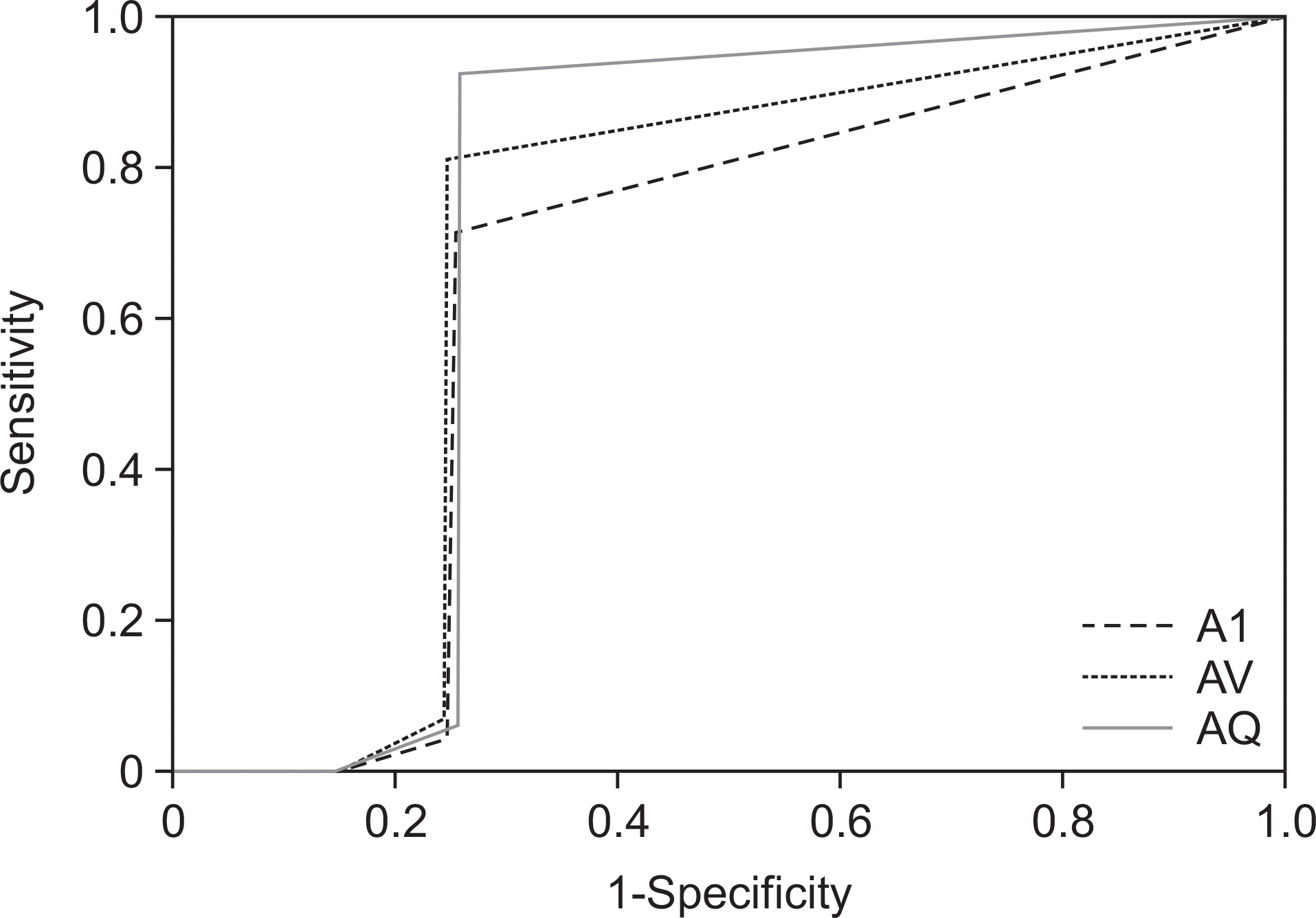
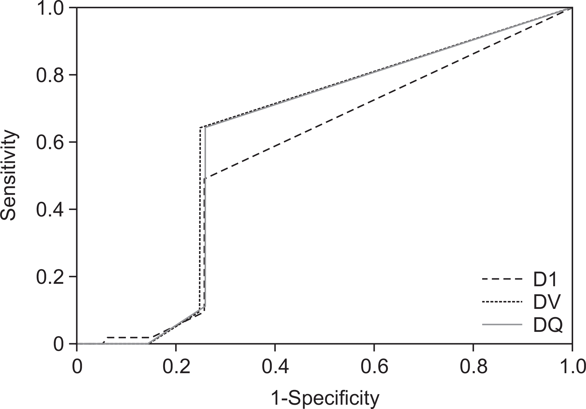
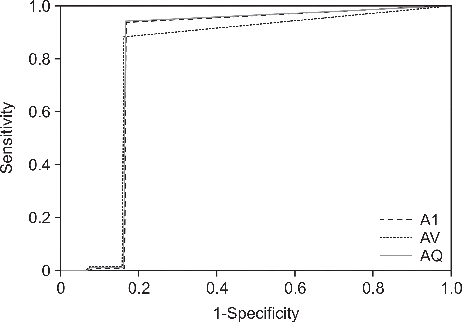
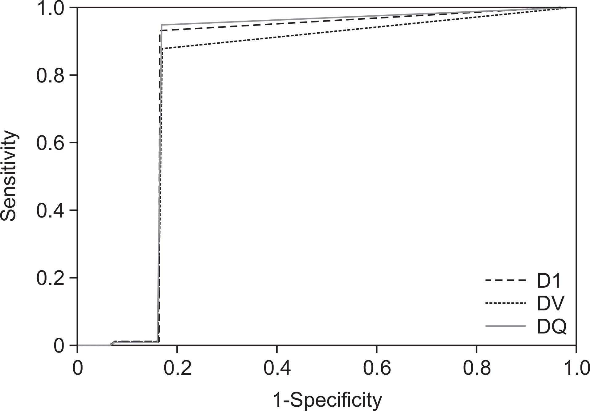
Table 1.
Observed agreement of examiners compared with gold standard (kappa value)
| Examiner | Optical examination | Visible ray image examination | 405 nm blue ray image examination |
|---|---|---|---|
| Examiner A (Oral epidemiologic expert) | 0.969 | 0.941 | 0.963 |
| Examiner D (Non expert) | 0.946 | 0.928 | 0.947 |
Table 2.
ROC curve analysis of caries detection in examiner A
Table 3.
ROC curve analysis of caries detection in examiner D
Table 4.
ROC curve analysis of filling body detection in examiner A
 XML Download