Abstract
Objectives
The purpose of this study was to understand the dental caries pattern in permanent dentition among Korean adolescents aged 12-16 years.
Methods
This study comprised 5,301 teenagers, aged 12-16 years. We analyzed the dental caries pattern in patients with permanent dentition using data from the 2006 Korean National Oral Health Survey. The methods used for data analysis included frequency analysis, correlation analysis, cluster analysis, and multidimensional scaling (MDS).
Results
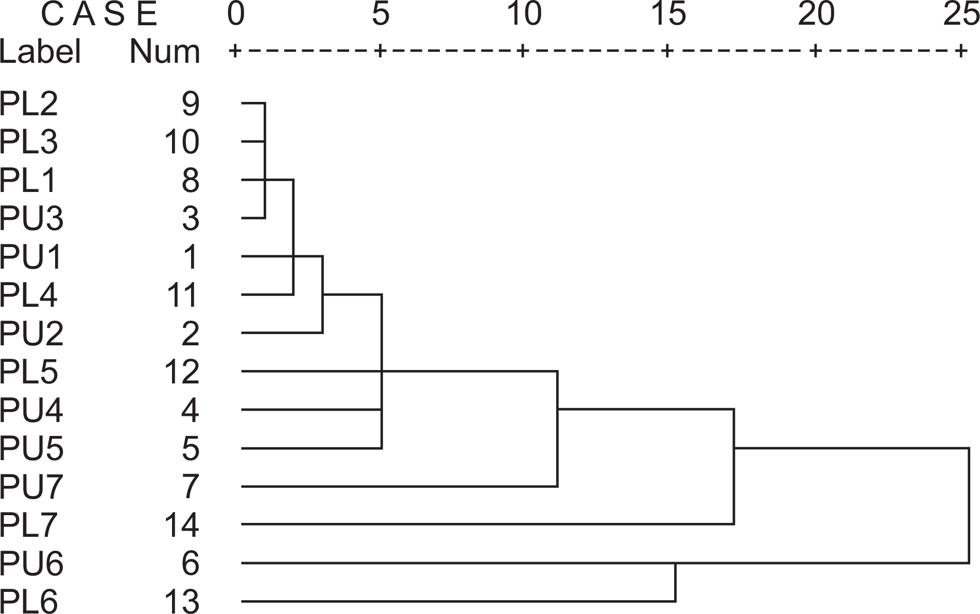
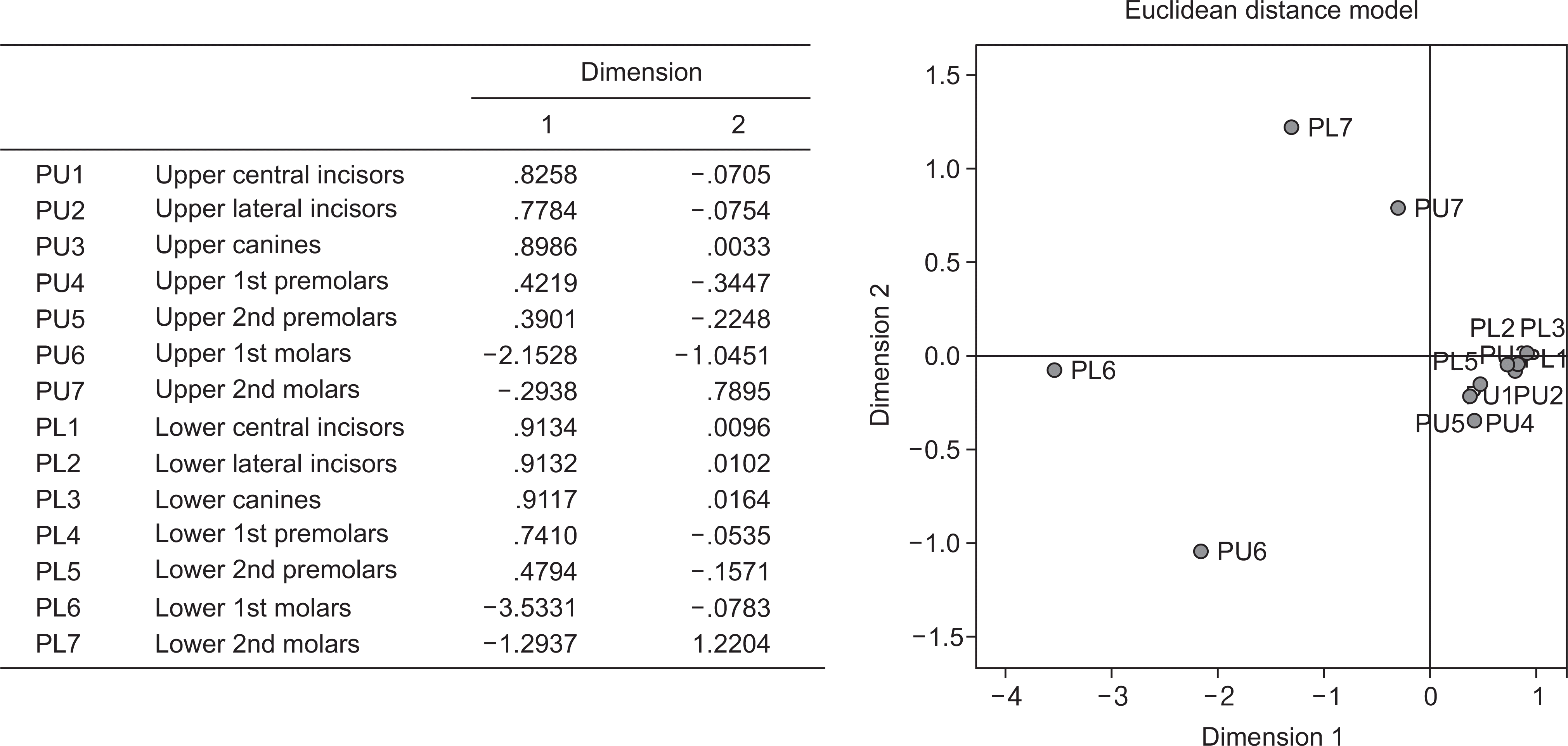
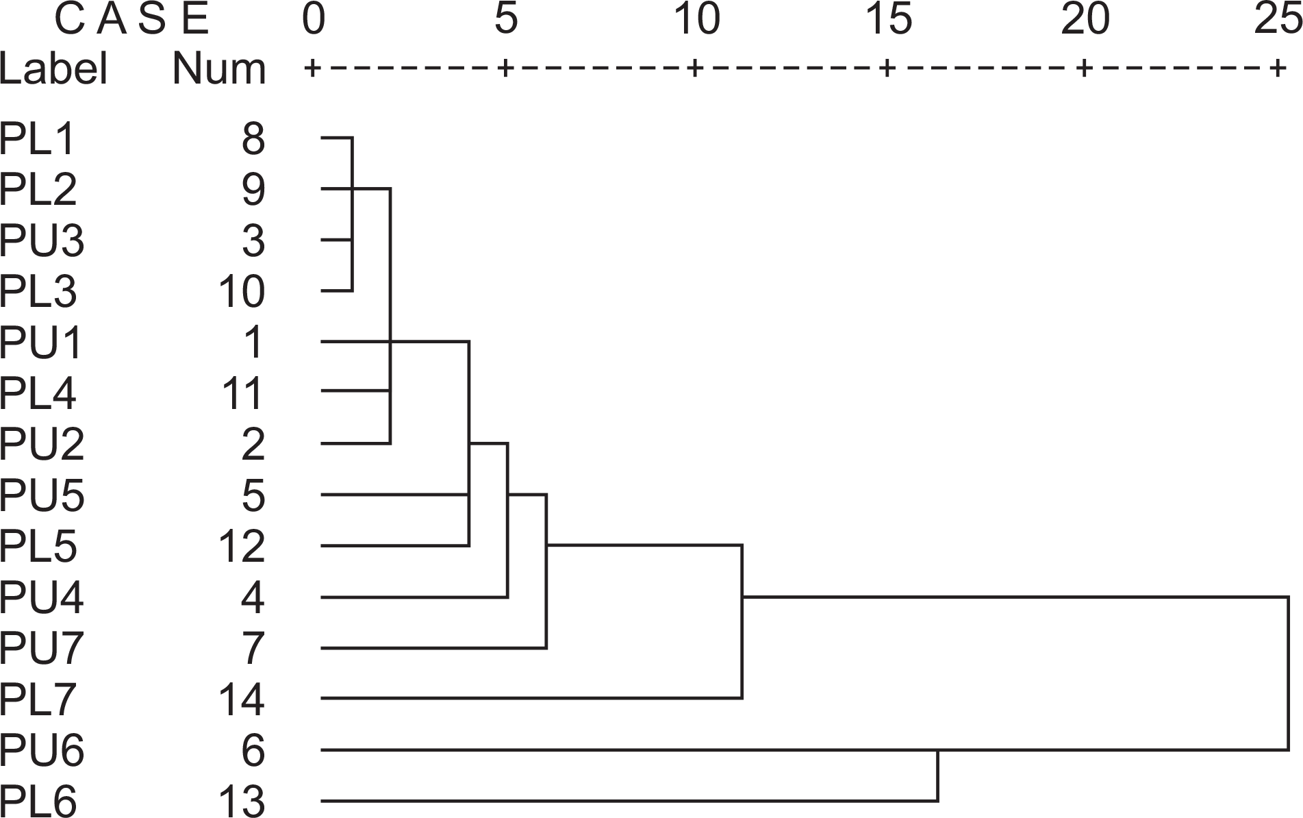
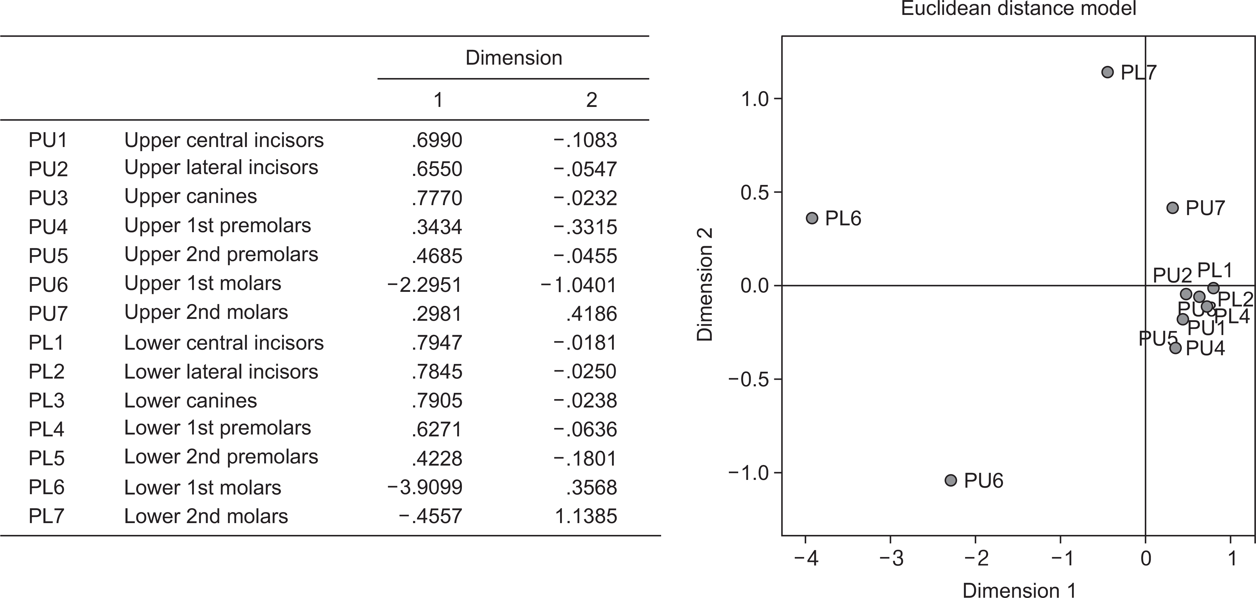
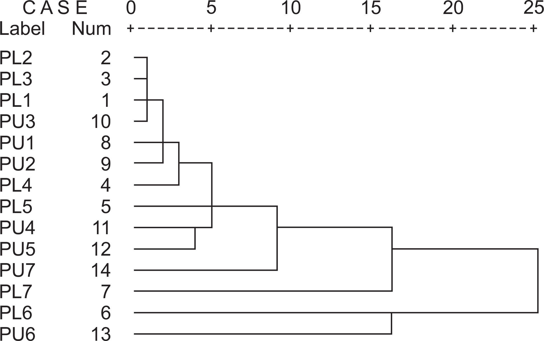
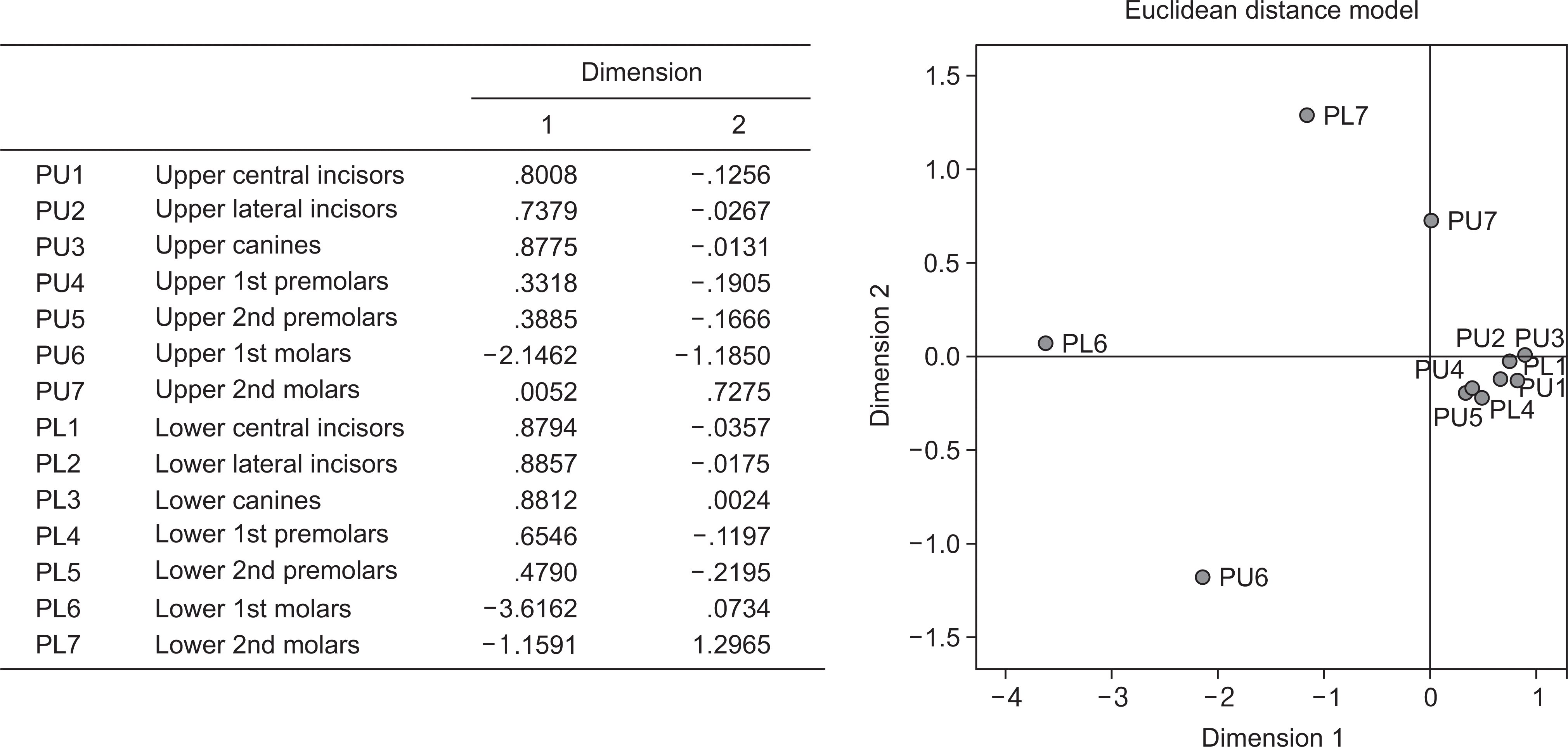
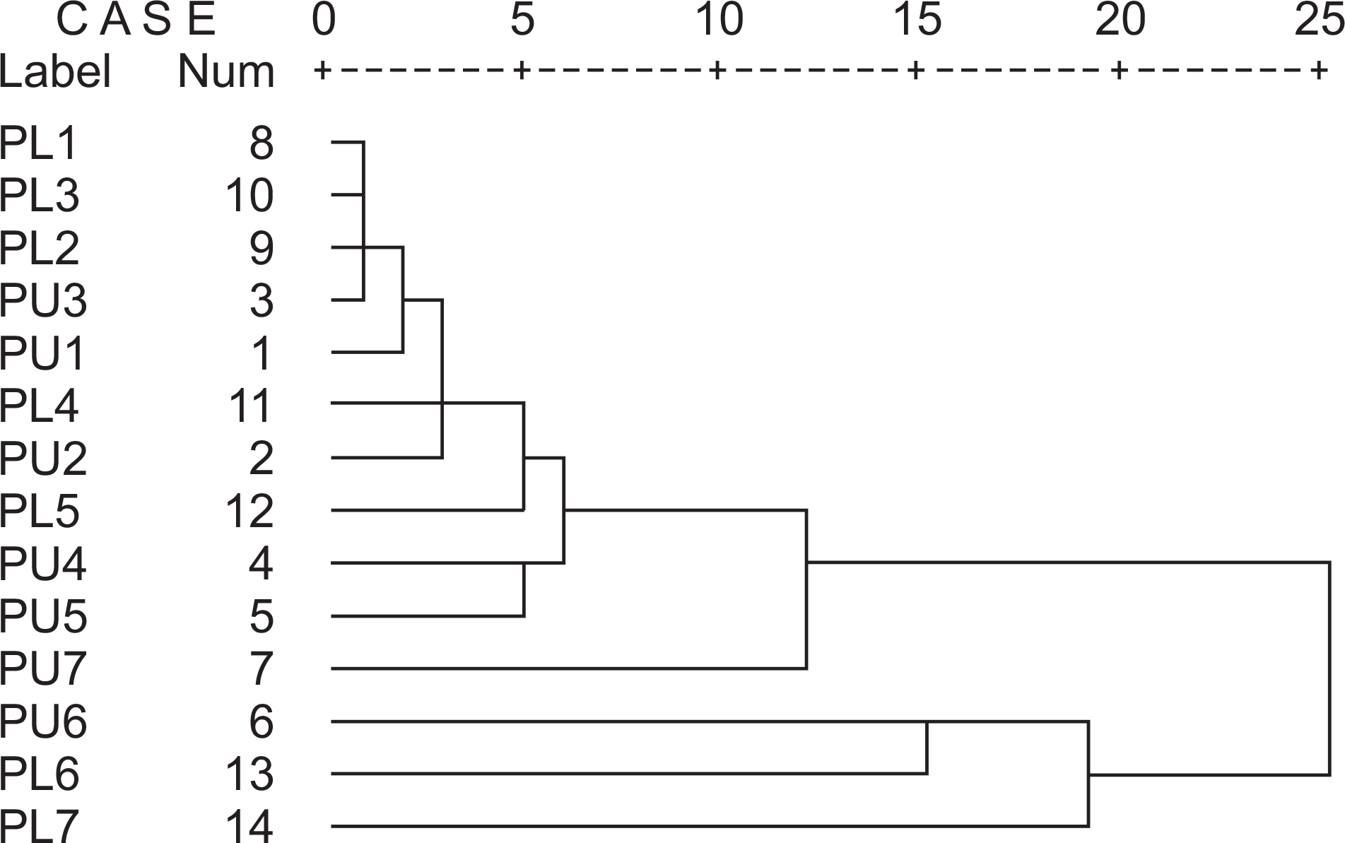
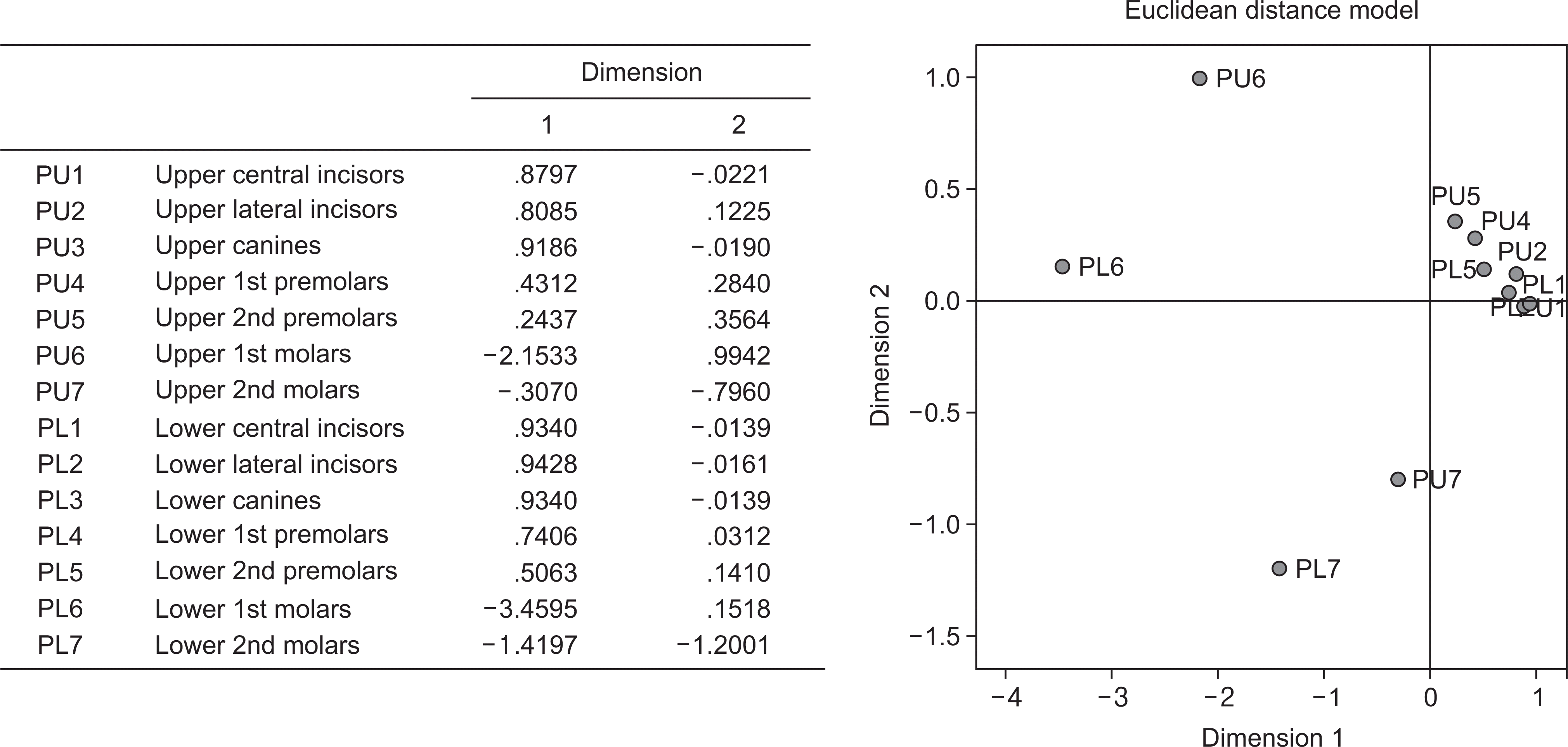
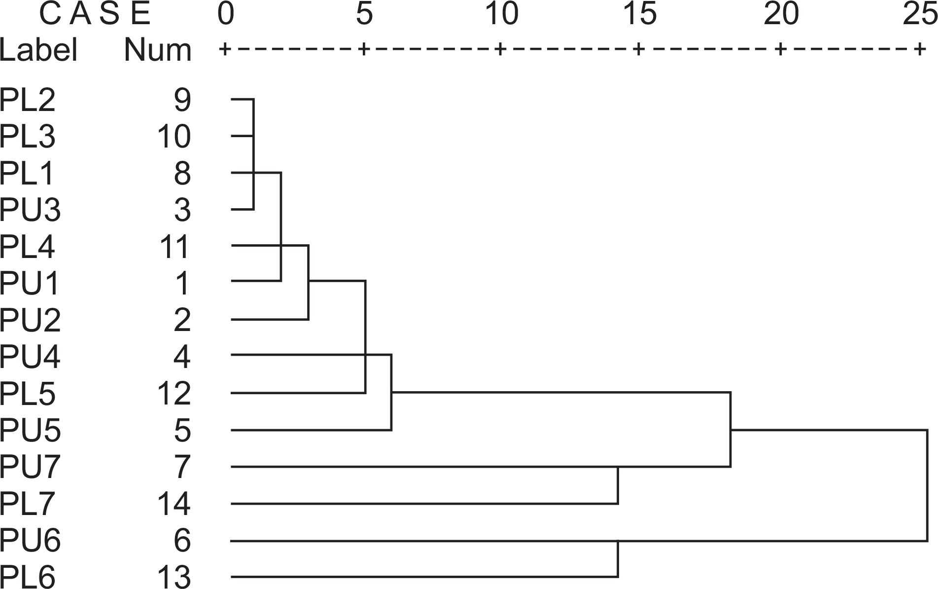
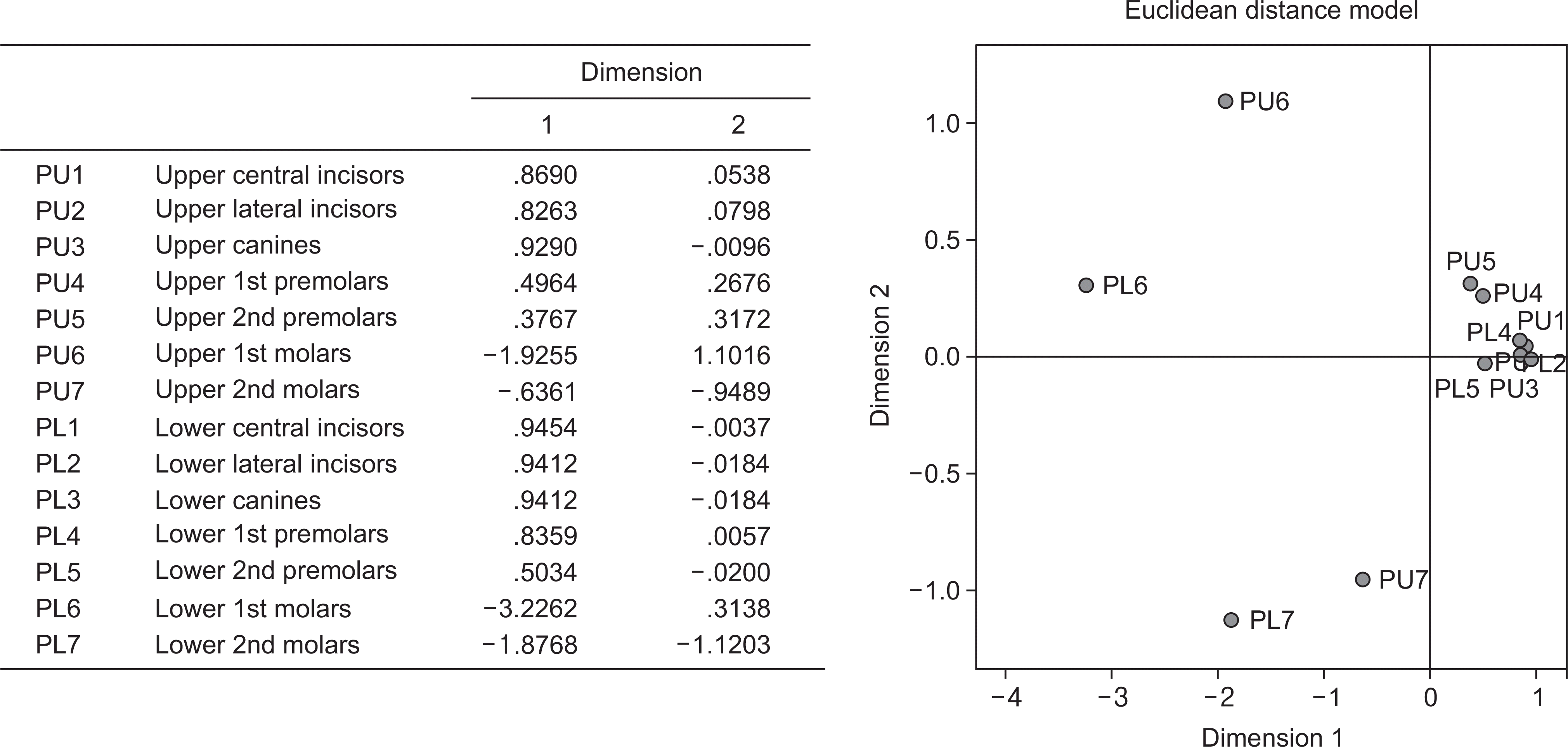
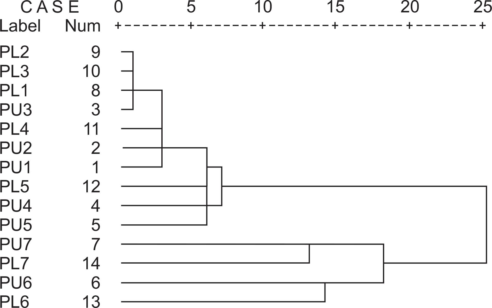
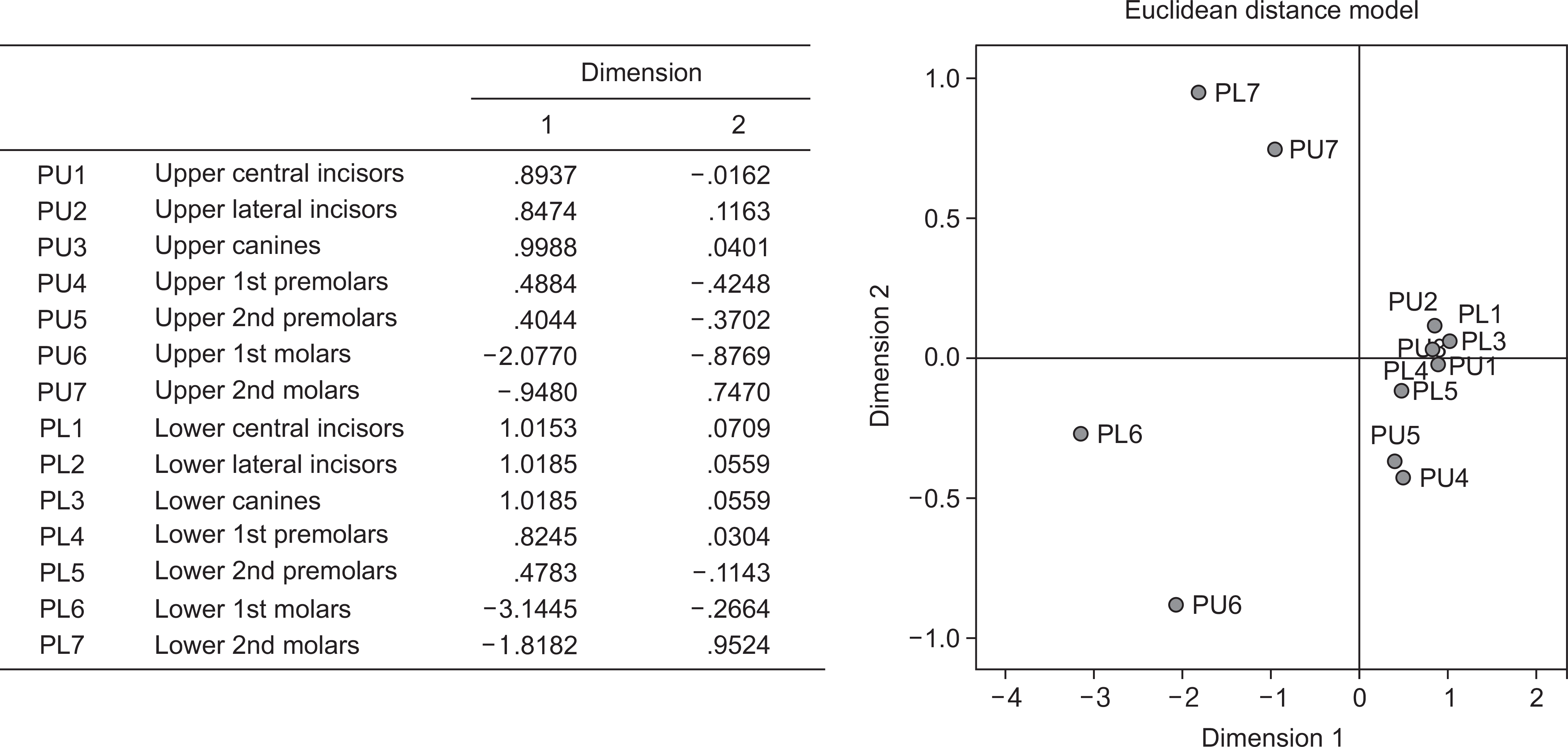
With cluster analysis, it was difficult to clearly distinguish between anterior and posterior caries, and categorization was difficult owing to the mandibular first premolar and maxillary lateral incisor. The molars had severe caries, and results of the cluster analysis categorized this as clusters independent from other teeth; therefore, efforts must be made to prevent dental caries in molars. The maxillary premolars had the highest incidence of caries followed by the molars, and accordingly, these formed independent clusters, with the exception of the molar. During the eruption stage, despite the secondary premolar erupting later than the first premolar, there was a higher caries incidence in the secondary premolar. Out of the anterior teeth, the maxillary later incisor had the highest incidence of caries and formed an independent cluster. The multidimensional scaling (MDS) results clearly showed the molar teeth cluster.
References
1. Psoter WJ, Morse DE, Pendrys DG, Zhang H, Mayne ST. Historical evolution of primary dentition caries pattern definitions. Pediatr Dent. 2004; 26:508–511.
2. Johnsen DC, Schubot D, Bhat M, Jones PK. Caries pattern identification in primary dentition: a comparison of clinician assignment and clinical analysis groupings. Pediatr Dent. 1993; 15:113–115.
3. Psoter WJ, Zhang H, Pendrys DG, Morse DE, Mayne ST. Classification of dental caries patterns in the primary dentition: a multidimensional scaling analysis. Community Dent Oral Epidemiol. 2003; 31:231–238.

4. Lee JS, Lee KH, Kim DE. Caries patterns in primary dentition by caries experience of individual teeth. J Korean Acad Pediatr Dent. 1999; 26:1–13.
5. Jeong SY, Lee KH, Ra JY, An SY, Kim YH. Dental caries patterns in the primary dentition: a cluster analysis and a multidimensional scaling analysis. J Korean Acad Pediatr Dent. 2010; 37:159–167.
6. Lee BG. Lee HS, Ju HJ, Oh HY. Dental caries experience pattern in permanent dentition among Korean children. J Korean Acad Oral Health. 2014; 38:95–104.
7. Shaffer JR, Feingold E, Wang X, Weeks DE, Weyant RJ, Crout R, et al. Clustering tooth surfaces into biologically informative caries outcomes. J Dent Res. 2013; 92:32–37.

8. Shaffer JR, Feingold E, Wang X, Tcuenco KT, Weeks DE, DeSensi RS, et al. Heritable patterns of tooth decay in the permanent dentition: principal components and factor analyses. BMC Oral Health. 2012; Mar 9. 12:7. DOI: 10.1186/1472-6831-12-7.

9. Vanobbergen J, Lesaffre E, Garcia-Zattera MJ, Jara A, Martens L, Declerck D. Caries patterns in primary dentition in 3-, 5- and 7-year-old children: spatial correlation and preventive consequences. Caries Res. 2007; 41:16–25.

10. Berman DS, Slack GL. Dental caries in English school children: a longitudinal study. Br Dent J. 1972; 133:529–538.

11. Hujoel PP, Lamont RJ, DeRouen TA, Davis S, Leroux BG. Within-subject coronal caries distribution patterns: an evaluation of randomness with respect to the midline. J Dent Res. 1994; 73:1575–1580.

12. Kutesa A, Mwanika A, Wandera M. Pattern of dental caries in Mu-lago Dental School clinic, Uganda. Afr Health Sci. 2005; 5:65–68.
13. Oulis CJ, Berdouses ED, Mamai-Homata E, Polychronopoulou A. Prevalence of sealants in relation to dental caries on the permanent molars of 12 and 15-year-old Greek adolescents. A national pathfinder survey. BMC Public Health. 2011; Feb 14. 11:100. DOI: 10.1186/1471-2458-11-100.

14. Adeniyi AA, Agbaje O, Onigbinde O, Ashiwaju O, Ogunbanjo O, Orebanjo O, et al. Prevalence and pattern of dental caries among a sample of nigerian public primary school children. Oral Health Prev Dent. 2012; 10:267–274.
15. Hashim R, Williams SM, Thomson WM, Awad MA. Caries prevalence and intra-oral pattern among young children in Ajman. Community Dent Health. 2010; 27:109–113.
16. Hopcraft MS, Morgan MV. Pattern of dental caries experience on tooth surfaces in an adult population. Community Dent Oral Epidemiol. 2006; 34:174–183.

17. Ministry of Health and Welfare. Korean national oral health survey 2006: III. Summary. Seoul: Ministry of Health and Welfare;2006. p. 3–10.
18. Agustsdottir H, Gudmundsdottir H, Eggertsson H, Jonsson SH, Gudlaugsson JO, Saemundsson SR, et al. Caries prevalence of permanent teeth: a national survey of children in Iceland using ICDAS. Community Dent Oral Epidemiol. 2010; 38:299–309.

19. Druy TF, Horrowitz AM, Ismail AI, Maertens MP, Rozier RG, Selwitz RH. Diagnosing and reporting early childhood caries for research purposes. A report of a workshop sponsored by the National Institute of Dental and Craniofacial Research, the Health Resources and Services Administration, and the Health Care Financing Administration. J Public Health Dent. 1999; 59:192–197.

20. Greenwell AL, Johnsen D, DiSantis TA, DiSantis TA, Gerstenmaier J, Limbert N. Longitudinal evaluation of caries patterns from the primary to the mixed dentition. Pediatr Dent. 1990; 12:278–282.
21. O’Sullivan DM, Tinanoff N. Social and biological factors contributing to caries of the maxillary anterior teeth. Pediatr Dent. 1993; 15:41–44.
22. Douglass JM, Wei Y, Zhang BX, Tinanoff N. Caries prevalence and patterns in 3-6-year-old Beijing children. Community Dent Oral Epidemiol. 1995; 23:340–343.

23. Douglass JM, Tinanoff N, Tnag JMW, Altman DS. Dental caries patterns and oral health behaviors in Arizona infants and toddlers. Community Dent Oral Epidermiol. 2001; 29:14–22.

24. Lee HS, Im JH. SPSS 14.0 manual. Seoul: bobmunsa;2008. p. 452–471.
25. Noh HJ. Multivariate statistical analysis by Hangeul SPSSWIN. Seoul: Sukjungbooks;1999. p. 559–590. 621-637.
26. Macek MD, Beltran-Anguilar ED, Lockwood SA, Malvitz DM. Updated comparison of the caries susceptibility of various morphological types of permanent teeth. J Public Health Dent. 2003; 63:174–182.

27. Lee KH, Ra JY, An SY, Kim YH. Degree of symmetry of dental caries in primary dentition. J Korean Acad Pediatr Dent. 2010; 37:453–460.
28. Burnside G, Pine CM, Williamson PR. Modelling the bilateral symmetry of caries incidence. Caries Res. 2008; 42:291–296.

29. Hannigan A, O’Mullane DM, Barry D, Schafer F, Roberts AJ. A caries susceptibility classification of tooth surfaces by survival time. Caries Res. 2000; 34:103–108.

30. Lucas JR, Largaespada LL. Explaining sex differences in dental caries prevalence: saliva, hormones, and “life-history” etiologies. Am J Hum Biology. 2006; 18:540–555.
Table 1.
Distribution of sample by age and gender unit: N (%)
Table 2.
Caries experience in the permanent dentition (12-16 year old, n=5,301) unit: N (%)
Table 3.
Caries experience by tooth type unit : N (%)
Table 4.
Correlation of DMFT indexes among the quadrant in permanent dentition†
| Side | Upper right | Upper left | Lower right | Upper | Left |
|---|---|---|---|---|---|
| Upper left | 0.733* | ||||
| Lower right | 0.595* | 0.579* | |||
| Lower left | 0.586* | 0.607* | 0.751* | ||
| Lower | 0.679* | ||||
| Right | 0.827* |


 PDF
PDF ePub
ePub Citation
 Print
Print


 XML Download