Abstract
Dysgonomonas capnocytophagoides is a gram-negative, facultatively anaerobic coccobacillus that was formerly designated CDC group dysgonic fermenter (DF)-3, occurring as a normal flora in human gut and rarely causing human infections such as bacteremia, abscess, diarrhea, and cholecystitis. In this study, we report a case of biliary sepsis caused by D. capnocytophagoides in a patient with biliary obstruction. A seventy four-year-old man, admitted to the hospital due to common bile-duct stone, also had cholangitis caused by D. capnocytophagoides and Enterococcus avium, which were isolated from his blood cultures. D. capnocytophagoides was initially identified as D. gadei by MALDI-TOF mass spectrometry, but later confirmed as D. capnocytophagoides by 16S rRNA gene sequencing. To the best of our knowledge, this is the first report of human infec-tion by D. capnocytophagoides in Korea.
REFERENCES
1.Hofstad T., Olsen I., Eribe ER., Falsen E., Collins MD., Lawson PA. Dysgonomonas gen. nov. to accommodate Dysgonomonas gadei sp. nov., an organism isolated from a human gall bladder, and Dysgonomonas captocytophagoides (formerly CDC group DF-3). Int J Syst Evol Microbiol. 2000. 50:2189–95.
2.Wallace PL., Hollis DG., Weaver RE., Moss CW. Characterization of CDC group DF-3 by cellular fatty acid analysis. J Clin Microbiol. 1989. 27:735–7.

3.Kita A., Miura T., Okamura Y., Aki T., Matsumura Y., Tajima T, et al. Dysgonomonas alginatilytica sp. nov., an alginate-degrading bacterium isolated from a microbial consortium. Int J Syst Evol Microbiol. 2015. 65:3570–5.

4.Hansen PS., Jensen TG., Gahrn-Hansen B. Dysgonomonas capnocytophagoides bacteraemia in a neutropenic patient treated for acute myeloid leukaemia. APMIS. 2005. 113:229–31.
5.Wagner DK., Wright JJ., Ansher AF., Gill VJ. Dysgonic fermenter 3-associated gastrointestinal disease in a patient with common variable hypogammaglobulinemia. Am J Med. 1988. 84:315–8.

6.Relman DA. Universal bacterial 16S rRNA amplifcation and sequencing. Persing DH, Smith TF, editors. eds.Diagnostic molecular microbiology principles and applications. 1st ed.Washington DC: ASM Press;1993. p. 489–95.
7.Clinical and Laboratory Standards Institute. Interpretive criteria for iden-tifcation of bacteria and fungi by DNA target sequencing; approved guideline. CLSI document MM18-A. Wayne, PA: Clinical and Laboratory Standards Institute. 2008.
8.Gill VJ., Travis LB., Williams DY. Clinical and microbiological observations on CDC group DF-3, a gram negative coccobacillus. J Clin Microbiol. 1991. 29:1589–92.
9.Blum RN., Berry CD., Phillips MG., Hamilos DL., Koneman EW. Clinical illnesses associated with isolation of dysgonic fermenter 3 from stool samples. J Clin Microbiol. 1992. 30:396–400.

10.Heiner AM., DiSario JA., Carroll K., Cohen S., Evans TG., Shigeoka AO. Dysgonic fermenter-3: a bacterium associated with diarrhea in immunocompromised hosts. Am J Gastroenterol. 1992. 87:1629–30.
11.Hironaga M., Yamane K., Inaba M., Haga Y., Arakawa Y. Characterization and antimicrobial susceptibility of Dysgonomonas capnocytophagoides isolated from human blood sample. Jpn J Infect Dis. 2008. 61:212–3.
12.Matsumoto T., Kawakami Y., Oana K., Honda T., Yamauchi K., Okimura Y, et al. First isolation of Dysgonomonas mossii from intestinal juice of a patient with pancreatic cancer. Arch Med Res. 2006. 37:914–6.

13.Kodama Y., Shimoyama T., Watanabe K. Dysgonomonas oryzarvi sp. nov., isolated from a microbial fuel cell. Int J Syst Evol Microbiol. 2012. 62:3055–9.

14.Zbinden R. Aggregatibacter, Capnocytophaga, Eikenella, Kingella, Pasteurella, and other fastidious or rarely encountered gram-negative rods. In: Jorgensen JH, Pfaller MA, Carroll KC, Landry ML, Guido Funke, Richter SS, et al. ed. Manual of Clinical Mcirobiology, 11th ed. Washington, DC. Amercian Society for Microbiology 2015.652–66.
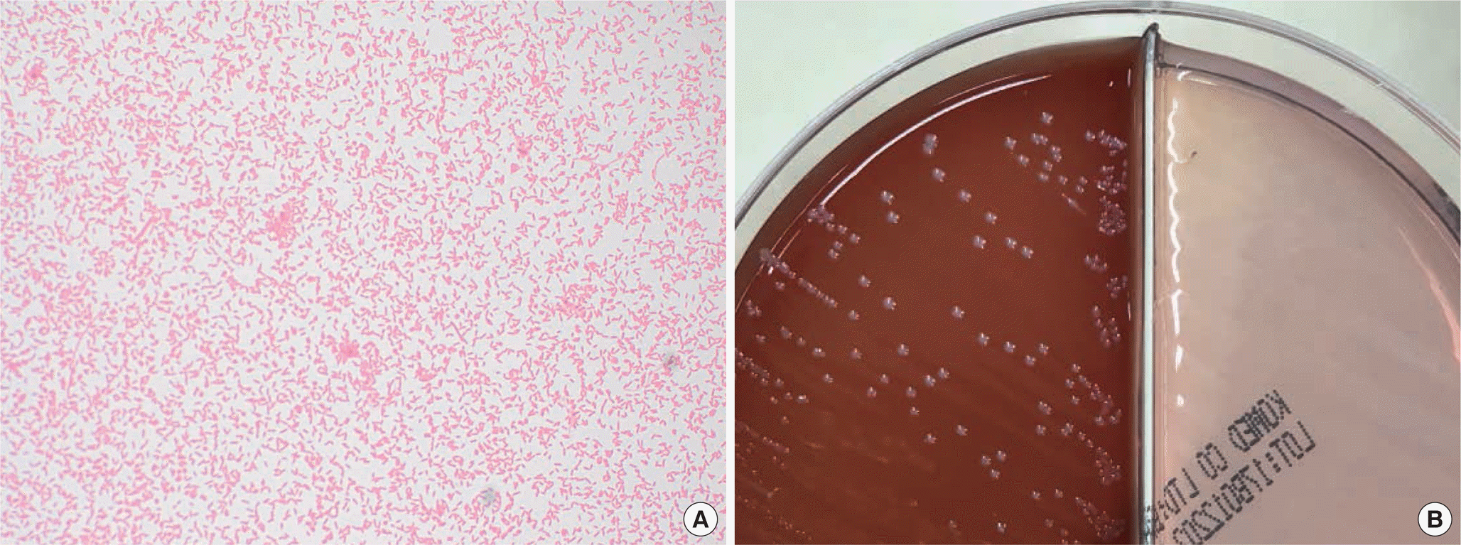
Fig. 1.
Microscopic findings and colony morphology of Dysgonomonas capnocytophagoides. (A) Gram-negative coccobacilli visualized under the microscope after 30.1 hr in the anaerobic culture vial (Gram stain, 1,000 ×). (B) Grey-white, smooth, and non-hemolytic colonies grew on blood agar, but none on MacConkey agar.

Table 1.
Biochemical test responses of the current isolate, compared to those of D. capnocytophagoides and Serratia plymuthica
|
Response of |
|||
|---|---|---|---|
| Biochemical test | This case | D. capnocytophagoides∗ | Serratia plymuthica†,‡ |
| Oxidase | Negative | Negative | Negative |
| β-galactosidase | Positive | Positive | Positive |
| Arginine dihydrolase | Negative | Negative | Negative |
| Lysine decarboxylase | Negative | Negative | Negative |
| Ornithine decarboxylase | Negative | Negative | Negative |
| H2S production | Negative | Negative | Negative |
| Urea hydrolysis | Negative | Negative | Negative |
| Tryptophan deamination | Negative | Negative | Negative |
| Indole production | Negative | Negative | Negative |
| Acetoin production | Positive | Negative | Positive |
| Gelatin hydrolysis | Positive | Negative | Positive |
| Carbohydrate fermentation | |||
| Glucose | Positive | Positive | Positive |
| Mannitol | Negative | Negative | Positive |
| Inositol | Negative | Negative | Negative |
| Sorbitol | Negative | Negative | Negative |
| Rhamnose | Negative | Negative | Negative |
| Sucrose | Positive | Positive | Positive |
| Melibiose | Positive | Positive | Positive |
| Amygdalin | Positive | Positive | Positive |
| Arabinose | Positive | Positive | Positive |


 PDF
PDF ePub
ePub Citation
 Print
Print


 XML Download