

 PDF
PDF ePub
ePub Citation
 Print
Print


Abstract
In patients with acute myeloid leukemia (AML), pleural effusion may be attributed to various factors, including infection, hypoalbuminemia, and renal failure. However, leukemic infiltration of the pleural fluid is rarely reported and poorly understood. Extramedullary diseases have been reported with increasing frequency as the survival rates of patients with AML have increased. However, the reported prognostic effects of leukemic pleural effusion in patients with AML range from none to a worse prognosis. Here, we report a case of acute promyelocytic leukemia (APL) in a patient exhibiting leukemic pleural effusion with fluorescence in situ hybridization (FISH) results indicating the presence of the PML-RARA fusion gene. A 52-year-old man presented with pancytopenia, dyspnea, and fever. He had a medical history of hypertension, end-stage renal disease, and hepatitis B virus-related liver cirrhosis. A peripheral blood smear revealed the presence of multiple abnormally hypergranular promyelocytes. White blood cell differential counts were not performed due to severe pancytopenia. A bone marrow examination, immunophenotyping analysis, and cytogenetic and molecular studies revealed APL. The patient was treated with all-trans retinoic acid immediately after abnormal promyelocytes were observed in the peripheral blood smear, but induction chemotherapy was delayed because of his poor condition. His persistent dyspnea and abdominal discomfort led to a thoracentesis and the observation of abnormal promyelocytes that were positive for PML-RARA fusion gene by FISH. To our knowledge, this is the first report of leukemic pleural infiltration with PML-RARA fusion gene-positivity via FISH.
Go to : 
REFERENCES
1.Muss HB., Moloney WC. Chloroma and other myeloblastic tumors. Blood. 1973. 42:721–8.

2.Chang H., Brandwein J., Yi QL., Chun K., Patterson B., Brien B. Extramedullary infltrates of AML are associated with CD56 expression, 11q23 abnormalities and inferior clinical outcome. Leuk Res. 2004. 28:1007–11.
3.Tallman MS., Kim HT., Paietta E., Bennett JM., Dewald G., Cassileth PA, et al. Acute monocytic leukemia (French-American-British classifcation M5) does not have a worse prognosis than other subtypes of acute myeloid leukemia: a report from the Eastern Cooperative Oncology Group. J Clin Oncol. 2004. 22:1276–86.
4.Disel U., Yavuz S., Paydas S., Sahin B., Zeren H. Extramedullary relapse in the pleura in acute promyelocytic leukemia. Leuk Lymphoma. 2003. 44:189–91.
5.Ganzel C., Manola J., Douer D., Rowe JM., Fernandez HF., Paietta EM, et al. Extramedullary disease in adult acute myeloid leukemia is common but lacks independent signifcance: analysis of patients in ECOG-ACRIN Cancer Research Group trials, 1980-2008. J Clin Oncol. 2016. 34:3544–53.
6.Kobayashi R., Tawa A., Hanada R., Horibe K., Tsuchida M., Tsukimoto I. Extramedullary infltration at diagnosis and prognosis in children with acute myelogenous leukemia. Pediatric Blood Cancer. 2007. 48:393–8.
7.Nasilowska-Adamska B., Majewski M., Seferynska I., Szczepinski A., To-maszewska A., Prochorec-Sobieszek M, et al. Predictive value of RT-PCR PML-RARA transcript monitoring for extramedullary relapse of acute promyelocytic leukemia in the pleura, heart and pericardium after allogeneic SCT. Ann Transplant. 2007. 12:33–8.
8.Ou MC., Hwang WL., Teng CL. Leukaemic pleural effusion in acute myeloid leukaemia. Br J Haematol. 2011. 154:669.
9.Hoffman LM., Gore L., Maloney KW. Pulmonary presentation of relapsed acute myeloid leukemia. J Pediatr Hematol Oncol. 2014. 36:228–30.
10.Lee DA., Harris CP., Gresik VM., Rao P., Lau CC. Granulocytic sarcoma presenting as pneumonia in a child with t(8;21) acute myelogenous leukemia: diagnosis by fuorescent in situ hybridization. J Pediatr He-matol Oncol. 2004. 26:431–4.
11.Ganzel C., Douer D. Extramedullary disease in APL: a real phenomenon to contend with or not? Best Pract Res Clin Haematol. 2014. 27:63–8.
12.Vega-Ruiz A., Faderl S., Estrov Z., Pierce S., Cortes J., Kantarjian H, et al. Incidence of extramedullary disease in patients with acute promyelocytic leukemia: a single-institution experience. Int J Hematol. 2009. 89:489–96.
13.Swerdlow SH., Campo E, et al. eds. WHO classifcation of tumours of haematopoietic and lymphoid tissues. 4th ed.Lyon, France: IARC;2008. p. 113–4.
14.Azoulay É. Fieux F., Moreau D., Thiery G., Rousselot P., Parrot A, et al. Acute monocytic leukemia presenting as acute respiratory failure. Am J Respir Crit Care Med. 2003. 167:1329–33.
15.Potenza L., Luppi M., Morselli M., Tonelli S., D'apollo N., Facchini L, et al. Leukaemic pulmonary infltrates in adult acute myeloid leukaemia: a high–resolution computerized tomography study. Br J Haematol. 2003. 120:1058–61.
16.De Botton S., Sanz MA., Chevret S., Dombret H., Martin G., Thomas X, et al. Extramedullary relapse in acute promyelocytic leukemia treated with all-trans retinoic acid and chemotherapy. Leukemia. 2006. 20:35–41.
17.Porcu P., Cripe LD., Ng EW., Bhatia S., Danielson CM., Orazi A, et al. Hy-perleukocytic leukemias and leukostasis: a review of pathophysiology, clinical presentation and management. Leuk Lymphoma. 2000. 39:1–18.
18.Ko BS., Tang JL., Chen YC., Yao M., Wang CH., Shen MC, et al. Extramedullary relapse after all-trans retinoic acid treatment in acute promyelocytic leukemia-the occurrence of retinoic acid syndrome is a risk factor. Leukemia. 1999. 13:1406–8.
Go to :
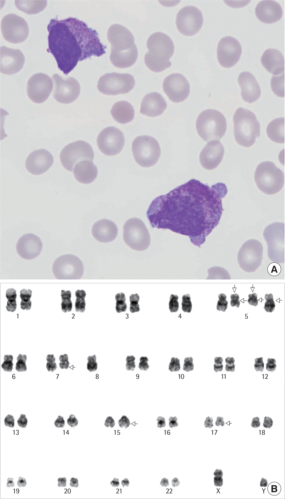 | Fig. 1.Abnormal promyelocytes with multiple Auer rods in the bone marrow (Wright's stain ×1,000) (A) and conventional bone marrow chromosome analysis result showing a 47,XY,+add(5)(q11.2)x2,der(5;8) (q10;p10),del(7)(q32), t(15;17)(q22;q21) [21] karyotype (B). |
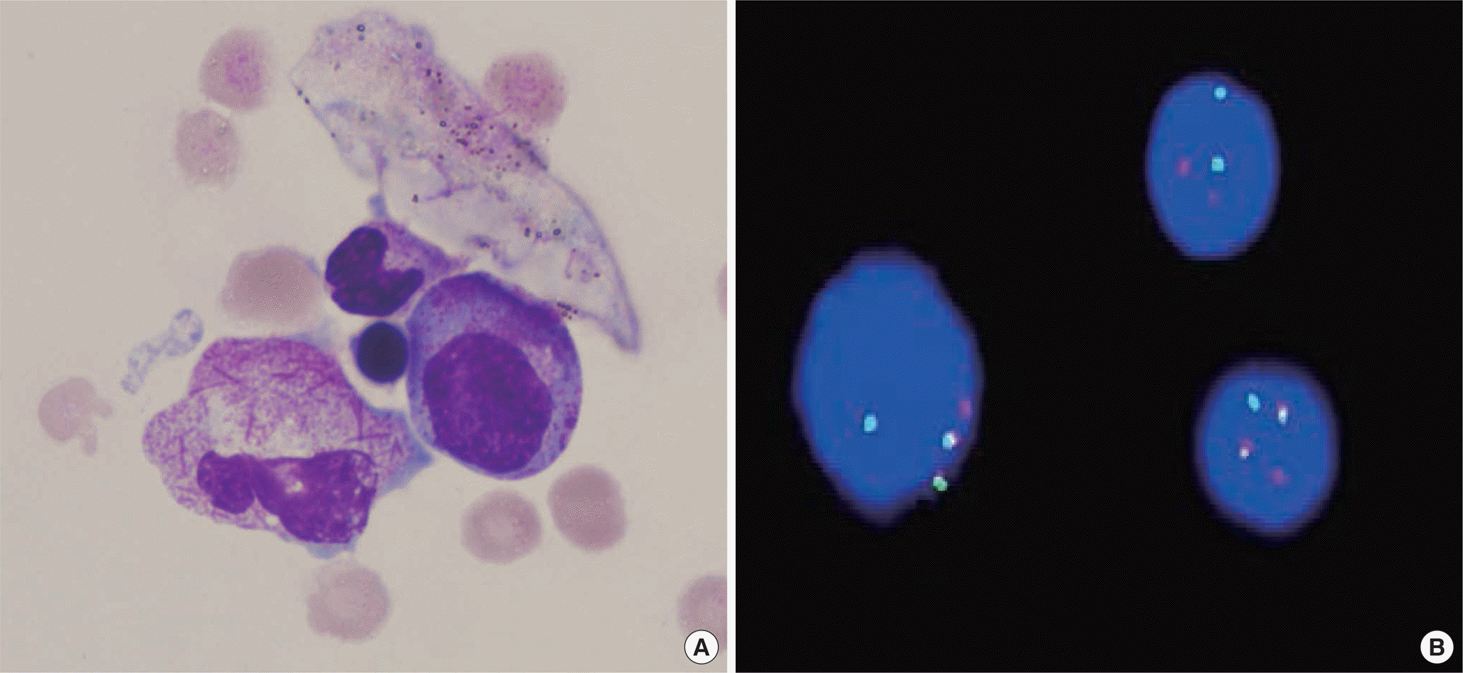 | Fig. 2.Abnormal promyelocytes with multiple Auer rods in pleural fluid (Wright's stain ×1,000) (A) and dual-color, dual-fusion fluorescence in situ hybridization analysis on interphase cells showing two green signals, indicating the t(15;17) translocation (B). |
Table 1.
Reported cases of pleural infiltration in AML
| Case No. | Age/Sex | WBC (×109/L) | Hb (g/dL) | Platelet (×109/L) | FAB | BM∗ | FCM | FISH or RT-PCR | EMD | P | R |
|---|---|---|---|---|---|---|---|---|---|---|---|
| 1 | 34/M | NA | NA | NA | M3 | N | ND | RT-PCR PML-RARA (+)† | Pleura, heart, pericardium | D | [7] |
| 2 | 39/M | 4.52 | 14 | 212 | M3 | N | PF | ND | Pleura | CR | [4] |
| 3 | 53/M | 11.5 | 8 | 103 | M2 | Y | ND | FISH RUNX1-RUNX1T1 (+) | Pleura | CR | [8] |
| 4 | 19/M | 6.9 | 9.5 | 94 | M5 | Y | BAL | ND | BAL, CSF | CR | [9] |
| 5 | 4/M | NA | NA | NA | M2 | Y | ND | FISH Lung Bx: RUNX1-RUNX1T1(+) | Lung | CR | [10] |
† PML-RARA (+) in peripheral blood Abbreviations: N, No; Y, Yes; ND, not done; NA, not available; CR, complete remission; FCM, flow cytometry; P, prognosis; R, reference; D, dead; PF, pleural fluid; BAL, bronchoalveolar lavage; EMD, extramedullary disease; FAB, French-American-British classification; Bx, biopsy.
 XML Download