

 PDF
PDF ePub
ePub Citation
 Print
Print


Neutrophilia can occur due to chronic neutrophilic leukemia (CNL) or neutrophilic leukemoid reaction (NLR) associated with infection, neoplasm, inflammation, drug use, and acute hemorrhaging, among other causes [12]. CNL is a subtype of myeloproliferative neoplasm associated with a mutation in the colony stimulating factor 3 receptor (CSF3R) gene [3]. The protein encoded by this gene is the receptor for colony stimulating factor 3 that produces a signal through downstream SRC family kinases and Janus kinase (JAK) pathways and plays an important role in the growth and differentiation of granulocytes [4]. There are two different classes of CSF3R mutation: One is a truncation mutation, which results in dysregulation of downstream SRC family kinases and leads to constitutive overexpression of the receptor and ligand hypersensitivity. The other is a membrane proximal mutation which results in dysregulation of the JAK family and constitutive activation of the receptor in the absence of granulocyte-colony stimulating factor (G-CSF) ligand [4]. An example of the membrane proximal mutation is on exon 14 of CSF3R resulting in T618I mutation, which has been identified in more than 80% of CNL cases [3].
CSF3R T618I could be used as a molecular marker to make a differential diagnosis between CNL and NLR. It has been reported that NLR can develop in 10% of patients with solid tumors (lung, genitourinary, gastrointestinal, bone metastases, breast cancer, etc.) [12], and may be due to the paraneoplastic production of G-CSF or other growth factors. Furthermore, NLR can be present years before the diagnosis of carcinoma [5]. A few studies have reported that CNL or NLR can occur in plasma cell neoplasms [67]. Here, we describe one CNL case and three NLR cases initially suspected of being CNL that were associated with solid cancers, plasma cell neoplasms or sepsis.
We retrospectively reviewed the database of bone marrow (BM) study patients from January 2014 to October 2016 and identified four patients with leukocytosis of more than 25×109/L and neutrophilia without left shift (more than 80% of segmented neutrophils). We performed a CSF3R mutation assay to help differentiate between CNL and NLR. DNA was extracted from BM aspirates, and a polymerase chain reaction (PCR) and Sanger sequencing were performed using primers for the T618I mutation on CSF3R exon 14 according to a previous study [3]: forward 5′-CCACGGAGGCAGCTTTAC-3′ and reverse 5′-AAATCAGCATCCTTTGGGTG-3′, which revealed an ACC to ATC mutation corresponding to the T618I mutation of CSF3R.
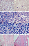
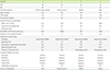
The clinical and laboratory findings of the four patients are shown in Table 1 and Fig. 1. The first patient (case 1) tested positive for CNL with CSF3R T618I. Three patients (cases 2 to 4) showed negative results for CSF3R T618I and the cause of NLR was associated with ovarian cancer, monoclonal gammopathy of undetermined significance (MGUS), and multiple myeloma (MM) with sepsis, respectively.
One patient (case 1) presented hypovolemic shock and had a history of chronic kidney disease, hypertension, and persistent leukocytosis, which had lasted for more than four months (22-61 ×109/L). Even after antibiotic treatment, his white blood cell (WBC) count had progressively increased up to 61×109/L. Three months after first admission, a BM study showed hypercellular marrow and granulocytic predominance with normal maturation (Fig. 1) along with the presence of CSF3R T618I. Taken together, he was diagnosed with CNL and was administered hydroxyurea. After 2.5 months of treatment, his WBC count normalized and remained in the normal range for a year, at the time of this study.
In case 2, diagnosed as having ovarian cancer, the patient showed uncontrolled leukocytosis and negative culture results except for Clostridium difficile. She underwent a total hysterectomy with both salpingo-oophorectomy and taxol-carboplatin chemotherapy. Although her WBC count decreased from 67×109/L to 12× 109/L immediately after surgery, it proceeded to increase up to 163.8×10⁹/L again in one month, and an abdominal computerized tomography (CT) scan revealed newly developed multiple hepatic metastases and metastatic lymph nodes at the small bowel mesentery. Despite continuous antibiotic treatment, extreme leukocytosis (more than 100×109/L) was persistent and she expired four months after the diagnosis of ovarian cancer. Her CSF3R T618I result was found to be negative.
With solid tumors, paraneoplastic autocrine production of G-CSF can induce NLR and increase proliferation of the carcinoma. Therefore, patients with solid tumors accompanied by paraneoplastic NLR are known to incur rapid tumor growth and have poor clinical prognosis [891011]. Our patient (case 2) with NLR also showed very poor prognosis. Previous studies have demonstrated that G-CSF levels decrease after treatment of the primary tumor and WBC counts go back to nearly normal levels [12131415]. As tumors recur, NLR redevelops with an increase in G-CSF production caused by solid tumors [121314].
The other patient (case 3, MGUS with IgG-lambda type) showed neutrophilia at the time of diagnosis (Fig. 1) and throughout the ten-month follow-up period (WBC count 24-40×109/L with 83-90% segmented neutrophils) without MGUS treatment. She did not show for CSF3R T618I mutation. However, CNL can be diagnosed even in cases without any clonal markers when other diagnostic criteria are met, such as persistent neutrophilia (at least three months) and splenomegaly when no identifiable cause of NLR is present. In case 3, clinical features such as splenomegaly and persistent neutrophilia showed the likelihood of diagnosing CNL, although CSF3R T618I was negative and a cytogenetic study showed a normal karyotype. Besides CSF3R T618I mutation, CSF3R M696T or the CSF3R mutation (2341_2342insC; cytoplasmic domain) on exon 17 could be associated with CNL [316]. In addition, in CNL patients without the CSF3R T618I mutation, mutations in other genes such as ASXL1, TET2, SRSF2, JAK2, and SETBP1 have also been reported [37]. It would be helpful to examine the mutations mentioned above as well as the CSF3R T618I mutation to determine clonality, but we could not perform them.
Some studies have reported that CNL can co-occur with plasma cell neoplasm [3617] although most cases did not confirm the clonality of neutrophils [67]. It remains unclear whether neutrophilic leukocytosis is a leukemoid response caused by cytokine release such as interleukin-6 by neoplastic plasma cells or independent clonal disorders. One study has shown that about a quarter of patients with neutrophilia have coexisting MM or MGUS [7].
In case 4, the patient was transferred to our emergency room because of septic shock accompanied by extreme leukocytosis (WBC count 150×109/L with 90% segmented neutrophils). He was initially thought to have CNL rather than NLR because of his extreme leukocytosis. A BM study showed hypercellularity with severe myeloid hyperplasia (myeloid: erythroid ratio, 25.4:1). The proportion of plasma cells was only 2.4% of the total BM-nucleated cells. Plasma cell neoplasm was not suspected at first, but unexpectedly, clonal plasma cells were found by an immunophenotyping analysis. Therefore, we performed an MM workup (serum protein electrophoresis and immunofixation, MRI whole body survey, etc.). Finally, the patient was diagnosed as having MM (IgA-lambda type). He received antibiotics. Two weeks later, his WBC count was normalized to 7.4×10⁹/L before MM treatment. Finally, the neutrophilia in this patient was thought to be caused by NLR associated with sepsis, rather than paraneoplastic syndrome, because the neutrophilia disappeared after antibiotic treatment.
In conclusion, making a diagnosis of CNL should be approached with caution in patients with coexisting solid cancer or plasma cell neoplasm, unless there is demonstrated clonality of myeloid cells, such as CSF3R T618I, other mutations, or chromosomal abnormalities. Furthermore, when infection is unlikely to be the only contributor in patients having NLR, the possibility of paraneoplastic syndromes caused by solid tumors or plasma cell neoplasms should be considered.
 XML Download