

 PDF
PDF ePub
ePub Citation
 Print
Print


Abstract
Paenibacillus urinalis was first isolated from the urine of a woman in 2008, and was reported to be a contaminant. Here, we report 5 cases of P. urinalis isolated over 5 months at a tertiary hospital. Using an API kit, 4 cases were classified as Cellulomonas species. Owing to the low reliability of API kit results and Gram stain results indicating gram variable bacilli for few specimens, MALDI-TOF MS and 16S rRNA gene sequencing were performed for identification. The last case showed Gram variable bacilli, and therefore, based on previous experience, 16S rRNA gene base sequence analysis was carried out without an additional API kit. All isolated strains were confirmed to be P. urinalis, and were judged to be contaminants. As for Gram variable bacteria, the use of current biochemical identification systems may lead to misidentification as other bacteria, which may cause unnecessary or improper use of antibiotics. Moreover, whereas most of the Paenibacillus species are reported to be contaminants, some of them are being reported as sources of infection. Therefore, more accurate identification will be necessary in the future. Accordingly, it is expected that accurate identification of this genus will help clinical physicians make decisions regarding appropriate treatment and use of antibiotics.
Figures and Tables
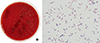 | Fig. 1(A) Blood agar plate after a 24-hour incubation at 37℃, 5% CO2; Colonies are non-hemolytic, circular, greyish, smooth and 1–3 mm in diameter, (B) Gram stain of the clinical isolate shows Gram variable rods.
|
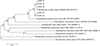 | Fig. 2The neighbor-joining phylogenetic tree based on 16S rRNA sequences showing the phylogenetic position of the isolates from the studied cases and related Paenibacillus species. Bootstrap tests (1000 replicates) are shown adjacent to the branches. The scale bar length of 0.01 indicates 1% sequence distance.
|
Table 1
Characteristics of patients with isolation of P. urinalis in 2016
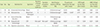
Reference range: Procalcitonin ~0.046 ng/mL; CRP, ~0.30 mg/L; WBC, 4.0–10.0 ×10³/µL.
*Presented as an unacceptable profile; †Culture performed using blood agar, chocolate agar, and thioglycolate broth; ‡As for the 5th case, as Gram variable bacilli were observed on the Gram stain, 16S rRNA gene capillary sequencing was carried out immediately without an API kit test, based on experience.
![]()
References
1. Roux V, Fenner L, Raoult D. Paenibacillus provencensis sp. nov., isolated from human cerebrospinal fluid, and Paenibacillus urinalis sp. nov., isolated from human urine. Int J Syst Evol Microbiol. 2008; 58:682–687.

2. Nasu Y, Nosaka Y, Otsuka Y, Tsuruga T, Nakajima M, Watanabe Y, et al. A case of Paenibacillus polymyxa bacteremia in a patient with cerebral infarction. Kansenshogaku Zasshi. 2003; 77:844–848.
3. Bosshard PP, Zbinden R, Altwegg M. Paenibacillus turicensis sp. nov., a novel bacterium harbouring heterogeneities between 16S rRNA genes. Int J Syst Evol Microbiol. 2002; 52:2241–2249.
4. Khosravi Y, Dieye Y, Poh BH, Ng CG, Loke MF, Goh KL, et al. Culturable bacterial microbiota of the stomach of Helicobacter pylori positive and negative gastric disease patients. Scientific World Journal. 2014; 2014:610421.
5. Yu W, Lee K, Hwang K. Resource Development of Unidentified Human Pathogens using Automated Identification Systems. Public Health Wkly Rep. 2015; 42:990–997.
6. Tamura K, Peterson D, Peterson N, Stecher G, Nei M, Kumar S. MEGA5: molecular evolutionary genetics analysis using maximum likelihood, evolutionary distance, and maximum parsimony methods. Mol Biol Evol. 2011; 28:2731–2739.
7. Priest FG. Paenibacillus. In : Whitman WB, editor. Bergey's manual of systematics of archaea and bacteria 2015. Hoboken: John Wiley & Sons Inc.;2015. p. 1–40.
8. Grady EN, MacDonald J, Liu L, Richman A, Yuan ZC. Current knowledge and perspective of Paenibacillus: a review. Microb Cell Fact. 2016; 15:203.
9. Salas NM, Prevost M, Hofinger D, Fleming H. Cellulomonas, an emerging pathogen: a case report and review of the literature. Scand J Infect Dis. 2014; 46:73–75.
10. Rieg S, Martin Bauer T, Peyerl-Hoffmann G, Held J, Ritter W, Wagner D, et al. Paenibacillus larvae Bacteremia in injection drug users. Emerg Infect Dis. 2010; 16:487–489.
11. Quénard F, Aubry C, Palmieri M, Edouard S, Parola P, Lagier JC. First case of bone infection caused by Paenibacillus turicensis. New Microbes New Infect. 2016; 11:45–46.
 XML Download