

 PDF
PDF ePub
ePub Citation
 Print
Print


INTRODUCTION
Cardiovascular disease is a major cause of death in adults and treatment modalities include medication and stent insertion. Inserted stents stimulate vessel walls and platelets, and release platelet-derived growth factors and thicken the intima of vessel walls, worsening symptoms [1]. It is important to prescribe antiplatelet agents to prevent thrombus formation in patients with a stent insertion. Aspirin and clopidogrel are the major antiplatelet agents used.
Aspirin is a cyclooxygenase (COX) inhibitor and prevents platelet aggregation by inhibiting the formation of thromboxane A2. Clopidogrel is a 5'-adenosine diphosphate (ADP) receptor blocker and inhibits platelet aggregation by increasing the amount of cyclic adenosine monophosphate (cAMP) in platelets. Recently, patients showing high post treatment platelet reactivity (HPPR) after taking antiplatelet agents have been reported to show worse prognoses. The frequency of HPPR is reported to be 5-45% in patients taking aspirin [2] and 4-30% in patients taking clopidogrel [3]. Methods for monitoring the effects of these medications have been developed using platelet aggregometry.
Among patients with stent insertions, the addition of cilostazol, which acts differently from aspirin and clopidogrel, resulted in a better prognosis than a dual therapy [4]. Cilostazol increases the level of cAMP by inhibiting phosphodiesterase 3 (PDE3), which degrades cAMP. Increased cAMP phosphorylates vasodilator-stimulated phosphoprotein (VASP) and inhibits platelet aggregation. Many efforts, such as measuring phosphorylated VASP and directly measuring cAMP, have been made to monitor the pharmacological effect of cilostazol [5]. However, resistance to cilostazol has not been reported since the methods for monitoring the effect of cilostazol are not widely used.
It is reported that the addition of prostaglandin E1 (PGE1) signicantly increases the phosphorylation of VASP resulting in decreased platelet aggregation compared to cilostazol alone [6]. PGE1 stimulates adenylate cyclase (AC) and increases the level of cAMP, strengthening its antiplatelet effect. We report the development of a method to monitor the effect of cilostazol using in vitro incubation of PGE1 and cilostazol.
METHODS
1. Subjects
Twenty healthy volunteers without any family history of bleeding disorders agreed to participate in the experiment. Blood was withdrawn in 3.2% sodium citrate tubes and all tests were carried out within 4 hours.
2. Reagents
1) Cilostazol
A stock solution of 1 mM cilostazol (Sigma Aldrich, MO, USA) was prepared using dimethyl sulfoxide (DMSO, Sigma Aldrich, MO, USA). The stock solution of cilostazol (C20H27N5O2) was diluted to yield final concentrations of 1 µM, 2 µM, 5 µM, and 10 µM.
3. Platelet Aggregometry
Platelet aggregometry was carried out with PACKS-4, a Platelet Aggregation Chromogenic Kinetic System (Helena Laboratories, TX, USA). The number of platelets was measured. The samples were centrifuged at 800 RPM (140 g) for 600 seconds and supernatants were collected as platelet rich plasma (PRP). The remaining samples were additionally centrifuged at 3,000 RPM (2,090 g) for 900 seconds and the supernatants were collected as platelet poor plasma (PPP). If necessary, the final number of platelets in PRP was diluted with PPP to set in the range of 200-250 K/µL. Light transmission of PPP was established as 100% and that of PRP without aggregation was set as 0%. 450 µL of PRP was pipetted into 4 glass tubes provided in PACKS-4 and 500 µL of PPP in one glass tube. ADP (50 µL) was added and maximum aggregation (MA) was measured after observing reactions for 15 minutes.
4. Establishing Concentrations
For establishing the appropriate concentration of ADP, 5 µM and 20 µM of ADP were used under different concentrations of PGE1 and cilostazol, and the MA of preliminary tests was measured with PACKS-4. For establishing the concentration of cilostazol, 10 µM, 5 µM, 2 µM, and 1 µM cilostazol was pipetted, respectively, and left to react at room temperature for 10 minutes. Various concentrations of PGE1 were added and left to react at room temperature for an additional 10 minutes. Aggregation was induced with 5 µM and 20 µM of ADP, and the MA was measured. For PGE1, various concentrations of cilostazol were added to 10 nM, 40 nM, 80 nM, and 100 nM PGE1 and the MA was measured.
5. Methods
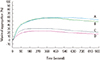
The appropriate concentrations of ADP, PGE1, and cilostazol to be used in screening tests were established as previously explained (4. Establishing concentrations). Cilostazol at 1 µM and 2 µM was added in channels 1 and 2. PGE1 was added to 1 µM and 2 µM cilostazol, in channels 3 and 4 (Fig. 1). The MA under established conditions was measured using blood samples obtained from 20 healthy volunteers. Statistical analysis was done using SPSS software (SPSS V.18.0 for Windows; SPSS Inc., Chicago, IL, USA).
RESULTS
1. Concentration of ADP
The average MA of samples induced with 20 µM ADP was 77.3% whereas that of the samples incubated with 2 µM cilostazol and 80 nM PGE1 was 18.6%. The average MA of samples induced with 5 µM ADP was 62.0% whereas that of the samples incubated with 2 µM cilostazol and 80 nM PGE1 was 5.6%, which provided a near baseline graph. We decided to use 20 µM ADP in our experiments.
2. Concentration of Cilostazol
When 10 µM cilostazol was added, the MA was 1.8% even in the absence of PGE1. The MA of samples containing 80 nM PGE1 and 1, 2, and 5 µM cilostazol were 13.1%, 4.8%, and 4.6%, respectively. Aggregation was inhibited signicantly at 2 µM cilostazol. The response to 1 µM cilostazol was expected to show individual variations. We decided to use 1 µM and 2 µM cilostazol in our experiments.
3. Concentration of PGE1
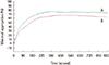
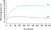
The average MA of samples induced by 20 µM ADP and 2 µM cilostazol, and with the addition of 10 nM, 40 nM, 70 nM, and 100 nM PGE1 was 35.2%, 24.4%, 14.3%, and 8.6%, respectively, and the rate varied among individuals. Aggregation decreased almost proportional to the concentration of PGE1 when more than 40 nM was added. We decided to use 80 nM PGE1 in our experiments. When two samples, one with 100 nM PGE1 and the other with none, were induced by 20 µM ADP, the MA showed no signicant difference (Fig. 2). Addition of PGE1 to cilostazol showed a marked effect on the inhibition of platelet aggregation (Fig. 3).
4. Frequency of Resistance to Cilostazol in Normal Population
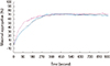
The 20 volunteers who participated in this experiment compris ed 13 women and 7 men. Their average age was 32 yr (range: 26-55 yr), average number of platelets was 160×103/µL (range: 83-249×103/µL), and the average number of platelets in PRP was 238×103/µL (range: 188-305×103/µL). The average MA of samples without any cilostazol was 59.0% (range: 28.6-82.7%), and that of samples with 1 µM cilostazol was 58.1% (range: 35-83%), which showed no statistically signicant difference (P=0.085). The average MA of samples with 1 µM cilostazol and 80 nM PGE1 was 22.0% (range: 4-72%), which showed a statistically signicant difference (P<0.001) from the average MA of samples incubated with 1 µM cilostazol only. The average MA of samples incubated with 2 µM cilostazol was 42.8% (range: 10.0-74.5%), which showed a statistically significant decrease (P=0.004). It decreased to 21.2% (range: 2.3-74.1%) when 80 nM PGE1 was added to 2 µM cilostazol (P<0.001) (Fig. 4).
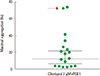
Two subjects (both women, aged 29 and 55 yr, with platelet counts of 178×103/µL, 150×103/µL, respectively) showed an MA of over 70% even with PGE1 and 2 µM cilostazol (Fig. 5). This decreased to nearly 30% when the amount of cilostazol was increas ed to 10 µM and less than 5% when PGE1 was added.
At 2 µM cilostazol and 80 nM PGE1, which is the theoretical condition for the maximum inhibition of aggregation in our experiment, MA was maximum 74.1%, minimum 2.3%, average 21.2% and standard deviation was 22.2% (Fig. 6). In conclusion, we propose incubation with 2 µM cilostazol and 80 nM PGE1 and induction with 20 µM ADP to monitor cilostazol resistance, and the cutoff for resistance to be 65.6% which is the average+2SD.
DISCUSSION
Monitoring the effect of cilostazol, which is prescribed together with aspirin and clopidogrel, is important. Although the response to cilostazol is expected to differ among individuals, there is no report on any methods of monitoring. We developed an experiment protocol using 1 µM and 2 µM cilostazol, and PGE1, to monitor the inhibition of platelet aggregation.
In a study by Yamamoto et al.[6], 10 healthy volunteers took 100 mg of cilostazol and had blood withdrawn hourly for several hours; thereafter, the level of VASP phosphorylation was measured by western blot analysis. PGE1 was added to the samples and LTA was measured after inducing aggregation with ADP and collagen, indicating that PGE1 is important in monitoring the effect of cilostazol.
PGE1, with adenosine, is an intrinsic agonist, which increases cAMP and inhibits the activation of platelets via the prostacyclin receptor. Cilostazol protects the cardiovascular system by extending and stimulating the effect of factors producing cAMP, and PGE1 increases cAMP by acting on Gαs-coupled receptors. It has been proven that cAMP induces nitric oxide synthase in endothelial cells and increases cGMP levels [7]. It is anticipated that the effect of cilostazol was absent without PGE1 since the half-lives of prostacyclin and nitric oxide are 6-7 minutes and a few seconds, respectively. The substances are regarded to be degraded within 20 minutes of preparing PRP.
In 2 volunteers, the activity of platelets was not inhibited even in the presence of 2 µM cilostazol and PGE1, and showed a response when cilostazol concentration was increased to 10 µM. This is expected to be the reaction in patients demonstrating resistance to cilostazol. Theoretically, increasing the dosage of cilostazol, elongating the time of exposure with platelets, or administration of PGE1, can be considered in patients with HPPR to cilostazol. One report showed that an increase in the incubation time of cilostazol had no effect on aggregation when induced by collagen but a significant decrease of aggregation was observed when it was induced by ADP [1]. It is thought that increasing the time of exposure to cilostazol increases the amount of cAMP, and compensates for the process where ADP blocks the production of cAMP via the P2Y12 receptor. Although the time of exposure of platelets to cilostazol will vary among individuals according to their metabolism rate, we carried out our experiment based on the hypothesis that an incubation of 10 minutes was enough.
Aggregation of platelets was observed to be significantly inhibited at 1 µM and 2 µM cilostazol, among various concentrations of cilostazol, and concentrations of 1 µM and 2 µM were used in experiments with volunteers. According to the literature, the peak level of cilostazol is achieved at 2-4 hr after the administration of 100 mg of cilostazol and its concentration is 2 µM [8], showing a similarity with the 1 µM and 2 µM concentrations used in our experiment.
Methods for monitoring the effects of aspirin and clopidogrel are developed and considerable research has been performed on the mechanisms of resistance of these two medications. When aspirin is taken with ibuprofen, the two forms of medication competitively interact with COX-1 receptors leading to a decreased effect [9]. Increased active oxidants may lessen the antiplatelet effect in hyperglycemic patients [10] and aspirin may have a decreased effect on thrombin in patients with high cholesterol levels [11]. In addition, increased secretion of catecholamine due to exercise and stress is suggested as a reason for the inhibited effects of aspirin [12].
Cilostazol is presently used for peripheral artery diseases especially in Asian countries such as Korea and Japan [13]. It not only inhibits PDE3 but also decreases platelets that are activated by interaction with stimulated endothelial cells and prevents restenosis of vessels by blocking the proliferation of smooth muscles of vessel walls [13]. About 4% of patients who took clopidogrel showed abnormal liver function within 52 weeks [14]. Antacids are recommended with clopidogrel and low-dose aspirin because gastric bleeding may occur [1516]. About 38% of patients taking low-dose aspirin develop ulcer and mucocutaneous erosion upon endoscopic examination whereas less than 20% of patients with clopidogrel and cilostazol showed such endoscopic findings [17]. In one study, 60 patients with HPPR to clopidogrel were divided into two groups, one with addition of cilostazol and the other with increased dose of clopidogrel. The group with the addition of cilostazol showed a significantly decreased platelet aggregation rate [18]. The use of cilostazol will increase due to its advantages and emerging problems of other antiplatelet agents, and as such, monitoring of its effects will be necessary.
We developed a convenient in vitro incubation method of monitoring the effect of cilostazol without having patients take cilostazol. Further research on whether the results reflect an accurate in vivo effect is necessary. Furthermore, the cilostazol used in our experiment was the pure form of cilostazol whereas patients take mixed forms of cilostazol, which may show different results. The same experiment was carried out with 100 mg of Pletaal (Otsuka Pharmaceutical Co. Limited, Tokyo, Japan) dissolved in DMSO, showing a relatively weak aggregation compared to the experiment with pure form but its difference was not significant (data not shown).
Additional study on the clinical correlation and evidence for the interpretation of results are necessary for the practical application of monitoring the antiplatelet effect of cilostazol, as is currently in place with aspirin and clopidogrel. Adding cilostazol to patients showing resistance to aspirin or clopidogrel is helpful and choosing different antiplatelet agents for patients showing resistance to cilostazol is important. We developed an in vitro incubation method of monitoring resistance to cilostazol using a small amount of PGE1 and ADP.

 XML Download