

 PDF
PDF ePub
ePub Citation
 Print
Print


Uterine rupture is defined as a full-thickness separation of the uterine wall and overlying visceral peritoneum.1 According to a systematic review of maternal morbidity and mortality by the World Health Organization in 2005, the median incidence of uterine rupture is 5.3 per 10000 deliveries. A major risk factor for uterine rupture is previous cesarean section, including a short duration (less than 18 months) since the last cesarean delivery, the number of previous cesarean sections and single layer closure instead of two-layer closure. Other risk factors include previous scarring of the uterus after myomectomy, placenta previa, uterine trauma, uterine instrumentation and congenital uterine anomaly.2
Uterine rupture presents with fetal heart rate change, most commonly bradycardia. Mother's clinical features include abdominal pain, vaginal bleeding and altered uterine contraction. More severe conditions of uterine rupture are presented as hypotension, shock, hematuria, scar tenderness and maternal tachycardia.2 For differential diagnosis of uterine rupture, placental abruption, placenta previa, uterine inversion, cervical tear, vaginal tear, coagulopathy, uterine atony and uterine artery rupture may be considered.3 Placental invasion anomalies refer to the abnormal adherence of the placenta to the uterine wall, resulting in detachment failure after delivery.4 Placental invasion anomalies are classified according to invasion depth. Placenta accreta occurs when the villi penetrate the decidua but not the myometrium. Placenta increta occurs when the villi penetrate the myometrium. Placenta percreta occurs when these villi penetrate the serosa and occasionally invade into adjacent organs such as the bladder.5
Here we present the case of a woman who developed uterine rupture during due to placenta percreta during the second trimester, after two previous cesarean sections.
CASE
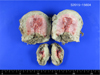
A 32-year-old woman gravida 2, para 2 at 21 weeks' gestation presented to the emergency room with acute, moderate abdominal pain. She was drowsy and had a history of two previous cesarean sections. Physical examination revealed a systolic blood pressure of 40 mmHg and that she was in hypovolemic shock. Her abdomen was soft but distended. Massive hemoperitoneum and fetal bradycardia were detected with ultrasound examination. Hematoma was also found posterior to the placenta. Central venous catheter placement on the jugular vein and arterial line placement were performed. Emergency transfusion was commenced. She was transported to the operating room for an emergency cesarean section. A midline laparotomy was performed, and the peritoneal cavity was filled with the massive hematoma. The hematoma was being expelled through the incision and an extended incision was required. Uterine rupture (approximately 1-2 cm) was detected between the left lower segment of the uterus and the borderline of the cervix in the anterior wall. A male neonate weighing 232 g, was delivered but was stillborn without spontaneous breathing. The placenta was lying over the anterior lower segment uterine wall covering the cervical orifice, and it was suspected to total or partial placenta previa. The placenta was manually removed but immediate complete removal was infeasible. Even after suturing the tear in the uterine wall, active bleeding continued from the inner orifice of the cervix which was indicative of placenta increta or percreta. Subtotal hysterectomy was performed without removing both ovaries because the bleeding site was not clear inside the uterus. After subtotal hysterectomy, however, bleeding was persistent, especially from near the inner orifice. She was also bleeding from a laceration between the bladder dome and cervix. It was concluded that removal of the cervix was necessary for bleeding control. Once the retroperitoneal bleeding was controlled and the removal of the hematoma was completed, the tear in the bladder dome was sutured and total hysterectomy was performed. The estimated blood loss during the surgery was 19,000 ml. She received 36 units of packed red blood cells, 13 units of fresh frozen plasma and 16 units of platelet concentrate during the operation. The uterus was sent for biopsy to have the pathology confirmed (Fig. 1), and the pathology report revealed placenta percreta (Fig. 2). She was then admitted to the intensive care unit and was discharged from the hospital in good general condition in 12 days.
DISCUSSION
In our case, the patient had two previous cesarean sections, abnormal placental invasion is diagnosed either clinically during cesarean delivery or histopathologically after hysterectomy. As in case, uterine rupture due to placenta percreta during the second trimester, is a rare, but life-threating complication. Ulkumen BA et al.6 published a similar report as our case. A 25-year-old woman had acute abdominal pain at 24 gestational weeks and had emergency cesarean section performed. Placental biopsy after the operation confirmed placenta percreta. In another case, a woman at 16 weeks gestation experienced uterine rupture due to placenta percreta. She also had a previous history of cesarean section as well as dilation and curettage.7 In Korea, Park SH et al.8 reported that women with a history of previous cesarean section and placenta previa were treated with cesarean hysterectomy because of spontaneous uterine rupture with placenta percreta at 37weeks. Another reported the spontaneous rupture of the uterus at 14 weeks of pregnancy with
Uterine rupture is an uncommon but serious obstetrical complication.10 The overall incidence of uterine rupture is estimated at 0.05% of total deliveries.4 Furthermore, spontaneous uterine rupture due to placenta accreta or percreta is a rare obstetric emergency with an estimated incidence of 1/5000 pregnancies.5 90% of cases occur in women with a uterine scar, most commonly from a previous cesarean section.46 92.3% of ruptures of scarred uterus involved the lower uterus, and 3.8% of uterine ruptures were located in the parametrium.11 The majority of uterine ruptures occur during the third trimester due to a thinned uterine lower segment. Mothers with uterine rupture present with hypovolemic shock secondary to hemorrhage, disseminated intravascular coagulation (DIC), genitourinary injury, a potential need for hysterectomy and maternal death.12 Uterine rupture results in change in fetal heart three previous cesarean sections.9 Few case reports have been published after that report. rate, bradycardia and late deceleration, fetal hypoxia, fetal acidosis and fetal or neonatal death.13 Patients with clinical shock need resuscitation and immediate surgical intervention. After urgent evaluation, clinicians need to decide if the rupture is surgically repairable or hysterectomy is needed. The choice of the surgical procedure depends on the type, location, and extent of uterine tear.14 If a pregnant woman has acute abdominal pain or distension, the doctor should consider the differential diagnosis, including acute appendicitis, ovarian torsion or cyst rupture and hepatic or splenic rupture.
Uterine rupture can also occur in an unscarred uterus. It is more common in older, multiparous women, compared to rupture of the scarred uterus. Thus, if a pregnant woman has acute abdominal pain, vaginal bleeding, fetal deceleration or fetal bradycardia, the differential diagnosis should include uterine rupture, placental abruption, placenta previa and uterine atony. The abnormal placental implantation at the uterine scar of our patient may have increased the risk of uterine rupture. A history of dilatation and curettage, cesarean section, manual removal of the placenta, uterine sepsis and previous uterine surgery can cause the deficiency of decidua, which predisposes to abnormal placental invasion.12
Although uterine rupture during pregnancy is a rare event, it should be considered in pregnant women with hemoperitoneum, particularly when the patient has a history of previous cesarean section. In patients with atypical histories, diagnosis maybe delayed or may even be established at the time of laparotomy, thereby increasing maternal and fetal morbidity and mortality. Important steps for the successful management of uterine rupture include prompt diagnosis followed by definitive surgical management.

 XML Download