Abstract
Introduction
Bronchial carcinoid tumors seldom occur in children, sometimes mistaken for a minor disease and diagnosed slowly. Materials and Methods: We report on a patient who diagnose tumors slowly because confused with asthma.
Results
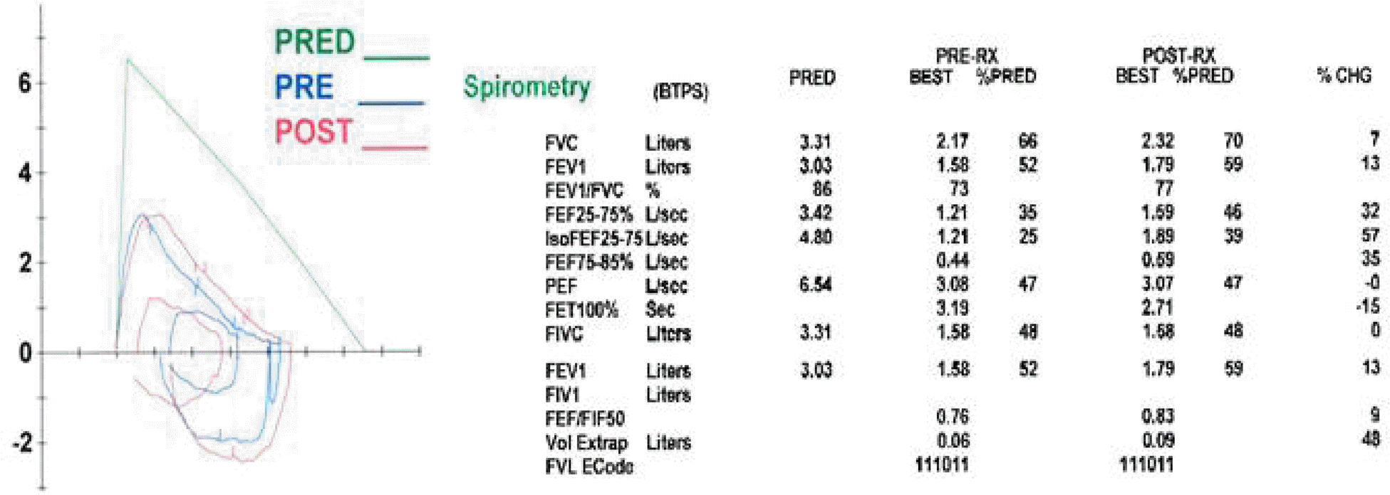
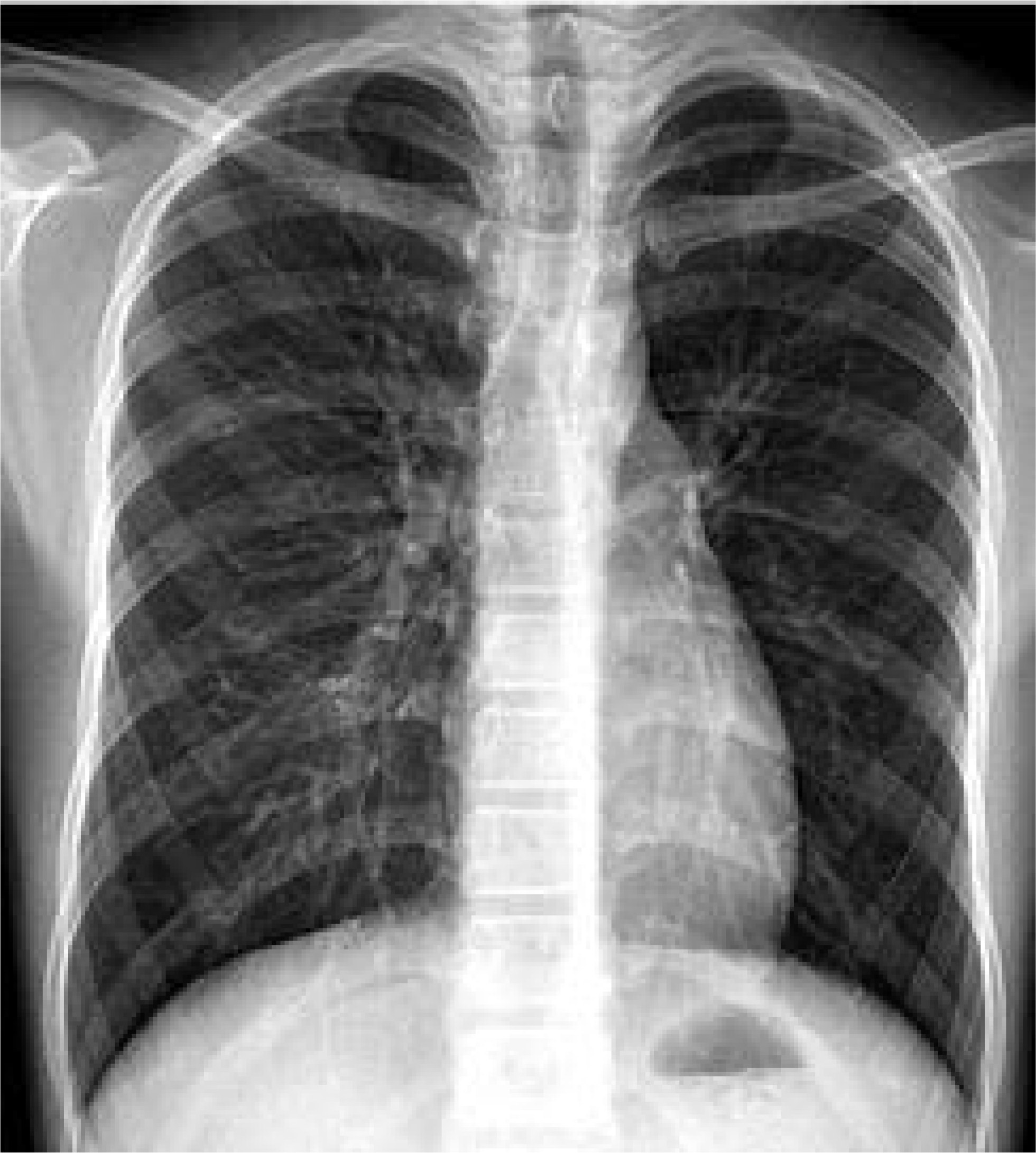
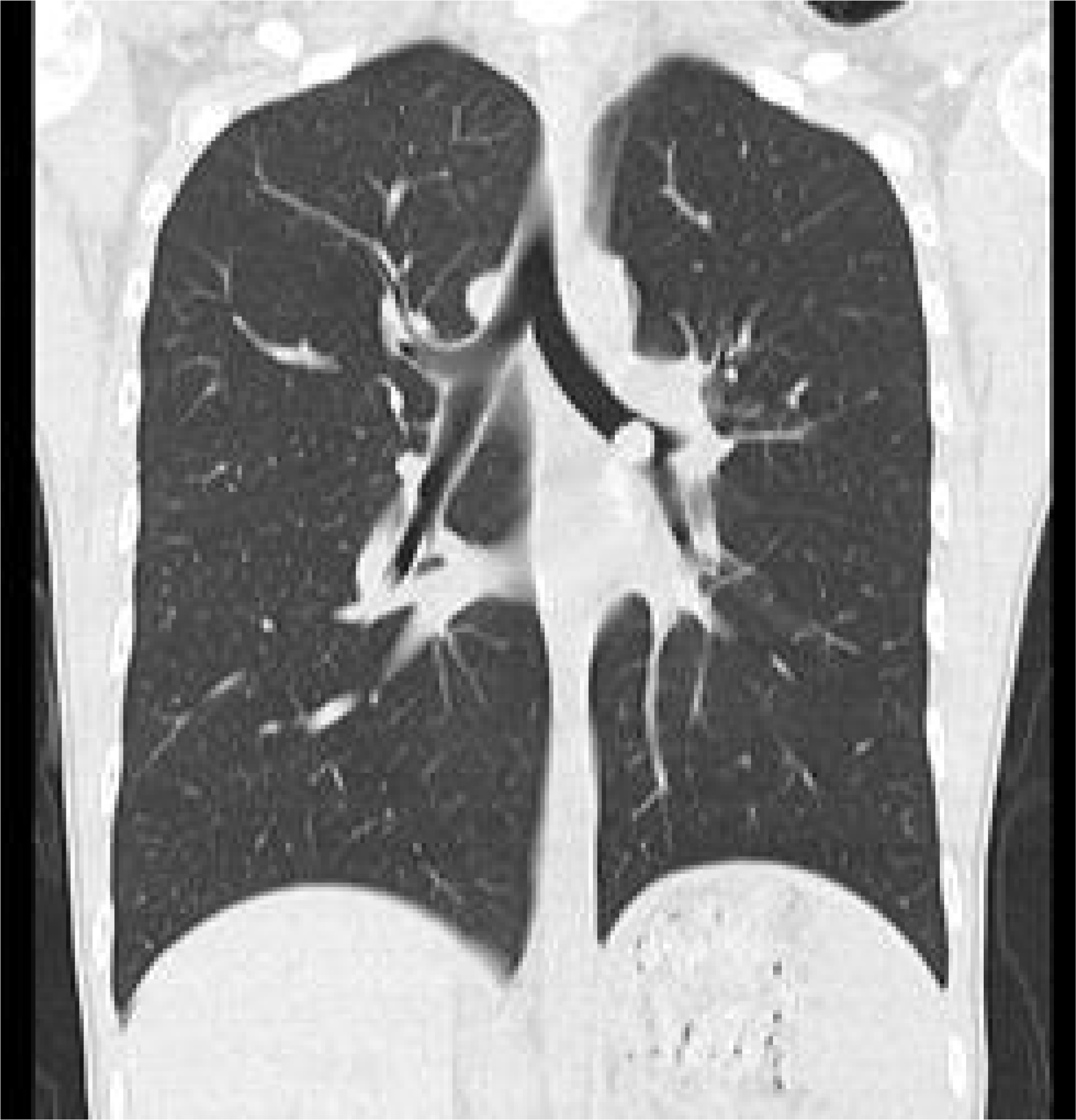
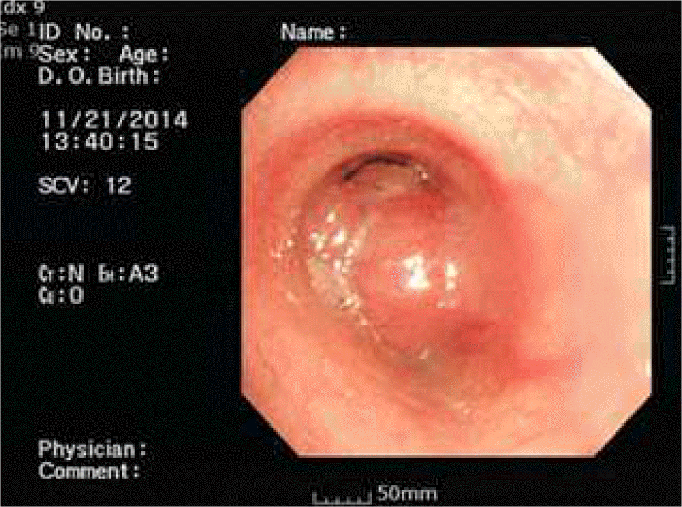
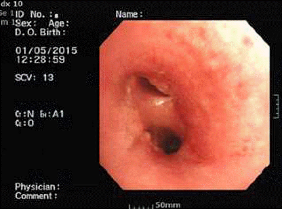
This case describes a 14-year-old boy, presenting with asthma-like symptoms throughout 3 years. He was treated as asthma but wax and wane. Chest x-ray showed an hyperlucent left lung, so we rechecked high resolution computed tomography (HRCT) for unilateral hyperinflation diseases diagnosis. It was found 1×1㎝ nodule in left main bronchus. We did bronchoscopy and discovered a round mass in the left bronchus, 2∼3㎝ away from carina. In the biopsy, it was bronchial carcinoid tumor, so we resected tumor.
REFERENCES
1.Lal DR., Clark I., Shalkow J., Downey RJ., Shorter NA., Klimstra DS, et al. Primary epithelial lung malignancies in the pediatric population. Pediatr Blood Cancer. 2005. 45:683–6.

2.Hauso O., Gustafsson BI., Kidd M., Waldum HL., Drozdov I., Chan AKC, et al. Neuroendocrine tumor epidemiology: contrasting Norway and North America. Cancer. 2008. 113:2655–64.
3.Wang LT., Wilkins EW Jr., Bode HH. Bronchial carcinoid tumors in pediatric patients. chest. 1993. 103:1426–8.

4.Eyssartier E., Ang P., Bonnemaison E., Gibertini I., Diot P., Carpentier E, et al. Characteristics of Endobronchial Primitive Tumors in Children. Pediatric Pulmonol. 2014. 49:121–5.

5.Skuladottir H., Hirsch FR., Hansen HH., Olsen JH. Pulmonary neuroendocrine tumors: incidence and prognosis of histological subtypes. A population-based study in Denmark. Lung Cancer. 2002. 37:127–35.

6.Divisi D., Crisci R. Carcinoid tumors of the lung and multimodal therapy. Thorac Cardiovasc Surg. 2005. 53:168–72.

7.Fink G., Krelbaum T., Yellin A., Bendayan D., Saute M., Glazer M, et al. Pulmonary carcinoid: pre- sentation, diagnosis, and outcome in 142 cases in Israel and review of 640 cases from the literature. Chest. 2001. 119:1647–51.
9.Travis WB. The concept of pulmonary neuroendocrine tumors. Travis WB, Brambilla E, Muller-Hermeli HK, Harris CC, editors. editors.Pathology and genetics of tumours of lung, pleura, thymus, and heart. Lyon: IARC Press;2004. p. 19–20.
10.Steven RB. Glenna BW. Emphysema and Overinflation. Robert MK, Bonita FS, Joseph WS, Nina FS, Richard EB, editors. editors.Nelson Textbook of Pediatrics. 19th ed.Philadelphia: Elsevier saunders;2011. p. 1460–1461.


 PDF
PDF ePub
ePub Citation
 Print
Print


 XML Download