

 PDF
PDF ePub
ePub Citation
 Print
Print


The prevalence of myopia, which is one of the most common ocular diseases, is increasing in both children and adults,12 and the rate is particularly high in Asian regions such as South Korea, Japan, Singapore, and Hong Kong. Kim and Koo3 reported that the incidence of myopia was 23% among elementary school students, 47.3% among middle school students, and 66.9% among high school students in 1998. Han et al.4 reported that the incidence of myopia was 50.1% among fourth graders in elementary schools in 2007. Lin et al.5 reported that 84% of Taiwanese adolescents had myopia in 2001.
Myopia, once it occurs, progresses in most cases; in some instances, its severity can be high, increasing the risk of retinal detachment, macular degeneration, choroidal neovascularization, and glaucoma, which may lead to blindness.67 To avoid these problems, many efforts have been made to halt or prevent the progress of myopia in children.8 Suggested methods include treatment using medicated eye drops such as tropicamide, atropine, and pirenzepine, as well as the use of intraocular pressure-lowering agents, under-correcting eye glasses, the wearing of bifocal or multifocal eye glasses, and the wearing of oxygen-permeating spherical lenses. However, none of these methods have been recognized in terms of their effects and stability when preventing myopic progression.910111213
Since Cheung and Cho14 first introduced the possibility of preventing myopic progression with the use of orthokeratology lenses, many studies have been conducted on these lenses. Orthokeratology is a methodology in which soft contact lenses with reverse geometry are used to change the curvature of the cornea in order to reduce or remove the refractive error.151617 Since the curvature of the orthokeratology lens is flatter than the curvature of the central part of the cornea, the lens presses the cornea and thus temporarily decreases the myopia. Recent reports indicate that orthokeratology lenses may prevent myopic progression by restraining optic axis elongation.171819 In South Korea, despite the increasing prescription of orthokeratology lenses, there are not many reports on the effect of orthokeratology lenses on optic axis elongation and the prevention of myopic progression. To date, no studies have investigated variations in optic axis length following the introduction of IOL Master® (Carl Zeiss Jena GmbH, Jena, Germany), which enables one to elongate the optic axis in an accurate manner. Therefore, to investigate the effect of an orthokeratology lens on the prevention of myopic progression, as well as on optic axis elongation, we employed the IOL Master® to analyze changes in patients' optic axis length, which occurred before and after wearing orthokeratology lenses for more than 1 year; the results were compared with control group, who wore general eye glasses.
MATERIALS AND METHODS
This study investigated 37 eyes in 19 children who visited our institution between January 2012 and September 2014; these children were diagnosed with myopia and had worn orthokeratology lenses for more than 1 year during the follow-up period. The control group was 46 eyes in 23 children who were diagnosed with myopia during the same period of time. The prescribed orthokeratology lenses were the Lucid Korea Lens® (Lucid Korea, Seoul, South Korea). The orthokeratology lenses were basically worn for 8 hours per day, on average, spanning from the period of sleep the night before to the next morning. The refractive error was measured prior to the prescription via a refraction test under accommodative palsy, and the curvature of the cornea was measured using an automated refraction tester (RK-F1, Cannon, Japan). The optic axis length was measured using an IOL Master® (Carl Zeiss Jena GmbH, Jena, Germany), and the difference in the optic axis length measurements before and after the prescription was compared.
Measurements using the IOL Master® were performed between 3 p.m. and 6 p.m. in the afternoon to minimize the corneal flattening effect, which occurred after wearing the lenses during the night. The mean of the three measurements obtained from each person was calculated for all the subjects. An unpaired t-test was performed to compare the two groups with respect to age, the severity of their myopia and astigmatism, the spherical equivalent (SE), and the length of the optic axis. A nonparametric test (Mann–Whitney U test) was performed to compare the two groups in terms of gender. One year after the prescription of the orthokeratology lens, an unpaired t-test was performed to compare the change in the optic axis length. A P-value < 0.05 was considered statistically significant for all cases.
RESULTS
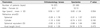
The orthokeratology lens-wearing group included 37 eyes from 19 individuals, including seven males and 12 females with an average age of 9.56 ± 1.67 years. The eyeglass-wearing group included 46 eyes from 23 individuals including ten males and 13 females with an average age of 9.83 ± 1.34 years. There were no significant differences in age between the two groups (P = 0.554). At the initial examination, the refractive error test results were as follows for the lens-wearing group: myopia of –3.26 ± 1.78 diopters (D), astigmatism of –1.10 ± 0.82 D, and SE of –3.82 ± 2.10. For the eyeglass-wearing group, the results were as follows: myopia of –3.31 ± 1.57 D, astigmatism of –0.74 ± 0.51 D, and SE of –3.68 ± 1.64 D; this indicated that there were no differences between the two groups in terms of the refractive error (P = 0.915, P = 0.182, and P = 1.000). The optic axis length was 24.62 ± 1.39 mm in the lens-wearing group and 24.59 ± 0.74 mm in the eyeglass-wearing group, with no significant difference between the two groups (P = 0.473) (Table 1).
After 1 year of follow-up, the refractive error test results were as follows: myopia of –0.13 ± 0.27 D, astigmatism of –0.10 ± 0.38 D, and SE of –0.22 ± 1.10 D in the lens-wearing group, and myopia of –3.58 ± 2.15 D, astigmatism of –0.94 ± 0.83 D, and SE of –4.14 ± 1.95 D in the eyeglass-wearing group, indicating that the refractive index significantly decreased in the lens-wearing group (all P < 0.001). The optic axis length was changed to 24.73 ± 1.28 mm by 0.11 ± 0.12 mm in the lens-wearing group and to 24.80 ± 0.71 mm by 0.21 ± 0.07 mm in the eyeglass-wearing group, indicating that the elongation of the optic axis length was significantly less in the lens-wearing group during the same period (P < 0.001) (Table 2).
DISCUSSION
Orthokeratology lenses have been applied since the early 1960s, when Jensen11 introduced the idea that wearing soft contact lenses may flatten the cornea to decrease myopia and astigmatism. Orthokeratology is a method in which soft contact lenses are used to change the curvature of the cornea to reduce or remove the refractive error. The efficacy of orthokeratology lenses for correcting vision has been reported in many previous studies, which indicates that wearing orthokeratology lenses has excellent vision correction effects in mild myopia and astigmatism.20212223 However, since orthokeratology lenses flatten the cornea within a short period of time following the start of treatment, and given that they turn myopia to emmetropia, the actual therapeutic effect on the refractive error is not well reflected. Therefore, measuring the changes in the optic axis length may be a critical factor when determining myopic progress.24
Cheung and Cho14 reported a case of a 13-year-old boy whose refractive state was changed by a decrease in his myopia of 0.25 D and a decrease in his astigmatism of 0.5 D. This was accompanied by an increase in his optic axis length of 0.1 mm within 2 years, after wearing an orthokeratology lens on the left eye. Conversely, when the orthokeratology lens was not worn, the patient exhibited an increase in the optic axis length by 0.34 mm, as well as an increase in his myopia of 0.75 D in the right eye, suggesting that the use of an orthokeratology lens has the potential to correct myopia and astigmatism, and it can also prevent myopic progression. Cho et al.17 reported that the optic axis length increased by 0.29 ± 0.27 mm in 2 years in the orthokeratology lens-wearing group and by 0.54 ± 0.27 mm in the eyeglass-wearing group, indicating that elongation of the optic axis length was less in the orthokeratology lens-wearing group. Similarly, Walline et al.19 reported that the optic axis length increased by 0.25 mm in 2 years in the orthokeratology lens-wearing group and by 0.57 mm in the eyeglass-wearing group. To investigate whether orthokeratology lenses have a myopia-repressing effect, Kakita et al.25 conducted a long-term follow-up study for 5 years with an orthokeratology lens-wearing group and an eyeglass-wearing control group; the authors reported that the optic axis length was increased by 0.99 ± 0.47 mm in the orthokeratology lens-wearing group and by 1.41 ± 0.68 mm in the control group. These previous studies show that orthokeratology lenses have an effect of preventing myopic progress.2122232425 In this study, the optic axis length was increased over the course of 1 year of follow-up by 0.11 ± 0.12 mm in the lens-wearing group and by 0.21 ± 0.07 mm in the eyeglass-wearing group, indicating that the elongation of the optic axis length was significantly less in the lens-wearing group (P < 0.001). Notably, in previous studies where the optic axis length was measured using contact-type ultrasonic wave A scanning, the accuracy of the measurement results was controversial due to the difficulties experienced when trying to achieve an accurate gaze, as well as when measuring the optic axis length without pressing on the children's eyeballs. On the other hand, in this study (as well as in the study conducted by Kakita et al.2) where the IOL Master® was used to measure the optic axis length in a non-contact and noninvasive manner, more accurate and objective results were rapidly obtained.
The mechanism that slows optic axis elongation in myopic children has not been accurately identified, but some recent experimental studies conducted with primates have found that the peripheral retina affects the growth of the eyeballs, and also results in changes in refractive power.262728 Since a normal cornea is typically steep around the center, but becomes flatter as one moves toward the peripheral region, the light focus is located in front of the retina in the central macula, but behind the retina in the peripheral retina, forming peripheral hyperopia. In a study conducted with monkeys, the fovea centralis and the macula were eliminated using a laser in some subjects, while the peripheral retina was eliminated in other subjects. The comparison showed that the optic axis length was increased only in those monkeys whose peripheral retinas were eliminated, and where the damage to the central retina did not affect their optical axis length. Likewise, a severe refractive error found in children with retinopathy of prematurity who had undergone laser photocoagulation in the peripheral retina is a finding that suggests that the peripheral retina may affect the elongation of the optic axis length and the refractive error.29 As it is different from a normal hard lens, an orthokeratology lens basically features a flat curvature in the center, which makes the central cornea flat, so that the light may be focused on the macula, not on the region in front of the macula. In addition, the force that pushes the central cornea and the force that pulls the peripheral retina of the lenses change the retina in a plateau shape, enabling the light to be refracted not only on the macula, but also on the peripheral retina. This may correct the peripheral hyperopia and thus have the effect of retarding the elongation of the optic axis length, which is one of the major causes of myopia.30 In other words, while the hyperopic change of the peripheral retina stimulates axial myopia, wearing an orthokeratology lens may change the shape of the cornea, leading to a decrease in the hyperopic change of the peripheral retina, thus repressing myopic progress.
In conclusion, the elongation of the optic axis length was significantly less in the orthokeratology lens-wearing group than in the eyeglass-wearing group, indicating that wearing orthokeratology lenses may contribute to the repression of myopia. This result suggests that wearing orthokeratology lenses may slow or halt myopic progress in Koreans, as the lens is able to repress the elongation of the optic axis length.
However, the interpretation of the results of this study is limited given that the number of subjects was small and the follow-up period was short. In addition, this study is limited in the sense that the effect of corneal flattening on the optic axis length, as caused by the wearing of orthokeratology lenses, was not completely excluded. Therefore, a long-term and large-scale follow-up study may need to be conducted to investigate the effect of wearing orthokeratology lenses, as well as to determine the relationship between peripheral hyperopia and the peripheral cornea.

 XML Download