Abstract
Peritoneal and gastrointestinal metastasis from breast cancer is very rare. We report here a rare case of metastatic peritoneal and gastric cancer from breast lobular carcinoma after modified radical mastectomy. A 65-year old woman presented with anorexia, nausea, vomiting and dyspepsia for several weeks at 44 months after surgery. Radiologic study showed peritoneal metastasis, and surgical histopathology reported peritoneal and omental metastatic carcinoma. Esophagogastroduodenoscopic (EGD) biopsy also confirmed metastatic carcinoma originated from breast primary.
References
1. Ciulla A, Castronovo G, Tomasello G, Maiorana AM, Russo L, Daniele E, et al. Gastric metastases originating from occult breast lobular carcinoma: diagnostic and therapeutic problems. World J Surg. 2008; 6:78.

2. Ayantunde AA, Agrawal A, Parsons SL, Welch NT. Esophagogastric cancers secondary to a breast primary tumor do not require resection. World J Surg. 2007; 31:1597–601.

3. Choi JE, Park SY, Jeon MH, Kang SH, Lee SJ, Bae YK, et al. Solitary small bowel metastasis from breast cancer. J Breast Cancer. 2011; 14:69–71.

4. Tremblay F, Jamison B, Meterissian S. Breast cancer masquerading as a primary gastric carcinoma. J Gastrointest Surg. 2002; 6:614–6.

5. Nazareno J, Taves D, Preiksaitis HG. Metastatic breast cancer to the gastrointestinal tract: a case series and review of the literature. World J Gastroenterol. 2006; 12:6219–24.

6. Pectasides D, Psyrri A, Pliarchopoulou K, Floros T, Papaxoinis G, Skondra M, et al. Gastric metastases originating from breast cancer: report of 8 cases and review of the literature. Anticancer Res. 2009; 29:4759–64.
7. Jones GE, Strauss DC, Forshaw MJ, Deere H, Mahedeva U, Mason RC. Breast cancer metastasis to the stomach may mimic primary gastric cancer: report of two cases and review of literature. World J Surg Oncol. 2007; 5:75.

8. Ambroggi M, Stroppa EM, Mordenti P, Biasini C, Zangrandi A, Michieletti E, et al. Metastatic breast cancer to the gastrointestinal tract: report of five cases and review of the literature. Int J Breast Cancer. 2012; 2012:439023.

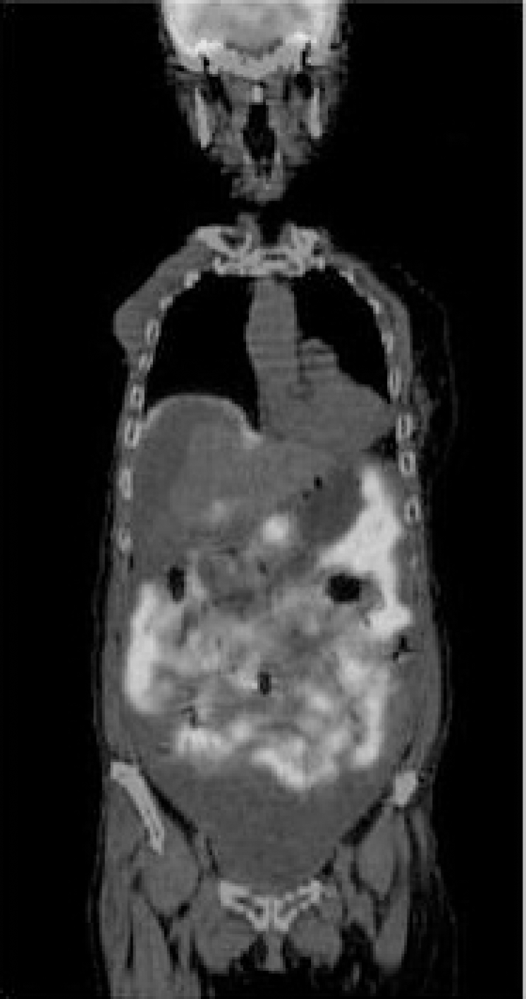
Fig. 1.
PET scan showed glucose hypermetabolism of abdominal cavity consistent with cancer peritonei. PET: positive emission tomography.

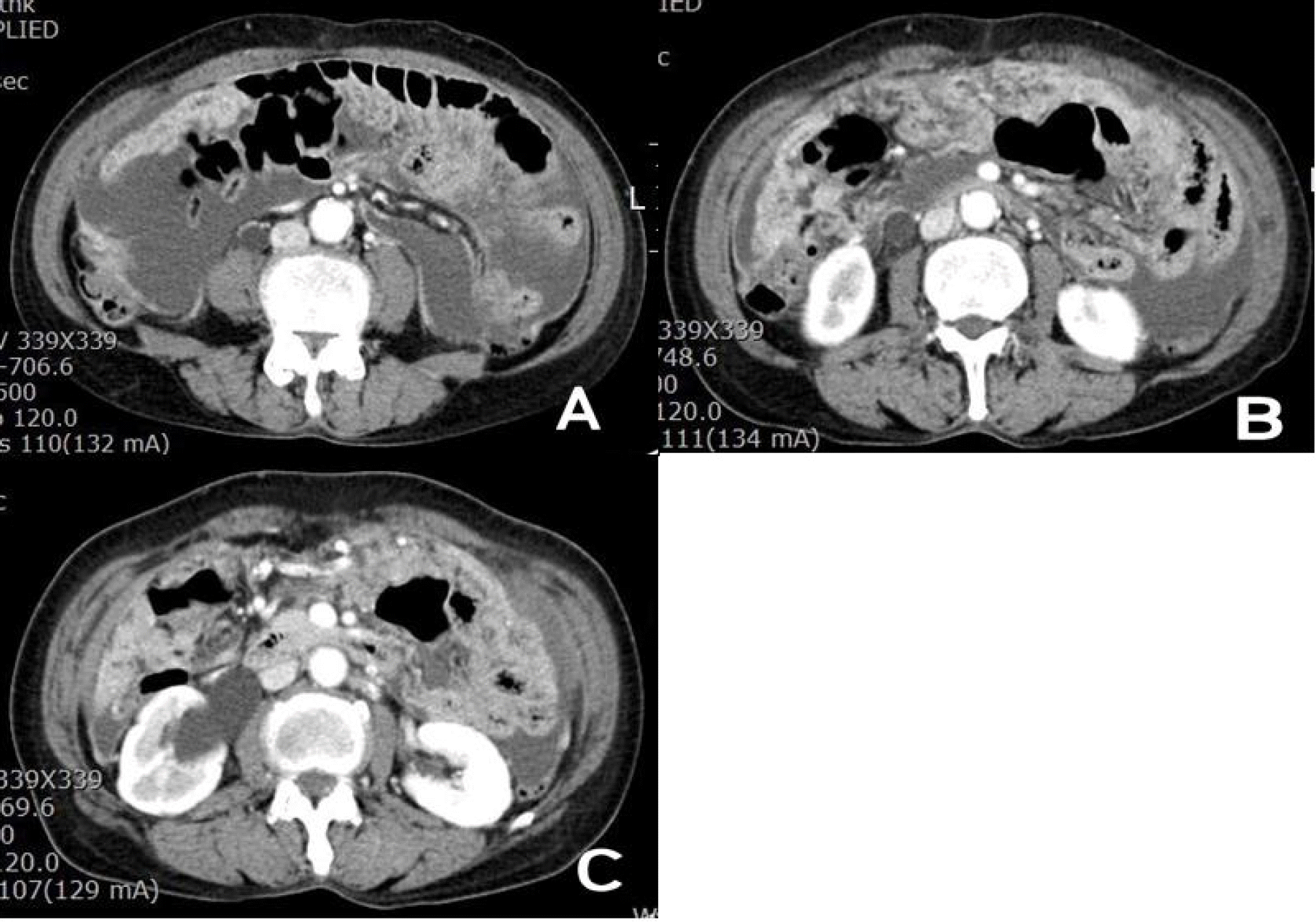
Fig. 2.
(A, B) Abdominal CT scans showed ascites, peritoneal thickening, omental smudge and cakes consistent with cancer peritonei. (C) Hydronephrotic change at the right kidney also presented. CT: computed tomography.


 PDF
PDF ePub
ePub Citation
 Print
Print


 XML Download