

 PDF
PDF ePub
ePub Citation
 Print
Print


Abstract
Objectives
This study analyzed clinical and radiologic results, and complications of minimally invasive plate osteosynthesis(MIPO) for distal tibia fracture.
Methods
16 patients (17 cases) who were performed MIPO for distal tibia fractures between January 2007 and June 2011, postoperative followed up at least for one year, were selected for this study. The average age was 55.0(26-76) years old and the average period of followup was 15.1(6-27) months. Most of the patients were encouraged to perform ankle dorsiflexion and straight leg raising exercise on splints, from the next day of the operation. As radiologic evaluation, we checked period until bone union, degree of angulation. And postoperative complications were also checked. For functional evaluation of the ankles, American Orthopaedic Foot and Ankle Society(AOFAS) score was used.
Results
16 of the 17 cases were achieved primary bone union, and average period of bone union in all the cases was 17.4 (12-42) weeks. Mean varus/valgus angulation after the bone union was 0.8 degrees and mean anteroposterior(AP) angulation was 1.8 degrees. Mean AOFAS score was 85.2(71-95) points; 5 cases of excellent, 10 cases of good, 1 case of fair, showing that 93.8% of the patients represented at least good AOFAS scores. As complications, there were 2 cases of superficial infection, and each 1 case of nonunion and skin irritation. There were no cases of deep infection, metal breakage, nor limb length discrepancy.
Conclusions
MIPO for distal tibia fracture is considered to be an effective operative method, because of its high bone union rate and low complications by minimal disruption of soft tissue and improved bone fixation strength. Also, for earlier return to daily life, ankle joint exercise should be started as soon as possible after the operation.
Go to : 
REFERENCES
1. Collinge C, Protzman R. Outcomes of minimally invasive plate osteosynthesis for metaphyseal distal tibia fractures. J Orthop Trauma. 2010; 24:24–9.

2. Teeny SM, Wiss DA. Open reduction and internal fixation of tibial plafond fractures. Variables contributing to poor results and complications. Clin Orthop Relat Res. 1993; 292:108–17.
3. Lau TW, Leung F, Chan CF, Chow SP. Wound complication of minimally invasive plate osteosynthesis in distal tibia fractures. Int Orthop. 2008; 32:697–703.
4. Redfern DJ, Syed SU, Davies SJ. Fractures of the distal tibia: Minimally invasive plate osteosynthesis. Injury. 2004; 35:615–20.
5. Holbrook JL, Swiontkowski MF, Sanders R. Treatment of open fractures of the tibial shaft: Ender nailing versus external fixation. A randomized, prospective comparison. J Bone Joint Surg Am. 1989; 71:1231–8.
6. Brumback RJ, McGarvey WC. Fractures of the tibial plafond. Evolving treatment concepts for the pilon fracture. Orthop Clin North Am. 1995; 26:273–85.
7. Hasenboehler E, Rikli D, Babst R. Locking compression plate with minimally invasive plate osteosynthesis in diaphyseal and distal tibial fracture: A retrospective study of 32 patients. Injury. 2007; 38:365–70.
8. Borrelli J Jr, Prickett W, Song E, Becker D, Ricci W. Extraosseous blood supply of the tibia and the effects of different plating techniques: A human cadaveric study. J Orthop Trauma. 2002; 16:691–5.
9. Helfet DL, Suk M. Minimally invasive percutaneous plate osteosynthesis of fractures of the distal tibia. Instr Course Lect. 2004; 53:471–5.
10. Lee KB. Distal Tibia Fracture. Plate Osteosynthesis. J Korean Fract Soc. 2009; 22:306–13.
11. Kim SK, Lee KB, Lim KY, Moon ES. Minimally invasive osteosynthesis with locking compression plate for distal tibia fractures. J Korean Fract Soc. 2011; 24:33–40.
12. Yoo JS, Park HW. Clinical outcomes of locking compression plate fixation through minimally invasive percutaneous plate osteosynthesis in the treatment of distal tibia fracture. J Korean Fracture Soc. 2012; 25:117–22.
13. Wilber MC, Evans EB. Fractures of the femoral shaft treated surgically. Comparative results of early and delayed operative stabilization. J Bone Joint Surg Am. 1978; 60:489–91.
14. Segal D, Wiss DA, Whitelaw GP. Functional bracing and rehabilitation of ankle fractures. Clin Orthop. 1985; 199:39–45.
15. Vander Griend R, Michelson JD, Bone LB. Instructional course lectures, the american academy of otrhopaedic surgeons. Fractures of the ankle and the distal part of the tibia. J Bone Joint Surg Am. 1996; 78:1772–83.
16. Gul A, Batra S, Mehmood S, Gillham N. Immediate unprotected weight-bearing of operatively treated ankle fracture. Acta Orthop Belg. 2007; 73:360–5.
17. Egol KA, Dolan R, Koval KJ. Functional outcome of surgery for fractures of the ankle. A prospective, randomised comparison of management in a cast or a finctional brace. J Bone Joint Surg Br. 2000; 82:246–9.
18. Ahmad MA, Sivaraman A, Zia A, Rai A, Patel AD. Percutaneous locking plates for fractures of the distal tibia: Our experience and a review of the literature. J Trauma Acute Care Surg. 2012; 72:E81–7.
19. Hazarika S, Chakravarthy J, Cooper J. Minimally invasive locking plate osteosynthesis for fractures of the distal tibia-Results in 20 patients. Injury. 2006; 37:877–87.
20. Hong KD, Ha SS, Chung NS, Sim JC, Ahn SC. Lateral plate fixation of distal tibial metaphyseal fracture using minimally invasive plate osteosynthesis technique. J Korean Fract Soc. 2006; 19:24–8.
21. Shon OJ, Park CH. Minimally invasive plate osteosynthesis of distal tibial fractures: A comparison of medial and lateral plating. J Orthop Sci. 2012; 17:562–6.
Go to :
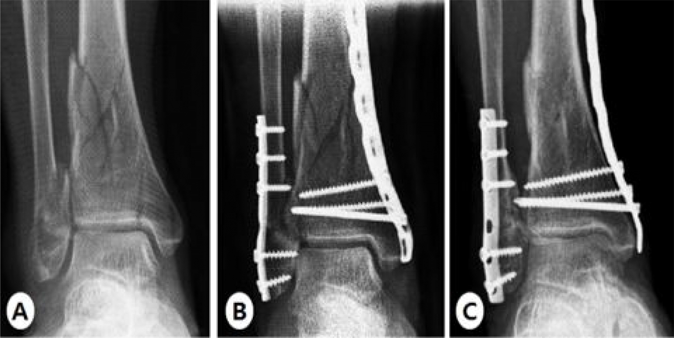 | Fig. 1.(A) A 73 yeared-old woman was sustained distal tibiofibular comminuted fracture by slip down. (B) The distal tibia fracture was stabilized with LCP metaphyseal plate using minimal invasive technique. (C) At 16 weeks postoperatively, radiographs shows bone union. |
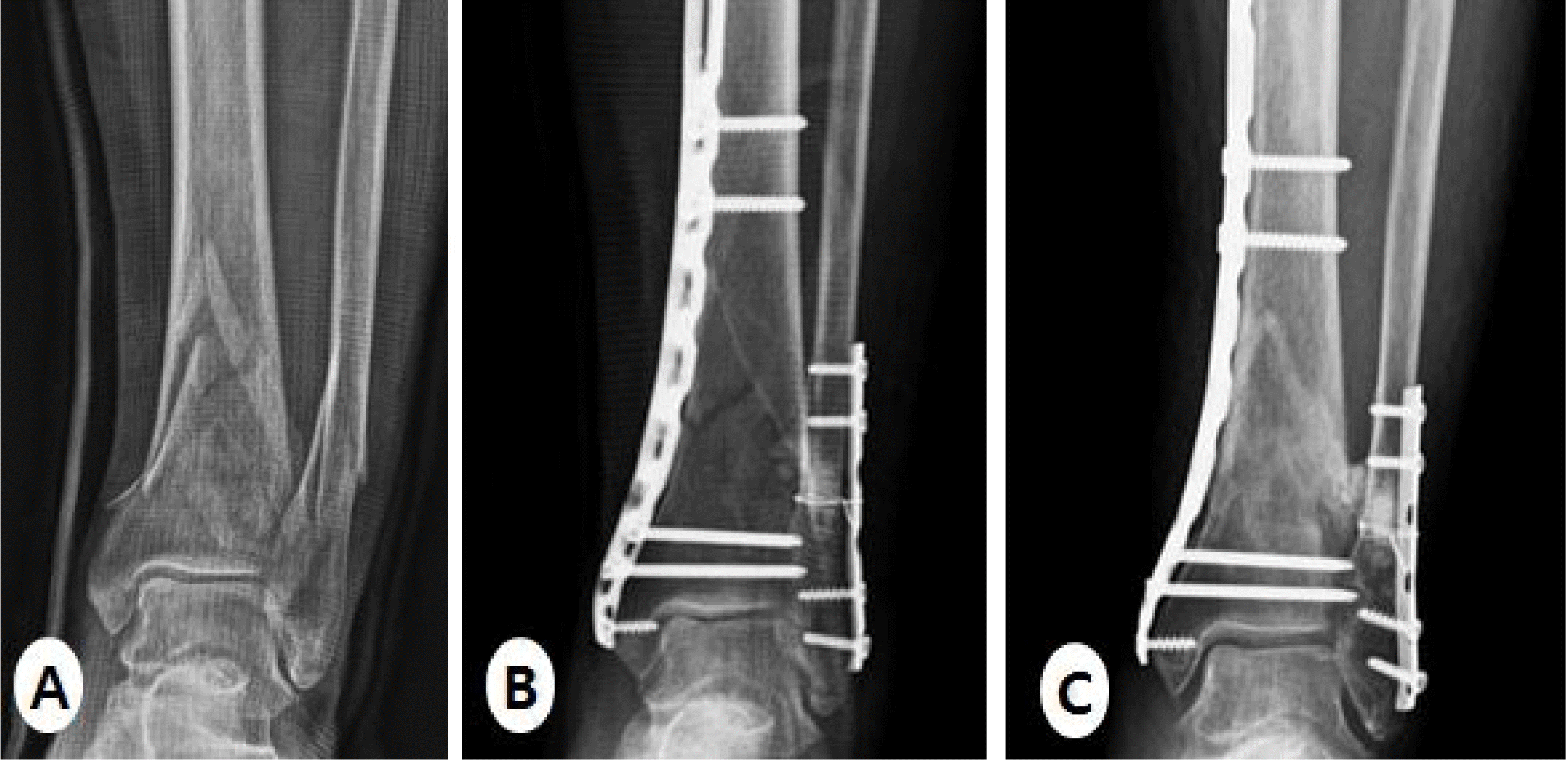 | Fig. 2.(A) A 65-yeared old woman was sustained distal tibiofibular fracture by slip down. (B) The distal tibia fracture was treated with MIPO technique using LCP metaphyseal plate on medial side. (C) At 12 weeks postoperatively, radiograph shows bone union. |
Table 1.
Patient data
| Case | Age/Sex | Injury mechanism | AO/OTA ⋆ classification | G-A† classification | Union time (week) | Ankle‡ score | Complication |
|---|---|---|---|---|---|---|---|
| 1 2 | 62/F | SD§ | A2 A1 | Closed Closed | 13 17 | 86 81 | No |
| 3 | 49/F | TA∥ | A1 | Closed | 21 | 80 | Metal irritation |
| 4 | 37/M | TA | A2 | Type I | 42 | 82 | Nonunion |
| 5 | 30/M | TA | A1 | Type I | 17 | 85 | No |
| 6 | 65/F | SD | A2 | Closed | 12 | 84 | No |
| 7 | 53/M | SD | A1 | Closed | 14 | 91 | No |
| 8 | 50/F | SD | A1 | Closed | 17 | 91 | No |
| 9 | 73/F | SD | B1 | Closed | 18 | 81 | No |
| 10 | 76/M | FD¶ | A1 | Closed | 15 | 88 | No |
| 11 | 26/M | FD | A1 | Closed | 12 | 95 | No |
| 12 | 64/F | SD | A2 | Closed | 14 | 81 | No |
| 13 | 51/M | TA | A1 | Type II | 24 | 71 | Skin infection |
| 14 | 54/F | SD | A1 | Type I | 14 | 92 | No |
| 15 | 48/M | SD | A1 | Closed | 13 | 95 | No |
| 16 | 73/F | SD | A3 | Closed | 16 | 85 | No |
| 17 | 69/M | FD | A3 | Closed | 18 | 81 | Skin infection |
 XML Download