

 PDF
PDF ePub
ePub Citation
 Print
Print


Abstract
Objective:
The ᅵink among carotid intimamedia thickness (IMT), vascular elastic property and the disease activity of systemic lupus erythematosus (SLE) is not well defined. We investigated the association between carotid atherosclerosis, elastic properties of the carotid arterial wall and clinical parameters of SLE.
Methods:
Fifty-one SLE patients and fifty healthy controls were included. Peak systolic global circumferential and posterior radial strains of carotid artery were measured to assess the elastic properties. Beta stiffness index was used as conventional method for the distensibility of the carotid artery. Information concerning SLE duration, cumulative dose of steroids and/or immunosuppressive drug intake was recorded, and SLE activity was assessed by SLE disease activity index (SLEDAI) score.
Results:
Carotid plaques were more common in SLE patients. SLE patients with plaques were 㢌der and showed the increased mean ᅵMT, high sensitivity C-reactive protein (hs CRP), IgG anti-cardi㢌ipin antibody (aCL), and longer disease duration compared with those without plaques. Peaksyst㢌ic global circumferential and posterior radial strain as well β stiffness index were significantly lower in SLE group. Age, disease duration, hsCRP, IgG aCL showed significant correlations with mean ᅵMT and parameters of carotid elastic property (all P's<0.05).
Conclusions:
Carotid atherosclerosis was more common in SLE patients, and carotid arterial stiffness had significant correlation with disease duration, hsCRP and IgG aCL level. Speckle tracking strain imaging is a comparative method for the assessment of elastic properties of carotid artery of SLE patients.
REFERENCES
1.J. Wajed., Y. Ahmad., P.N. Durrington., Bruce IN. Prevention of cardiovascular disease in systemic lupus erythematosus? proposed guidelines for risk management. Rheumatology. 2004. 43:7–12.
2.Manzi S., Meilahn EN., Rairie JE., Conte CG., Medsger TA Jr., Jansen-McWilliams L, et al. Age-specific incidence rates of myocardial infarction and angina in women with systemic lupus erythematosus: comparison with the Framingham Study. Am J Epidemiol. 1997. 145:408–15.

3.Roman MJ., Shanker BA., Davis A., Lockshin MD., Sammaritano L., Simantov R, et al. Prevalence and correlates of accelerated atherosclerosis in systemic lupus erythematosus. N Engl J Med. 2003. 349:2399–406.
4.Bjomadal L., Yin L., Granath F., Klareskog L., Ekborn A. Cardiovascular disease a hazard despite improved prognosis in patients with systemic lupus erythematosus: results from a Swedish population based study. J Rheumatol. 2004. 31:713–9.
5.Tassiulas IO., Boumpas DT. Textbook of Rheumatology. 8th ed.p. 1429–51. Philadelphia, Saunders;2009.
6.Alam TA., Seifalian AM., Baker D. A review of methods currently used for assessment of in vivo endothelial function. Eur J Vase Endovasc Surg. 2005. 29:269–76.
7.Sohn IS., Lee JB., Cho BH., Park JH., Jin ES., Cho JM, et al. Carotid intimamedia thickness and arterial stiffness in hypertensive patients with first attack of ischemic stroke. J Korean Soc Hypertens. 2010. 16:14–21.
8.Hoeks AP., Brands PJ., Smeets FA., Reneman RS. Assessment of the distensibility of superficial arteries Ultrasound Med Biol. 1990. 16:121–8.
9.Selzer RH., Mack WJ., Lee PL., Kwong-Fu H., Hodis HN. Improved common carotid elasticity and intimamedia thickness measurements from computer analysis of sequential ultrasound frames. Atherosclerosis. 2001. 154:185–93.
10.El-Magdmi M., Bodill H., Ahmad Y., Durrington PN., Mackness M., Walker M, et al. Systemic lupus erythematosus. An independent risk factor for endothelial dysfunction in women. Circulation. 2004. 110:399–404.
11.Svenuungsson E., Jensen-Urstad K., Heimburger M., Silveira A., Hamsten A., de Faire U, et al. Risk factors for cardiovascular disease in systemic lupus erythematosus. Circulation. 2001. 104:1887–93.
12.Greene ER., Lanphere KR., Sharrar J., Roldan CA. Arterial distensibility in systemic lupus erythematosus. Conf Proc IEEE Eng Med Biol Soc. 2009. 2009:1109–12.
13.Cacciapaglia F., Zardi EM., Coppolino G., Buzzelini F., Margiotta D., Arcarese L, et al. Stiffness parameters, intimamedia thickness and early atherosclerosis in systemic lupus erythematosus patients. Lupus. 2009. 18:249–56.
14.Barenbrock M., Kosch M., Joster E., Kisters K., Rahn KH., Hausberg M. Reduced arterial distensibility is a predictor of cardiovascular disease in patients after renal transplantation. J Hypertens. 2002. 20:79–84.
15.Van Popele NM., Grobbee DE., Bots ML., Asmar R., Topouchian J., Reneman RS, et al. Association between arterial stiffness and atherosclerosis: the Rotterdam Study. Stroke. 2001. 32:454–60.
16.Harloff A., Strecker C., Reinhard M., Kollum M., Handke M., Olschewski M, et al. Combined measurement of carotid stiffness and intimamedia thickness improves prediction of complex aortic plaques in pa仕ents with ischemic stroke. Stroke. 2006. 37:2708–12.
17.Manzi S. Systemic lupus erythematosus; a model for atherogenesis? Rheumatology. 2000. 39:353–9.
18.Ames PR., Margarita A., Alves JD. Antiphospholipid antibodies and atherosclerosis: insights from systemic lupus erythematosus and primary antiphospholipid syndrome. Clin Rev Allergy Immunol. 2009. 37:29–35.
19.Bots ML., Dijk JM., Oren A., Grobbee DE. Carotid intimamedia thickness, arterial stiffness and risk of cardiovascular disease: current evidence. J Hypertens. 2002. 20:2317–25.
20.Greenland P., Abrams J., Aurigemma GP., Bond MG., Clark LT., Criqui MH, et al. Prevention Conference V: Beyond secondary prevention: identifying the high-risk patient for primary prevention: noninvasive tests of atherosclerotic burden: Writing Group III. Circulation. 2000. 101:E16–22.
21.de Leeuw K., Smit AJ., de Groot E., van Roon AM., Kallenberg CG, Bijl M. Longitudinal study on premature atherosclerosis in patients with systemic lupus erythematosus. Atherosclerosis. 2009. 206:546–50.
22.Manzi S., Selzer F., Sutton-Tyrrell K., Fitzgerald SG., Rairie JE., Tracy RP, et al. Prevalence and risk factors of carotid plaque in women with systemic lupus erythematosus. Arthritis Rheum. 1999. 42:51–60.
23.Roman MJ., Shanker BA., Davis A., Lockshin MD., Sammaritano L., Simantov R, et al. Prevalence and correlates of accelerated atherosclerosis in systemic lupus erythematosus. N Engl J Med. 2003. 349:2399–406.
24.Gilman G., Khandheria BK., Hagen ME., Abraham TP., Seward JB., Belohlavek M. Strain rate and strain: a step-by-step approach to image and data acquisition. J Am Soc Echocardiogr. 2004. 17:1011–20.
25.Laurent S., Boutouyne P., Lacolley P. Structural and genetic bases of arterial stiffness. Hypertension. 2005. 45:1050–5.
26.Golemati S., Sassano A., Lever MJ., Bharath AA., Dhanjil S., Nicolaides AN. Carotid artery wall motion estimated from B-mode ultrasound using region tracking and block matching. Ultrasound Med Biol. 2003. 29:387–99.
27.Lee JH., Cho KI., Kim SM. Carotid arterial stiffness in pa仕ents with rheumatoid arthritis assessed by speckle tracking strain imaging: its association with carotid atherosclerosis Clin Exp Rheumatol. 2012. 30:720–8.
Figure 1.
An automated tracking algorithm outlined the intima- media complex of common carotid artery (a). Systolic and diastolic diameters of common carotid artery were obtained from short axis view (b). Regions of interest with computation area of 1x1 mm were placed in the intimamedia complex from the short axis view of the common carotid artery (c and d). Peak systolic global circumferential strain (c) and posterior radial strain (d) and during systole were used for the analysis.
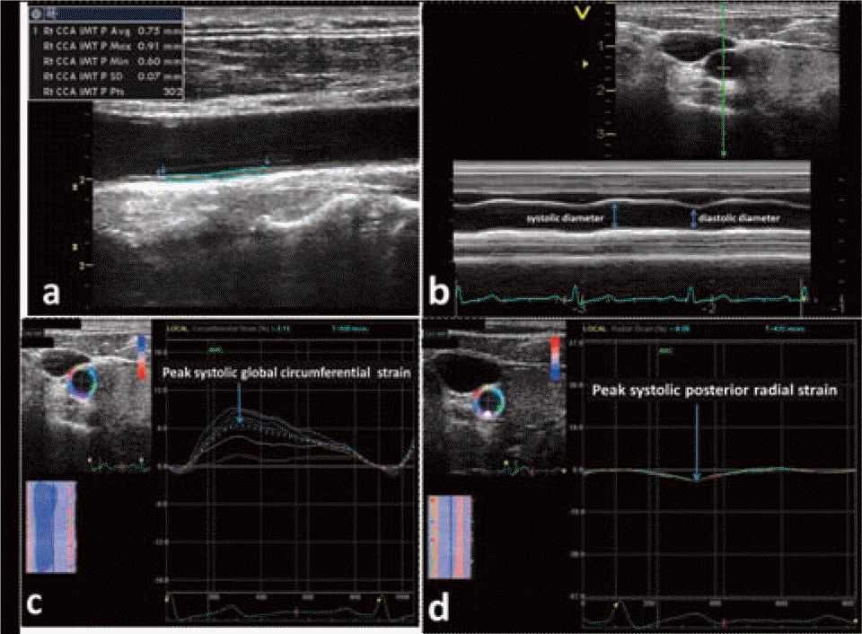
Table 1.
Clinical characteristics and parameters of the carotid artery of the study population
Table 2.
Characteristics of SLE patients with and without carotid arterial plaque
Table 3.
Correlation coefficients between the parameters of carotid artery and clinical parameters of SLE group (n=51)
Table 4.
Multiple linear regression analysis of mean IMT and parameters of arterial stiffness of common carotid artery in SLE groups.
 XML Download