

 PDF
PDF ePub
ePub Citation
 Print
Print


Abstract
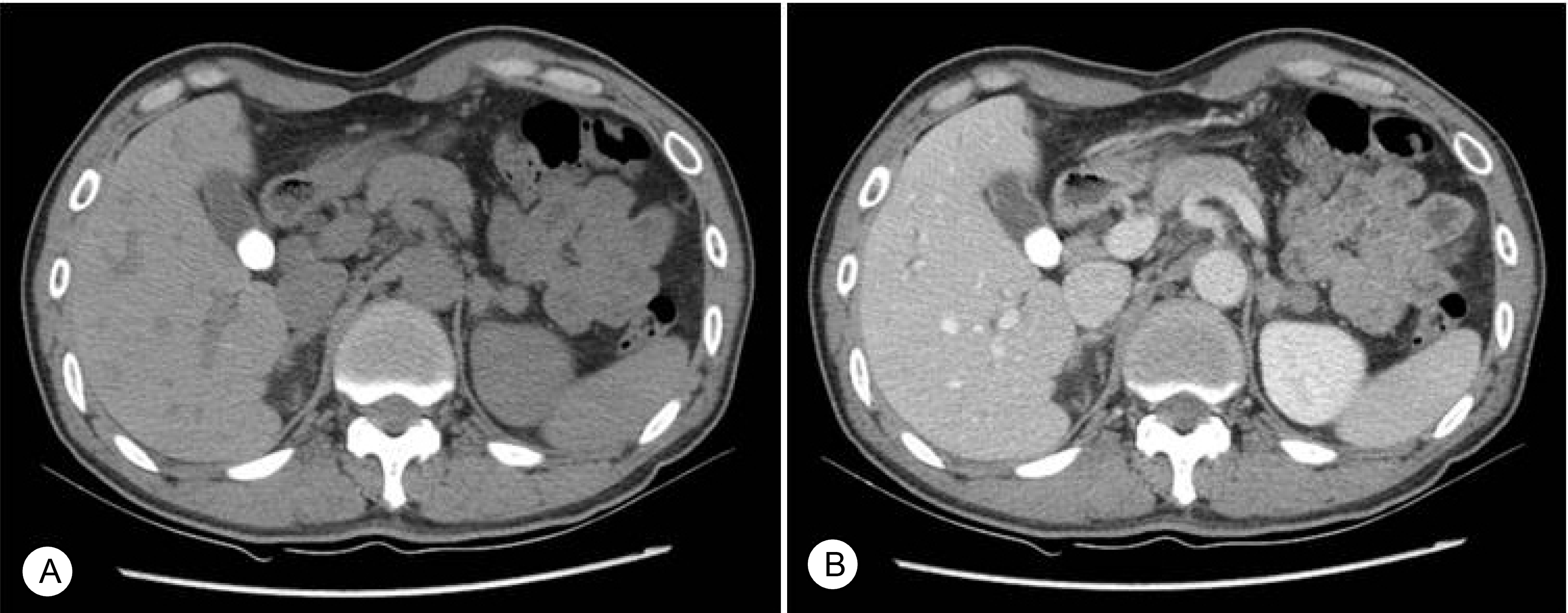
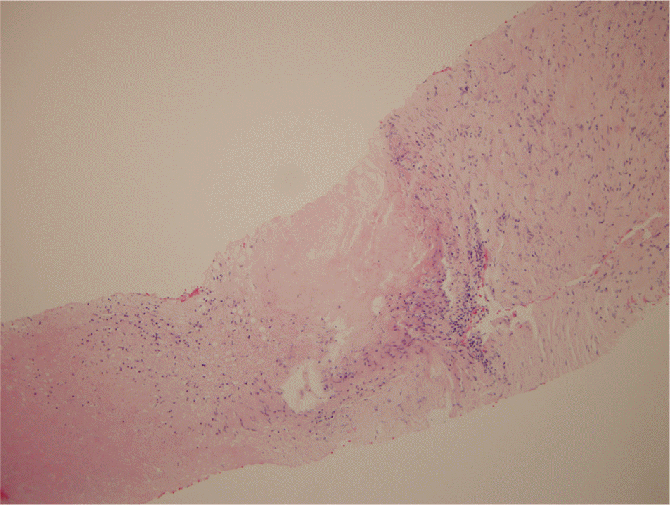
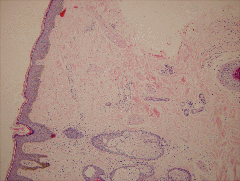
Addison’s disease is a rare disorder that causes fatigue, genral weakness, weight loss, pigmentation due to adrenal hypofunction and it’s underlying causes are various. We report a case of 42-year-old man with fatigue, generalized cutaneous pigmentation. Computed tomography showed bilateral adrenal enlargement, but no calcification. Adrenal tuberculosis was established by ultrasound-guided fine needle aspiration biopsy.
REFERENCES
1.Choi DY., Kim HS., Lee CK., Lim BS., Jung SJ., Tae CH. A case report of Addison's disease. Korean J Med. 1968. 11:455–60.
2.Park WY., Kee CS., Cho SK., Choi YK. A case report of Addison's disease. Korean J Med. 1971. 14-3:63–7.
3.Oelkers W. Adrenal insufficiency. N Engl J Med. 1996. 335:1206–12.

4.Sung SK., Kwon YJ., Lee BW., Kim DM., Yoo HJ. Clinical Review of Addison's Disease in Korea Previously Reported 14 Cases in Korea and 6 New Cases at National Medical Center. J Korean Soc Endocrinol. 1987. 2:189–93.
5.Ja Young Lee, Jee Hee Kim, Dong Joon Lim, Sung Dae Moon, Je Ho Han. A case of Addison's disease due to tuberculosis: pathologic confirmation by laparoscopic biopsy. Korean J Med. 2008. 75:704–8.
6.Kim JY., Jeon HC., Kim KY., Cha SE., Cha SE., Choi HS, et al. A case of Addison's disease due to tuberculosis: Pathological confirmation by fine-needle aspiration biopsy. J Korean Soc Endocrinol. 1995. 10:306–10.
7.Schultz CL. CT and MR of the adrenal glands. Seminars in ultrasound CT and MRI. 1986. 7:219–33.
8.Lam KY., Lo CY. A critical examination of adrenal tuberculosis and a 28-year autopsy experience of active tuberculosis. Clin Endocrinol (Oxf). 2001. 54:633–9.
9.Post FA., Soule SG., Willcox PA., Levitt NS. The spectrum of endocrine dysfunction in active pulmonary tuberculosis. Clin Endocrinol (Oxf). 1994. 40:367–71.
10.Vita JA., Silverberg SJ., Goland RS., Austin JHM., Knowlton AI. Clinical clues to the cause of Addison's disease. Am J Med. 1985. 78:461–6.
 XML Download