This article has been
cited by other articles in ScienceCentral.
Abstract
Anisakidosis is caused by Anisakis simplex and other anisakids larvae parasitizing marine fish and cephalopods. A lot of case reports about anisakidosis have been published in Korea because of raw fish eating habits. Recently seafood consumption has continued to increase due to health concerns and thus, it increases the risk for infectious diseases including anisakidosis. The aim of this review is to analyze the clinical and epidemiological characteristics of anisakidosis during the last 10 years in Korea, based on the case reports published from 2000 to 2010. The incidence age was changed from 30s and 40s to 50s. The young generation was considered to consume seafood in various ways, including raw fish as well. The most noticeable change was the appearance of Anisakis allergy patients over the last decade. The patients showed abdominal pain, urticaria after eating sea food. It reaffirmed that anisakid infection induces not only gastric and intestinal anisakidosis but also cause allergic reaction. Anisakid should be considered as a possible causative food allergen provoking allergic responses after eating raw fish.
Keywords: Anisakidosis, Anisakis allergy, Anisakis simplex, Case reports, Review
Anisakidosis (previously known as anisakiasis) refers to human infection with 3rd stage larva of ascaridoid nematodes (family Anisakidae). Ascaridoid worms, which are commonly called anisakids, normally parasitize adult marine mammals, such as sea whales, dolphins etc. These worms utilize cyclops as 1st intermediate host and marine fish and cephalopods as 2nd intermediate hosts or paratenic hosts. The family Anisakidae includes genus Contracaecum, Pseudoterranova and Raphidascaris, as well as genus Anisakis. The most commonly involved anisakid species in human infections is Anisakis simplex, less frequently Pseudoterranova decipens. When anisakids larvae are ingested by humans, they can penetrate into the stomach wall causing acute abdominal pain. When the larvae invade gastric or intestinal mucosa, inflammatory reaction often results in ulcer or eosinophilic granuloma associated with clinical symptoms. Recently it has been clear that anisakidosis is associated with allergic reactions.
Paratenic hosts of marine fish and cephalopods are particularly susceptible to infection with larval stage of anisakids. There have been many reports on the infection status of
A. simplex larva in fish since Chun's report.
1 Recently Choi et al., (2011) investigated more than 2,000 specimen including 43 species of fish and cephalopods, to study the infection status of
A. simplex larvae.
2 The overall infection rate was 34.3% and the average infection rate was 17.1 larvae per infected fish. These reports confirmed that infection rate of
A. simplex were still high in paratenic hosts.
The consumption per capita of seafood has increased continuously over the past decade because of health consciousness (Fisheries production survey, Statistics Korea, 2007). This increase can bring about a rise of infectious diseases by eating seafood. One of the most important infectious diseases will be the larval infection of anisakids, by ingestion of raw or inadequately salted, pickled, smoked or cooked fish or cephalopods.
Although there are many case reports on anisakidosis in Korea since the first case report in 1971,
3 the analysis of changing pattern of anisakidosis during the last 10 years is lacking. The objective of this study was to analyze the clinical and epidemiological characteristics of anisakidosis during the last 10 years in Korea, based on the case reports from 2000 to 2010. A review of the recent literature can provide opportunity of understanding the changing patterns of this zoonotic disease in Korea.
1. Data sources and syudy selection
The PubMed-Medline and KoreaMed database were searched for case reports published from 2000 to 2010. The following search strings were used: "anisakiasis and Korea" or "anisakidosis and Korea" or "aniskiasis and case report" or "anisakidosis and case report". Titles, abstracts and full-text publications were obtained and screened for original case report data. The review articles or case reports that were missing important data were excluded.
2. Data extraction
The following data about risk factors were extracted from the selected articles: age, gender, ingested paratenic host species, chief complaints, species identification of anisakids larvae, tests done at the hospitals, location of larvae or eosinophilic granuloma and treatment.
3. Case presentation
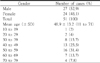
The Pubmed-Medline and KoreaMed search resulted in 30 case reports. The characteristics of the study population are shown in
Table 1. Fifty-three percent of the patients were male (
N=27) and 47.1% female (
N=24). The mean age was 48.9 year (range: 11 years to 71 years). Patients with age in their fifties were most abundant.
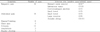
Table 2 provides the main clinical symptoms of the patients. Generally, symptoms of anisakidosis include severe epigastric pain, abdominal pain, nausea and vomiting. Forty out of 51 cases (78.4%) developed gastric pain, such as epigastric pain, nausea, vomiting and abdominal pain. In addition, urticaria and anaphylaxis were also revealed in allergic anisakidosis cases.
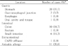
Table 3 demonstrates the location of larvae or eosinophilic granuloma in patients. The worms were most frequently located in the stomach (20 cases), followed by the colon (10 cases) and the small intestine (6 cases). It is noteworthy that allergic anisakidosis cases started to be reported in Korean patients since 2006. Allergic cases failed to find larva with gastrofiberscopy, although all patients recalled that they had eaten raw fish. But they showed specific IgE response to
A. simplex antigen.
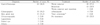
Table 4 shows parasitological factors of 51 Korean cases of anisakidosis. Sea eels were most commonly ingested fish by the patients, followed by squid and flatfish. More than half of the cases, however, were found eating mixed raw marine fish. The identified anisakid species was almost
Anisakis species, except 3 cases, which were diagnosed to
Pseudoterranova species. The species identification was not available in allergic cases and some eosinophilic granuloma cases.
Table 5 defines diagnostic tools and treatment for anisakidosis. The gastroscopy is the most widely used tool in anisakidosis. The colonoscopy was also applied frequently. The treatment was accomplished simply with the removal of worms in 19 cases (37.3%), followed by a resection of the affected lesion (13 cases). No treatment was done in allergic cases. Some cases were treated with albendazole (4 cases).
DISCUSSION
The aim of the present study is to review the characteristics of anisakidosis which was reported in the past 10 years in Korea. The most prominent change was the appearance of the Anisakis allergy cases, which have not been reported in Korea before 2006. All anisakid allergy patients showed symptoms of urticaria, without worm detection with gastrofiberscopy.
Anisakis species was reconfirmed as the most frequent species in human infection. Only 3 cases were identified as Pseudoterranova species among these 51 cases. Most of case reports, however, described briefly about anisakid larva species. It can be attributed that the case reports were written by clinicians and the focus was fixed on the disease itself. The detailed description of epidemiological factors, such as consumed fish, and the parasite species identification can bring deeper understanding of the relationship between anisakid species and anisakidosis.
The two most commonly consumed fish in anisakidosis cases in Korea were sea eel and squid. The present finding also showed a similar preference for fish eating. Because most of the fish and cephalopods were infected with A. simplex, eating any of these species of fish is likely to be infected with anisakids.
Age distribution revealed a difference compared to those found in the previous reports. The peak age of anisakid infection was 30s and 40s in the previous reports.
4-
7 The present study, however, demonstrated that 50s was the peak age of infection. This shift considered that the pattern of fish consumption is changing in young generation. Fisheries production survey (Statistics Korea, 2007) revealed that young generation consumes fish products in various ways, in addition to raw fish eating. No significant difference between the number of male and female was observed.
Sixteen out of 21 gastric cases complained epigastric pain. The epigastric pain is well-known as the chief complaints of gastric anisakidosis. We also confirmed epigastric pain to be the most common symptom in gastric anisakidosis. Almost 70% of gastric anisakidosis patients showed epigastric pain (
Table 2). However, intestinal anisakidosis developed various symptoms. Especially cases showing large intestine involvement produced a variety of symptoms. This finding implies that the diagnosis of intestinal anisakidosis would be difficult without specific information, such as raw fish eating habit.
The most widely used diagnostic tool is gastrofiberscopy. Since the first Korean case of anisakidosis was accomplished with gastrofiberscopy, it became widely used since it provides both diseases to be diagnosed and treated simultaneously. Anisakidosis cases rapidly increased after frequent use of gastrofiberscopy.
7 The most effective treatment of anisakidosis has been known to remove the larvae. We also confirmed that 64% of patients were treated by worm removal. Albendazole was treated in some cases. But the effect of albendazole against anisakid infection was not known.
The most noticeable change in ansakidosis over the last decade was that allergic patients began to appear. Most of anisakis allergic patients in Korea showed urticaria symptoms. But larvae were not found, despite their consumption of raw fish. Since allergic responses, including urticaria, anaphylaxis and angioedema, were reported after eating raw fish in Japan and Spain,
8-
10 many allergic cases have been reported throughout the world.
11-
13 In Korea, anisakis allergy cases were reported since 2006.
14,
15 The patients were admitted due to abdominal pain and allergic responses, such as urticaria and some other allergic symptoms after eating raw fish. The specific IgE to Anisakis were positive. Recently Kim et al., (2011) reported a case of anisakidosis seroprevalence of 5.0-6.6% in residents of southern part of Korea.
16 This study also implies that there is the possibility of Anisakis allergy patient in the Korean population. Anisakis allergy can be suspected in case of allergy patient who has raw fish eating habit.
In summary, the most significant change of anisakidosis in Korea over the last decade was that anisakis allergy patients had started to be reported in Korea. This finding reaffirmed that anisakid infection induces not only gastric and intestinal anisakidosis but allergic responses. Anisakid should be considered as a causative allergen which provokes allergic responses after eating raw fish.
CONCLUSION
We reviewed the case reports published from 2000 to 2010 to analyze the changing patterns of anisakidosis during the last 10 years in Korea. A total of 30 case reports were selected for review. We concluded several findings through a review of these articles and have summarized them below.
The peak age was delayed to age 50s compared to that of the previous reports
The most frequent symptom of gastric anisakidosis was epigastric pain. But symptoms of intestinal anisakidosis were variable. Anisakis allergic patients showed abdominal pain and urticaria symptom in common.
The anisakids were most frequently located in the stomach, followed by colon and small intestine. The most obvious change was the appearance of allergy patients since 2006. Allergic cases failed to find larva with gastrofiberscopy, although all patients had eaten raw fish. But they showed specific IgE response to A. simplex antigen.
Sea eels and squid were still the most common species to cause infection. In more than half of the cases, however, the Anisakis species could not be identified. The identified anisakid species was almost Anisakis species, except 3 cases which were diagnosed to Pseudoterranova species. The species identification was not available in allergic cases and some eosinophilic granuloma cases.
The gastrofiberscopy was the most widely used tool in anisakidosis. The colonoscopy was also applied frequently in intestinal anisakidosis. The treatment was accomplished simply by removing the worms in 19 cases, followed by a resection of affected lesion. No treatment was done in allergic cases.
As the conclusion, we found that the incidence age of anisakidosis was delayed to their 50s. The most remarkable change was that Anisakis allergy patient appeared since 2006. Anisakid larvae should be considered as a food allergen, as well as the cause of gastric or intestinal anisakidosis.
Anisakidosis is caused by Anisakis simplex and other anisakids larvae parasitizing marine fish and cephalopods. Recently seafood consumption has continued to increase due to health concerns and thus, it increases the risk for infectious diseases including anisakidosis. The aim of this review was to analyze the clinical and epidemiological characteristics of anisakidosis during the last 10 years in Korea. The most prominent change was the appearance of the Anisakis allergy cases, which have not been reported in Korea before 2006. All anisakid allergy patients showed symptoms of urticaria. In this review, new diagnostic methods in patients with Anisakidosis infection to make it easier to understand the summary and, in the early diagnosis in clinical practice is thought to be very helpful.


 PDF
PDF ePub
ePub Citation
 Print
Print


 XML Download