

 PDF
PDF ePub
ePub Citation
 Print
Print


Abstract
Objectives
The purpose of this study is to compare the apical transportation and working length change in curved root canals created in resin blocks, using 3 geometrically different types of Ni-Ti files, K3, NRT, and Profile.
Materials and Methods
The curvature of 30 resin blocks was measured by Schneider technique and each groups of Ni-Ti files were allocated with 10 resin blocks at random. The canals were shaped with Ni-Ti files by Crown-down technique. It was analyzed by Double radiograph superimposition method (Backman CA 1992), and for the accuracy and consistency, specially designed jig, digital X-ray, and CAD/CAM software for measurement of apical transportation were used. The amount of apical transportation was measured at 0, 1, 3, 5 mm from 'apical foramen - 0.5 mm' area, and the alteration of the working length before and after canal shaping was also measured. For statistics, Kruskal-Wallis One Way Analysis was used.
Figures and Tables
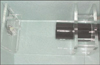 | Figure 1Photograph of a jig that maintains a constant distance between a X-ray tube and a resin block.
|
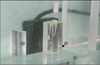 | Figure 2Photograph of a resin block and a digital sensor. (a) Holding part of digital radiographic sensor. (b) Cartesian system. (c) Table for constant position of resin block.
|


References
1. Schilder H. Cleaning and shaping the root canal. Dent Clin North Am. 1974. 18(2):269–296.
2. al-Omari MA, Dummer PM, Newcombe RG, Doller R. Comparison of six files to prepare simulated root canals. 2. Int Endod J. 1992. 25(2):67–81.

3. Schafer E, Tepel J, Hoppe W. Properties of endodontic hand instruments used in rotary motion. Part 2. Instrumentation of curved canals. J Endod. 1995. 21(10):493–497.
4. Roane JB, Sabala CL, Duncanson MG Jr. The "balanced force" concept for instrumentation of curved canals. J Endod. 1985. 11(5):203–211.
5. Abou-Rass M, Frank AL, Glick DH. The anticurvature filing method to prepare the curved root canal. J Am Dent Assoc. 1980. 101(5):792–794.
6. Caldwell JL. Change in working length following instrumentation of molar canals. Oral Surg Oral Med Oral Pathol. 1976. 41(1):114–118.
7. Civjan S, Huget EF, DeSimon LB. Potential applications of certain nickel-titanium (nitinol) alloys. J Dent Res. 1975. 54(1):89–96.
8. Walia HM, Brantley WA, Gerstein H. An initial investigation of the bending and torsional properties of Nitinol root canal files. J Endod. 1988. 14(7):346–351.
9. Gambill JM, Alder M, del Rio CE. Comparison of nickel-titanium and stainless steel hand-file instrumentation using computed tomography. J Endod. 1996. 22(7):369–375.
10. Glossen CR, Haller RH, Dove SB, del Rio CE. A comparison of root canal preparations using Ni-Ti hand, Ni-Ti engine-driven, and K-Flex endodontic instruments. J Endod. 1995. 21(3):146–151.
11. Elliott LM, Curtis RV, Pitt Ford TR. Cutting pattern of nickel-titanium files using two preparation techniques. Endod Dent Traumatol. 1998. 14(1):10–15.
12. Bergmans L, Van Cleynenbreugel J, Wevers M, Lambrechts P. Mechanical root canal preparation with NiTi rotary instruments: rationale, performance and safety. Status report for the American Journal of Dentistry. Am J Dent. 2001. 14(5):324–333.
13. Bryant ST, Thompson SA, al-Omari MA, Dummer PM. Shaping ability of Profile rotary nickel-titanium instruments with ISO sized tips in simulated root canals: Part 1. Int Endod J. 1998. 31(4):275–281.
14. Bramante CM, Berbert A, Borges RP. A methodology for evaluation of root canal instrumentation. J Endod. 1987. 13(5):243–245.
15. Garip Y, Gunday M. The use of computed tomography when comparing nickel-titanium and stainless steel files during preparation of simulated curved canals. Int Endod J. 2001. 34(6):452–457.
16. Backman CA, Oswald RJ, Pitts DL. A radiographic comparison of two root canal instrumentation techniques. J Endod. 1992. 18(1):19–24.
17. Glickman GN, Koch KA. 21st-century endodontics. J Am Dent Assoc. 2000. 131:Suppl. 39S–46S.
18. Pruett JP, Clement DJ, Carnes DL Jr. Cyclic fatigue testing of nickel-titanium endodontic instruments. J Endod. 1997. 23(2):77–85.
19. Bonetti Filho I, Miranda Esberard R, de Toledo Leonardo R, del Rio CE. Microscopic evaluation of three endodontic files pre- and postinstrumentation. J Endod. 1998. 24(7):461–464.
20. Weine FS, Kelly RF, Lio PJ. The effect of preparation procedures on original canal shape and on apical foramen shape. J Endod. 1975. 1(8):255–262.
21. Lim KC, Webber J. The validity of simulated root canals for the investigation of the prepared root canal shape. Int Endod J. 1985. 18(4):240–246.
22. Dummer PM, Alodeh MH, al-Omari MA. A method for the construction of simulated root canals in clear resin blocks. Int Endod J. 1991. 24(2):63–66.
23. Cha YB, Jung IY, Lee SJ, et al. Shaping ability of three ProFile rotary instrumentation techniques in simulated resin root canals. J Endod. 2000. 26(12):719–723.
24. Thompson SA, Dummer PM. Shaping ability of ProFile.04 Taper Series 29 rotary nickel-titanium instruments in simulated root canals. Part 1. Int Endod J. 1997. 30(1):1–7.
25. Schafer E, Florek H. Efficiency of rotary nickel-titanium K3 instruments compared with stainless steel hand K-Flexofile. Part 1. Shaping ability in simulated curved canals. Int Endod J. 2003. 36(3):199–207.
26. Iqbal MK, Maggiore F, Suh B, Edwards KR, Kang J, Kim S. Comparison of apical transportation in four Ni-Ti rotary instrumentation techniques. J Endod. 2003. 29(9):587–591.
 XML Download