

 PDF
PDF ePub
ePub Citation
 Print
Print


INTRODUCTION
Posterior composite restorations have risen in popularity as a result of the development of improved resin composites, bonding systems and operating techniques.1 Direct composite restorations are preferred to indirect composite restorations because they require minimal intervention during cavity preparation.2 A major limitation of direct restoration is the difficulty of controlling the degree of conversion and polymerization shrinkage.3 To overcome these limitations, the indirect fabrication of a composite inlay and cementation with a resin cement has been advocated. Unfortunately, the current available resin cements with indirect restorations do not always bond to dentin as strongly as dentin adhesive systems bond with direct resin composite restorations.4,5 A relatively weak bond to the tooth structure leads to a host of problems: marginal gap formation, postoperative sensitivity, premature failure, and secondary caries formation.5,6
Regarding an indirect composite restoration, successful dentin bonding is of particular clinical importance for inlays because the final strength of the tooth-restoration complex is highly dependent on the dentin adhesive procedures.7-10 For the cementation of an indirect composite restoration, several techniques have been proposed for a dentin bonding procedure. In the most commonly used method, the application of a dentin adhesive with light-curing on the treated dentin surface is followed by the application of resin cement. Nevertheless, the cured dentin adhesive could pool at the margin or inner line angles of the cavity due to the effect of gravity and the relatively low viscosity, which could hinder seating of the restoration.11-14 These concerns regarding the possible detrimental effects of pre-curing of dentin adhesive have led to another method, where the dentin adhesive is not polymerized beforehand but is polymerized with resin cement.13-15 In comparative studies of the bond strength, the tooth-restoration complex by dentin adhesive pre-curing was reported to have superior bond strength compared to when the dentin adhesive and resin cement had been cured simultaneously.11,12,16,17 Only a small number of reports have explained the reason for the higher bond strength of the former method, even though there are many comparative reports of the bond strength according to the application technique of dentin adhesive.
The introduction of confocal laser scanning microscopy (CLSM) provides a valuable new technique for visualizing the bonding structures, such as the hybrid layer and resin tags in dentin. The advantages of confocal microscopy include a non-destructive examination because the layer visualized can be up to 100 µm below the surface and drying of samples is unnecessary, meaning there is less risk of shrinkage or other drying artifacts.17-21
This study examined the effect of the light-curing of dentin adhesive on the bond interface when luting a resin inlay to dentin using microtensile bond strength tests and confocal microscopy.
MATERIALS AND METHODS
Twenty four extracted permanent molars without cavities, cracks or other defects were stored in a physiological saline solution. After embedding the teeth in a self-cure acrylic resin, the occlusal surfaces were ground flat to expose the dentin surface under water irrigation using a low speed diamond saw (MetSAW, MSH-04-112, R&B Inc., Daejeon, Korea). The hydrophilic primer of 3-step etch-and-rinse adhesive system (OptiBond FL, Kerr, Orange, CA, USA) and the bonding agent of 2-step etch-and-rinse adhesive system (One-Step, Bisco, Schaumburg, IL, USA) were labeled with 0.1% Rhodamine B isothiocyanate (R1755, batch No. 114F0376, Sigma, St. Louis, MO, USA) (Table 1). The fluorescent dye was applied directly to the supplied packaged bottles. The teeth were divided into 2 groups, according to the restoration methods; direct and indirect resin restoration (Figure 1).
For direct resin restoration groups, which served as the control groups, the exposed dentin surface was etched with 37% phosphoric acid (ETCH-37, Bisco) for 20 seconds, washed with water for 15 seconds and the surface was gently air dried. Subsequently, dentin adhesive was applied according to the manufacturer's recommendation. The entire surfaces were built with composite resin (EsteliteΣ, Tokuyama dental Corp, Tokyo, Japan) up to 3 mm, and each increment of 1 mm thick was light polymerized (Demetron LC, Kerr) at 600 mW/cm2 for 20 seconds.
For the indirect resin restoration groups, the exposed dentin surfaces were restored using a provisional restoration material (Quicks, Denkist, Seoul, Korea), 2 mm in height, and light polymerized for 20 seconds. After 1 week storage in saline, the provisional restoration was removed and dentin was cleaned with pumice. The experimental groups were varied according to the sequence and mode of application of the dentin adhesives.
In Groups OB-C and OB-NC, the exposed dentin surface was etched and washed in the same manner as in the control groups. The OptiBond FL primer was applied to the dentin surface, and air dried thoroughly for 5 seconds to evaporate the solvent. The adhesive resin was applied and air dried slightly. Light-curing of the adhesive resin was followed for 20 seconds for Group OB-C. On the other hand, Group OB-NC, the applied adhesive resin was left uncured until the application of the luting material. In Group OS-C and OS-NC, the exposed dentin surface was treated in a similar manner to previous groups. One-Step was applied to the surface, and air dried thoroughly for 5 seconds. Light-curing of the bonding agent was followed for 20 seconds for Group OS-C. In Group OS-NC, the applied bonding agent was left uncured until the luting material was applied (Table 2).
Sixteen indirect resin blocks, 3 mm in height and 10 mm in diameter (Tescera, Bisco), were prepared to simulate the laboratory-processed resin composite restorations. The surface of each composite disc was sandblasted with 50 µm aluminum oxide particles for 10 seconds from a distance of approximately 5 mm. The composite disc surface to be cemented was silanized with a Monobond-S (Ivoclar vivadent) for 1 minute, and then air dried. Variolink II (Ivoclar vivadent) luting resin cement was mixed according to the manufacturer's instructions, and applied to the tooth specimen with the composite disc. The excess resin cement was removed with a probe. The resin cement was light polymerized for 60 seconds.
The test specimens were then stored in 100% humidity for 24 hours at 37℃. The teeth of each group were divided randomly into two sub-groups for microtensile bond strength (µTBS) testing and CLSM (LSM510, Carl Zeiss, Oberkochen, Germary) observations.
Microtensile bond strength test
After storing in distilled water for 24 hours, the teeth were sectioned in the X and Y perpendicular directions with a low speed diamond saw to obtain 20 specimens in each group, 1 × 1 mm in size and 8 mm in length. The ends of each specimen were fixed to the Microtensile tester (Bisco) with cyanoacrylate adhesive (ZAPIT base, Dental Ventures of America Inc., Corona, CA, USA) plus an accelerator (ZAPIT accelerator, Dental Ventures of America Inc.). The results were obtained at the moment of the specimen fracture and calculated in MPa.
One-way ANOVA followed by Student-Newman-Keuls test was used to determine any differences between the groups using same material. The differences between the whole experimental groups were statistically analyzed with the two-way ANOVA and SNK test at a 5% level of significance.
Confocal laser scanning microscope analysis
After 24 hours water immersion, three 500-µm thick bucco-lingual sectioned samples were obtained from each teeth by sectioning in the perpendicular directions with a low speed diamond saw (MetSAW, MSH-04-112). The sliced specimens were mounted on glass slides and analyzed. The resin/dentin interfacial image examination was performed with a Leica TCS SP5 II microscope (Leica, Heidelberg, Germany) using a 50× epiplan objective.
RESULTS
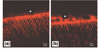
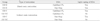
The results of the study are displayed in Table 3. The direct resin restoration groups, Groups OB-D and OS-D, showed higher µTBS than the indirect resin restoration groups. The mean µTBSs within the direct restoration groups (Groups OB-D and OS-D) were not statistically different, 24.37 and 21.94 MPa, respectively. The hybrid layer was definite and uniform, and some filler particles of the composite were also observed in these groups (Figure 2).
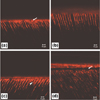
Light-curing of dentin adhesives and the use of three-step etch-and-rinse adhesives exhibited significantly higher µTBS in the indirect restoration groups (p < 0.05). Group OB-C, which showed the highest µTBS, contained a typical resin dentin interdiffusion zone (RDIZ, Figure 3a). The primer of OptiBond FL and fluorescent dye infiltrated the demineralized intertubular dentin, creating an evident and homogenous hybrid layer. Moreover, it is likely that the primer infiltrated a small layer of intertubular dentin from the lateral direction. Group OS-C also showed a clearly distinguishable hybrid layer but had a thinner hybrid layer than Group OB-C (Figure 3c). In this group, multiple lateral branches were observed along with resin tags as well.
In the uncured dentin adhesive groups, Groups OB-NC and OS-NC, the structural features of the RDIZ were different from those observed in the cured dentin adhesive groups. Group OB-NC showed an uncertain and blurred hybrid layer in RDIZ (Figure 3b). In Group OS-C, the hybrid layer was often discontinuous or was even absent (Figure 3d).
DISCUSSIONS
This in vitro study was designed to clarify the effect of light-curing of dentin adhesive when luting a resin inlay on the bond strength and micromorphological appearance of a RDIZ. The mean µTBSs of the cured dentin adhesives groups was higher than those of the uncured dentin adhesives, which is consistent with previous studies.11-15,22 The RDIZ of the cured adhesive groups, particularly Group OB-C (Figure 3a) showed an evident and homogenous hybrid layer along with the margin of the resin cement layer. Precuring of dentin adhesive prior to luting the resin inlay to dentin might improve the bond strength, and might be related to the formation of a thicker and more definite hybrid layer.
The µTBS of Group OB-NC was lower than that of Group OB-C. The CLSM image of this group showed a blurred and uncertain hybrid layer in RDIZ. This suggests that the uncured dentin adhesive might have spread and mixed with the resin cement during its application.
Group OS-NC exhibited the lowest bond strength (12.34 MPa) among the experimental groups, which was approximately half value of the control direct restoration groups (24.3 and 21.9 MPa). Surprisingly, the most common micromorphologic image of this group was debonding at the top of the hybrid layer in RDIZ (Figure 3d). It could be assumed that these defects and the discontinuity of the hybrid layer contribute to the lower bond strength compared to the other groups. Lee and Park reported that when dentin adhesives are not light cured prior to cementation, the exposed, decalcified collagen could collapse during the cementing procedure as a consequence of the pressure applied during the luting process, which can lead to the failure of an indirect restoration.12 However, from a practical point of view, it is recommended that the adhesive resin be kept unpolymerized before the restoration is fully seated because of interference of complete insertion, as mentioned previously. Considering the above, Magne et al. suggested the immediate dentin sealing (IDS) technique, that applying a dentin bonding agent (DBA) prior to impression taking.8,13 IDS technique provides the ideal condition for dentin bonding that has no contamination by temporary restoration and the freshly cut and clean dentin. Further, recent studies exhibited that prepolymerization of the DBA results in improved bond strength.1,8,12,13,23
The debonding of uncured dentin adhesive is also due to polymerization shrinkage of resin cement, which occurred during the light-curing procedure. Being unfilled or less highly-filled, the magnitude of polymerization shrinkage of resin cement is expected to be higher than direct restorative composites. This result confirmed previous reports that the bond between the resin inlay and resin cement was the weak link in indirect composite restorations.24,25 It is possible that (1) the uneven and ruptured hybrid layer and resin tags in the indirect resin restoration groups contributed to the lower bond strength, (2) polymerization shrinkage of resin cement occurring while light-curing weaken the bond strength. Variolink II used in the present study showed significant superior µTBS compared to the other composite luting cement, such as self-adhesive resin cement (e.g. Rely X Unicem). Variolink II luting cement is a dual-cure composite system that requires a total etching and bonding procedure. Owing to this distinctive dentin bonding procedure, it might exhibit an evident hybrid layer and thick resin tags (Figure 3a) when following the manufacturer`s instruction.26-28
In general, two-step etch-and-rinse adhesives performed less favorable clinically than conventional three-step etch-and-rinse adhesives, which is consistent with previous studies.29,30 Since simplified etch-and-rinse adhesive contain higher percentages of hydrophilic monomers, such as HEMA, compared to three-step adhesive, they exhibit more permeability after polymerization, which facilitates the presence of water-contained areas within the hybrid layer.31,32 On the other hand, in three-step adhesive, the hydrophobic coat of bonding resin may in part overcome the water movement, preserving the adhesive interface from hydrolysis. OptiBond FL, three step etch-and-rinse adhesive, also contains a barium glass filler with approximately 0.6 µm sized particles in its composition. The barium glass filler provides the bond gel consistency, which according to the theory of an 'elastic cavity wall', improves the bond strength and reduces polymerization shrinkage of the adhesive resin.
CONCLUSIONS
The µTBS of a resin inlay to dentin is improved by the light-curing of the dentin adhesive prior to the application of the cementing material. The hybrid layer was definite, homogenous in the RDIZ when the dentin adhesive was light cured before luting the resin inlay but not when it was cured with the resin cement simultaneously.


 XML Download