

 PDF
PDF ePub
ePub Citation
 Print
Print


Abstract
Objectives
The purpose of this study was to compare the microshear bond strength (µSBS) and bonding interfaces of two-step total-etching and self-etching adhesive systems to three etch types of dentin either the acid etched, laser etched or laser and acid etched.
Materials and Methods
The occlusal dentinal surfaces of thirty human molars were used. They were divided into six groups: group 1, 37% H3PO4 + Single Bond 2 (3M ESPE); group 2, Er:YAG laser (KEY Laser 3, KaVo) + Single Bond 2; group 3, Er:YAG laser + 37% H3PO4 + Single Bond 2; group 4, Clearfil SE Primer + Bond (Kuraray); group 5, Er:YAG laser + Clearfil SE Bond; group 6, Er:YAG laser + Clearfil SE Primer + Bond. The samples were subjected to µSBS testing 24 hr after bonding. Also scanning microscopic evaluations were made on the resin-dentin interfaces of six specimens.
Results
The µSBS of group 2 was significantly lower than that of groups 1 and 3 in Single Bond 2 (p < 0.05). There were significant differences among the uSBS of groups 4, 5, and 6 in Clearfil SE Bond (p < 0.05). Very short and slender resin tags were observed in groups 2 and 5. Long and slender resin tags and lateral branches of tags were observed in groups 3 and 6.
Figures and Tables
Figure 1
Scanning electron microscophic photograph showing the hybrid layer (H) and many taper resin tags with lateral branches (LB) in Group 1.
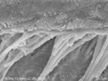
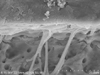
Figure 2
Scanning electron microscophic photograph showing the wide gap (G) and few short and slender resin tags in Group 2.
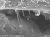
Figure 3
Scanning electron microscophic photograph showing the hybrid layer (H), slender and long rod shaped resin tags with lateral branches (LB) in Group 3.
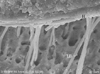
Figure 4
Scanning electron microscophic photograph showing the hybrid layer (H) and many long rod shaped resin tags with lateral branches (LB) in Group 4.
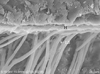
Figure 5
Scanning electron microscophic photograph showing the small gap (G) at the resin-dentin interface and short and slender resin tags in Group 5.
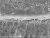
Figure 6
Scanning electron microscophic photograph showingthe hybrid layer (H) and few slender and long resin tags in Group 6.

References
1. Tay FR, Carvalho R, Sano H, Pashley DH. Effect of smear layers on the bonding of a self-etching primer to dentin. J Adhes Dent. 2000. 2:99–116.
2. Prati C, Chersoni S, Mongiorgi R, Pashley DH. Resin-infiltrated dentin layer formation of new bonding systems. Oper Dent. 1998. 23:185–194.
3. Hibst R, Keller U, Steiner R. Die wirkung gepulster Er:YAG Laserstrhlung auf zahngewebe. Lasers Sur Med. 1988. 4:163–165.
4. Sakakibara Y, Ishimaru K, Takamizu M. A study on bond strength to dentin irradiated by Erbium:YAG laser. Jpn J Conservative Dent. 1998. 41:207–219.
5. Barceleiro Mde O, de Mello JB, de Mello GS, Dias KR, de Miranda MS, Sampaio Filho HR. Hybrid layer thickness and morphology: the influence of cavity preparation with Er:YAG laser. Oper Dent. 2005. 30:304–310.
6. Gimbel CB. Hard tissue laser procedures. Dent Clin North Am. 2000. 44:931–953.
7. Keller U, Hibst R. Experimental studies of the application of the Er:YAG laser on dental hard substances:II. Light microscopic and SEM investigations. Lasers Surg Med. 1989. 9:345–351.

8. Visuri SR, Gilbert JL, Wright DD, Wigdor HA, Walsh JT Jr. Shear strength of composite bonded to Er:YAG laser-prepared dentin. J Dent Res. 1996. 75:599–605.
9. Sassi JF, Chimello DT, Borsatto MC, Corona SA, Pecora JD, Palma-Dibb RG. Comparative study of the dentin/adhesive systems interface after treatment with Er:YAG laser and acid etching using scanning electron microscope. Lasers Surg Med. 2004. 34:385–390.
10. Giachetti L, Scaminaci Russo D, Scrpelli F, Vitale M. SEM analysis of dentin treated with the Er:YAG laser: a pilot study of the consequences resulting from laser use on adhesion mechanism. J Clin Laser Med Surg. 2004. 22:35–41.
11. Dunn WJ, Davis JT, Bush AC. Shear bond strength and SEM evaluation of composite bonded to Er:YAG laser-prepared dentin and enamel. Dent Mater. 2005. 21:616–624.
12. Martínez-Insua A, Da Silva Dominguez L, Rivera FG, Santana-Penín UA. Differnces in bonding to acid-etched or Er:YAG-laser-trated enamel and dentin surfaces. J Prosthet Dent. 2000. 84:280–288.
13. Ramos RP, Chinelatti MA, Chimello DT, Borsatto MC, Pécora JD, Palma-Dibb RG. Bonding of self-etching and total-etch systems to Er:YAG laser-irradiated dentin. Tensile bond strength and scanning electron microscopy. Braz Dent J. 2004. 15(Spec No):SI9–SI20.
14. Bertrand MF, Semez G, Leforestier E, Muller-Bolla M, Nammour S, Rocca JP. Er:YAG laser cavity preparation and composite resin bonding with a single-component adhesive system: relationship between shear bond strength and microleakage. Lasers Surg Med. 2006. 38:615–623.
15. Van Meerbeek B, De Munk J, Mattar D, Van Landuyt K, Lambrechts P. Microtensile bond strength of an etch&rinse and self-etch adhesive to enamel and dentin as a function of surface treatment. Oper Dent. 2003. 28:647–660.
16. Eguro T, Maeda T, Otsuki M, Nishimura Y, Katsuumi I, Tanaka H. Adhesion of Er-YAG laser-irradiated dentin and composite resins: application of various treatments on irradiated surface. Lasers Surg Med. 2002. 30:267–272.
17. Bertrand MF, Rocca JP. Er:YAG laser and conservative dentistry. EMS-Stomatologie. 2005. 1:104–115.
18. Ceballos L, Osorio R, Toledane M, Marshall GW. Microleakage of composite restorations after acid or Er-YAG laser cavity treatments. Dent Mater. 2001. 17:340–346.
19. Moon YH, Kim JR, Choi KK, Park SJ. The Effect of thermocycling on the durability of dentin adhesive systems. J Korean Acad Conserv Dent. 2007. 32:222–235.
20. Kim CY, Shin DH. Microleakage of composite resin restoration according to the number of thermocycling. J Korean Acad Conserv Dent. 2007. 32:377–384.
21. Kim YM, Park JW, Lee CY, Song YJ, Seo DK, Rho BD. The influence of cavity configuration on the microtensile bond strength between composite resin and dentin. J Korean Acad Conserv Dent. 2008. 33:472–480.
22. Kameyama A, Aizawa K, Kato J, Hirai Y. Tensile bond strength of single-step self-etch adhesives to Er:YAG laser-irradiated dentin. Photomed Laser Surg. 2009. 27:3–10.
23. Oliveira DC, Manhães LA, Marques MM, Matos AB. Microtensile bond strength analysis of different adhesive systems and dentin prepared with high-speed and Er:YAG laser: a comparative study. Photomed Laser Surg. 2005. 23:219–224.
24. de Oliveira MT, de Freitas PM, de Paula Eduardo C, Ambrosano GM, Giannini M. Influence of diamond sono-abrasion, air-abrasion and Er:YAG laser irradiation on bonding of different adhesive systems to dentin. Eur J Dent. 2007. 1:158–166.
25. Gurgan S, Kiremitci A, Cakir FY, Yazici E, Gorucu J, Gutknecht N. Shear bond strength of composite bonded to erbium:yttrium -aluminum-garnet laser-prepared dentin. Lasers Med Sci. 2009. 24:117–122.
26. Bertrand MF, Hessleyer D, Muller-Bolla M, Nammour S, Rocca JP. Scanning electron microscopic evaluation of resin-dentin interface after Er:YAG laser preparation. Lasers Surg Med. 2004. 35:51–57.
 XML Download