

 PDF
PDF ePub
ePub Citation
 Print
Print


INTRODUCTION
The primary purpose of root canal filling is to establish a hermetic seal of the root canal system and to entomb any residual micro-organisms. The apical seal is critical for preventing periapical tissue-derived fluid from nourishing the remaining micro-organisms and for protecting the root canal from bacterial contamination. Failure to establish a hermetic seal in endodontically treated teeth and the presence of bacteria at the apical portion of the filled root canal have been cited as the major causes of post-endodontic problems.1,2 Therefore, root canal filling materials must establish a fluid-tight barrier that prevents apical leakage. Although gutta-percha is the most commonly used canal filling material, it fails to prevent leakage effectively, and there is still no obturation material and method that provides a perfect gap-free seal.3,4
Resilon (Resilon Research LLC, Madison, CT, USA) is a thermoplastic polyester-based root canal-filling material used with self-etching Epiphany primer and dual curable Epiphany resin composite sealer (Pentron Clinical Technologies Ltd, Wallingford, CT, USA).5,6 The Resilon-Epiphany system introduced a new adhesion concept to endodontics and offered improved sealing ability.5 Since the adhesion of the resin-based sealer to both Resilon filling material and root dentin creates a 'mono-block' obturation, it was suggested that this adhesion provides better sealing ability and greater resistance to microbial leakage than conventional methods, thereby improving the chances of root canal treatment success.5,7 Moreover, Resilon has user-friendly characteristics, such as handling properties similar to those of gutta-percha. It can be softened with heat or dissolved with solvents such as chloroform for retreatment.5 Like Epiphany, RealSeal (SybronEndo, Glendora, CA, USA) is a resin-based dual-curable adhesive sealer system that is also used with Resilon and consists of a self-etching RealSeal primer and a dual-curable resin-based RealSeal sealer. Like the adhesion procedures of composite resin restorations, this system adopts a 'self-etching concept' for priming root canal dentin and a 'dual-cure reaction' for the polymerization of the root canal adhesive sealer.
Adhesion efficiency can vary according to adhesion variables such as priming the root canal wall and adhesive curing. Priming of the root canal dentin surface can be accomplished by simultaneous self-etching/self-priming or traditional total-etching/wet-bonding protocols. Curing the adhesive, which corresponds to the RealSeal sealer in this system, can be performed with either dual-cure (usually starting with chemical-cure) or light-cure modes. For self-etching, Epiphany or RealSeal primers have acidic monomers, which hinder the self-cure portion of the polymerization reaction of the dual-cure root canal sealer.8 Dentists have already experienced these conditions during core build-up procedures with self-etching adhesives and self-cure composite resin core materials.9 The degree of conversion (DC) at the interface between the self-curing resin core material and the acidic self-etch adhesive was low, because the acidity of the oxygen-inhibited layer on the adhesive hindered the chemical cure of the core material at the interface.10,11 For adequate polymerization of the root canal sealer in the deepest apical part of the canal, dual curing of the sealer is required because of the limited penetration depth of the curing light through the opaque dentin and the Resilon filling material. However, the presence of a high percentage of hydrophilic acidic monomers and residual water in self-etching primers including the RealSeal primer can lead to sub-optimal DC, which results in poor mechanical properties and high permeability of the adhesive joint.12-15 Although the acidic monomers of self-etching adhesives need to be dissociated by water for curing, the dissociated acidic moieties hinder the activity of the amine accelerators of the adhesive.13,16 Therefore, in order to obtain a more fluid-tight apical seal, it can be suggested that after the etchant is thoroughly washed, primer and/or adhesive with a neutral pH be used and light-cured for an extended period of time.
The objective of this study was to evaluate the effects of adhesion variables on the sealing ability of a resin-based root canal filling system. The null hypothesis assumed in the present study was that there would be no difference in the apical microleakage between various combinations of the priming concepts of canal wall and the curing modes of adhesives used with a resin-based root canal filling system. For this purpose, the apical microleakages of the Resilon-RealSeal systems filled with 3 different combinations of adhesion variables were compared with the conventional gutta-percha filling as a control, using a dye penetration method. The results were evaluated under a stereomicroscope and a scanning electron microscope (SEM, S-4700, HITACHI High Technologies Co., Tokyo, Japan).
MATERIALS AND METHODS
Specimen preparation
A total of 40 straight and single-rooted (single canal) human premolars were used for the experiment. After extraction, the teeth were stored in a 0.5% aqueous solution of chloramine T for 2 hours to prevent surface infection. After removing soft tissue, they were stored in isotonic saline solution until used. At 10 mm from the root apex, all the teeth were cut perpendicularly to the long axis of the tooth with a high-speed diamond bur. Canal preparation was done with a conventional step-back technique with K-files (25 mm, Mani, Inc., Tochigiken, Japan). The working length was determined with bare eyes. Size 10 K-file was inserted into the root canal until the file tip was just beyond the apical foramen, and the entire length of the root canal was measured. The working length was established 0.5 mm short of the apical foramen.
Each canal was enlarged to a final apical size of 30 (ISO, International Organization for Standardization, 02 taper) to the working length and additionally to size 45 in a step-back mode by 1 mm with each increase in file size. After each filing, apical patency was confirmed with a size 10 K-file and the canal was irrigated with 1 mL of 5.25% sodium hypochlorite (NaOCl) solution.
After completing instrumentation, the canal was rinsed with 5 mL of 5.25% NaOCl followed by 5 mL of 17% ethylenediamine tetraacetic acid (EDTA) to remove dentin debris and smear layer and to eliminate the peroxide which might have a retarding effect on the setting of the resin-based adhesives. The canals were then thoroughly flushed with 10 mL of distilled water to avoid the prolonged effects of NaOCl and EDTA solution. The root canals were dried with sterile paper points before obturation. The samples were always kept in wet condition, except the root canal treatment procedures.
Obturation procedures
The prepared roots were randomly assigned into one control group and three experimental groups containing 10 roots (10 canals) each as shown in Table 1.
Group 1. GPCS, gutta-percha and conventional sealer, gutta-percha filling with AH26 plus sealer
An ISO size 30, 02 taper, gutta-percha master cone was inserted to the working length and its adaptation was confirmed with tug-back sensation. The master cone was coated with AH26 plus sealer (Dentsply International Inc., York, PA, USA) and placed into the canal. Lateral condensation of the accessory gutta-percha cones were performed using a finger spreader until they could no longer be placed more than 3 mm into the canal. Excess cones were removed at the level of 6 mm from the apex with a warmed instrument (Duo-Alpha, B&L Biotech Co. Ltd., Seoul, Korea) and the coronal 1 mm of the canal space was additionally filled with gutta-percha using a vertical condensation technique. The remaining root canal space was filled with Caviton (GC Corparation, Tokyo, Japan). The specimens were left under 100% relative humidity in a dark black jar containing distilled water for 24 hours at room temperature to allow the AH26 plus sealer to set.
Group 2. SEDC, self-etch and dual-cure, Resilon filling with RealSeal primer/RealSeal sealer
A size 30, 06 taper, Resilon master cone was inserted to the working length and its adaptation was confirmed with tug-back sensation. Self-etching RealSeal primer was applied onto the entire root canal wall using a microbrush. Excess primer was removed with paper points. The canal was dried with a gentle air stream from the coronal aspect. The Resilon master cone was coated with dual-curable RealSeal sealer and placed into the root canal. Lateral condensation of the accessory Resilon cones was performed using a finger spreader until they could no longer be placed more than 3 mm into the canal. The specimen was light-cured with an LED light-curing unit (Free-Light2, 3M ESPE, St. Paul, MN, USA; Light intensity, 900 mW/cm2) for 30 seconds from coronal aspect. By gripping the specimen with a wet tissue, the dual-cure sealer at the apical area was prevented from curing additionally by laterally-scattered light. Excess cones were removed at the level of 6 mm from the apex with a warmed instrument (Duo-Alpha) and the coronal 1 mm of the remaining root canal space was additionally filled with Resilon cones using a vertical condensation technique. The specimen was light-cured with the same light-curing unit for 30 seconds from the coronal aspect. The remaining root canal space was filled with Caviton. The specimens were left on the bench for appproximately 30 minutes to allow the RealSeal sealer to set in a self-cure mode5 and stored under 100% relative humidity in a dark black jar containing distilled water for 24 hours at room temperature.
Group 3. NELC, no etch and light-cure, Resilon filling with SBMP primer/SBMP adhesive
Instead of using the RealSeal self-etching primer and the dual-curable RealSeal sealer, Scotchbond Multi-Purpose (SBMP, 3M ESPE, St. Paul, MN, USA) primer and adhesive were used as the primer and as the sealer, respectively, in order to improve the integrity of the adhesive layer by increasing its DC with light-curing. However, except the removal of the smear layer by EDTA rinsing during canal irrigation, additional etching was not performed to omit surface priming, that is, we did not make any effort to create a relevant hybrid layer. The SBMP primer was applied twice onto the entire root canal wall using a microbrush. Excess primer was removed with paper points, and the canal was dried with a gentle air stream from the coronal aspect. The SBMP adhesive was then applied into the canal using a microbrush. Excess adhesive was removed with paper points. The adhesive coated on the canal wall was cured for 20 seconds with the light passing from the coronal aspect through the empty canal using the same light-curing unit. By gripping the specimen firmly with a wet tissue, the adhesive at the apical area was prevented from curing additionally by laterally-scattered light. After sufficient light-curing of SBMP adhesive, Resilon master cone was coated with the adhesive and placed into the root canal. Lateral condensation of accessory Resilon cones, removal of excess cones, vertical condensation, light-curing, occlusal sealing, and storage procedures were done in the same manner as in group 2.
Group 4. TELC, total-etch and light-cure, Resilon filling with SBMP primer/SBMP adhesive using total etch/wet-bonding and light-curing protocols
In this group, in order to maximize the effects of the priming root canal dentin and polymerization of the adhesive, the adhesion procedures were performed just like in composite resin restorations. To optimize priming, the root canal dentin surface was etched with 37% phosphoric acid (Phosphoric Acid Etchant Syringe, 3M ESPE) for 15 seconds and thoroughly rinsed with distilled water. Excess water in the canal was roughly absorbed with paper points. The hydrophilic and neutral SBMP primer was applied onto the wet decalcified dentin surface and dried completely. The SBMP adhesive was applied into the canal using a microbrush, and excess adhe sive was removed with paper points, so that only a thin coating of the adhesive was permitted to remain on the etched and primed dentinal wall within the root canal. With gripping the specimen as in group 2, the adhesive was then light-cured for 20 seconds with the light passing from the coronal aspect through the empty canal using the same light-curing unit. After sufficient light-curing of SBMP adhesive, Resilon master cone was coated with the adhesive and placed into the root canal. Lateral condensation of the accessory Resilon cones, and the rest of the canal filling procedures were done in the same manner as in group 2.
Evaluation of apical microleakage
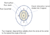
Nail varnish was applied twice to all external surfaces of the specimen except to the region 1 mm around the apical foramen. Specimens were immersed in 1% methylene blue dye solution for 12 hours at room temperature. Dye penetrated specimens were washed and mounted in an acrylic resin block with self-curing acrylic resin. Horizontal sections perpendicular to the long axis of the root were obtained every 1 mm from the apex up to 6 mm along the root with a low-speed diamond saw (Isomet, Buehler Ltd, Lake Bluff, IL, USA). Cross-sectioned surfaces were inspected under ×40 magnification with a stereomicroscope (SZ40, Olympus Corp., Tokyo, Japan) to evaluate the penetration length and the mean number of segments stained with dye. In order to compare the microleakage quantitatively, the first set of data was obtained by reading the dye penetration lengths longitudinally at one millimeter interval. The second set of data was obtained cross-sectionally. For each section at one millimeter interval, the cross-section of the root canal filling was divided into 5 segments and the number of segments stained was counted. By drawing an imaginary circle at the center corresponding to the canal filling material and four lines radiating from the circle to the nearest line angle, 5 separate segments were obtained from each cross-section (Figure 1). The stain at the interface between the dentinal wall and the canal filling material was regarded as belonging to the outer arc segment. If any trace of dye was present in a segment, the segment was counted as a dye-penetrated one. Therefore, the microleakage score of each section was graded from 0 to 5. For each group, the cross-sectional microleakage score was obtained at each level of the sections by dividing the total number of dye-penetrated segments at the level by the sample number (n = 10, Table 2).
Scanning electron microscopic observation
The interfaces between the root canal filling materials and the root canal dentin were also evaluated using a field-emission SEM operating at 15 kV. The sections randomly selected from the sections at 3 mm from the apex in each group were replicated with epoxy resin after treatment of the sectioned surface with 6N HCl for 3 seconds and 3.5% NaOCl for 5 minutes.
Statistical analysis
Statistical analysis of the data for longitudinal dye penetration lengths that were read at one millimeter intervals was performed with non-parametric Kruskal-Wallis test. The statistical differences in the cross-sectional microleakage scores, which were repeatedly measured along the distances from the apical foramen for the four groups, were evaluated with Friedman 2-way Analysis of Variance (ANOVA) and Kruskal-Wallis tests. All statistical analyses were performed at a 5% level of significance.
RESULTS
Under a stereomicroscope, dye penetration was observed mostly at the interface between the root canal dentinal wall and the filling material. Filling materials such as gutta-percha and Resilon cones did not fill the entire canal space, but the remaining portion was filled with corresponding sealers. Characteristic differences were not found in the leakage patterns among sealers. However, in severe cases with dense bands of dye staining, the stain radiated outwards from the interface along the dentinal tubules or spread inwards sparsely into the sealer occupying the remaining canal space between the cones and the canal wall. In Group 4 (TELC), sections having no stained segments were observed from 2 mm from the apical foramen.
The median values of the longitudinal dye penetration lengths read from each section at one millimeter intervals are summarized in Table 2. The median longitudinal dye penetration length of Group 4 (TELC) was significantly shorter than Groups 1 (GPCS) and 2 (SEDC) (Kruskal-Wallis test, p < 0.05). The longitudinal dye penetration length in Group 2, where the self-etching RealSeal primer and dual-curing RealSeal sealer were used according to the instructions of the Resilon-RealSeal system, was statistically not different from the values of Group 1, the conventional gutta-percha filling with AH26 plus sealer. Group 3 was not different from any other groups.
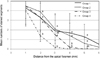
In Figure 2, The cross-sectional microleakage scores decreased significantly along the distance from the apical foramen (Friedman test, p < 0.05) and there was a significant interaction between the groups and the distances from the apical foramen (Friedman test, p = 0.042). The cross-sectional microleakage scores at each level of the sections also showed significant differences between groups. Group 4 (TELC) showed significant differences in the cross-sectional microleakage scores from all the other groups at 2 mm, from Groups 2 and 3 at 3 mm, and from Groups 1 and 2 at 4 and 5 mm from the apical foramen (Kruskal-Wallis test, p < 0.05).
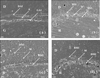
In Group 1, there was a layer of conventional AH26 plus root canal sealer between the root canal dentin wall and gutta-percha canal filling material. Continuous gaps were observed at the dentin side of the conventional root canal sealer (Figure 3a). In the other groups, both the self-etching RealSeal primer and the total-etch/wet bonding SBMP primer showed intimate contact with the root canal dentin wall, at which the junctions were not easily demarcated (Figures 3b, 3c and 3d). However, Group 2, where the dual-cure RealSeal sealer was used, showed a porous layer filled with a great number of small grains between the dentinal wall and the Resilon filling material (Figure 3b). In Groups 3 and 4, instead of using the RealSeal sealer, the SBMP adhesive developed a thick adhesive layer between the dentin wall and the Resilon core material (Figures 3c and 3d). The quality of its continuity and thickness was reliable for both groups.
DISCUSSION
Even with a number of testing methods to evaluate the quality of seal in root canals, such as radioactive isotope test, bacteria or bacterial metabolites leakage test, electrochemical technique, fluid filtration, and dye penetration, the conclusions of published reports on the apical sealing ability of canal filling materials have been still different or even conflicting.17 The dye penetration test was the most popular, but the method was characterized by a great deal of variation among results and regarded to be unreliable in some dental materials, especially in calcium hydroxide and mineral trioxide aggregate.18 However, even in the study, Fuji II did not show decoloration over time.18 In our study, we used resinous filling materials and sealers and conventional gutta-percha and AH26 sealer (Group 1) showed more leakage than the resinous materials. If the leakage is evaluated from just one section of each specimen, the assessment of dye penetration cannot represent the spatial distribution of the dye stain. The ordinal grades used in most leakage studies must also accept certain limitations in order to show the results quantitatively. In order to overcome the limitations of two-dimensional observations of dye penetration tests, in this study dye penetration was evaluated three-dimensionally along two axes, longitudinally and cross-sectionally. Longitudinal dye penetration length was read from each section obtained at one millimeter intervals starting from the root apex to 6 mm from the apex and the cross-sectional microleakage score was calculated as the mean number of the stained segments at each level of section in each group.
In this study, the longitudinal dye penetration length in Group 4 (TELC) was significantly less than those of Groups 1 (GPCS) and 2 (SEDC) (Kruskal-Wallis test, p < 0.05, Table 2). The values of the cross-sectional microleakage score of Group 4 (TELC) were significantly smaller than those of Groups 1 and 2 at each level of the sections from 2 mm to 5 mm from the apex (Friedman test, p < 0.05). Especially at the 2 mm sections, Group 4 showed significantly better sealing ability than all the other groups (Figure 2). According to the two sets of data from the longitudinal and the cross-sectional axes, the sealing ability of a resin-based Resilon-RealSeal system was not different from that of the conventional gutta-percha filling with AH26 sealer. However, when a total-etch 3-step SBMP adhesive system was used instead of the RealSeal primer / sealer (Group 4), the sealing ability of the canal filling was significantly improved (Kruskal- Wallis test and Friedman test, p < 0.05, Table 2 and Figure 2). Group 3, where SBMP adhesive was applied and light-cured without a pertinent priming procedure using total-etch and wet bonding procedures, exhibited intermediate dye penetration depth and microleakage scores. The group was statistically not different from both the groups showing worse sealing ability (Groups 1 and 2) and the best sealing ability (Group 4).
A number of studies have consistently criticized the sealing ability of conventional gutta-percha root canal filling material.3,4,19 Recently, there have been many reports on the improved sealing ability of the new resin-based Resilon root canal filling system.3,5,7,20 The improved sealing was attributed to 'mono-block' creation, which means an adhesion complex consisting of the dentin, closely adapted Resilon, and inter-posing Epiphany sealer.5 However, most of the reports focused on coronal leakage resulting from the light-curing step for coronal sealing5,7,20 and inferior, or at most similar, results have also been reported.21-23 Within the coronal portion of the canals, the polymerization shrinkage of the light-curing Resilon core material and inherent and highly unfavorable configuration factor (C-factor) of the canal were attributed to the deterioration in the adhesion of the sealer to the dentin wall and core material.5,24
Our study focused on the apical microleakage from the apex. In the case of the apical portion of the canals, the extremely high C-factor resulted in the poor sealing of the resinous sealer and core material system because it deprived them of the chances to relieve shrinkage stress,25 as did the less dense dentinal tubule configuration and dentinal sclerosis in the apical portion of adult teeth.22,26 In order to reduce the avenue for microleakage, 'mono-block' creation through the removal of smear layer and close adaptation between the sealer and core material was suggested for direct chemical bonding of the sealers to the dentin wall and to the core material, respectively.5 In group 2, considering the penetration depth of the curing light through the opaque root dentin and the Resilon core material, the polymerization of the RealSeal sealer and the Resilon core material within the apical portion of the canal must have predominantly occurred by a self-cure reaction. The Resilon cone coated with the dual-curable RealSeal sealer was delivered into the canal and condensed to the canal wall by lateral condensation. Therefore, the slow chemical reaction must have provided sufficient time to relieve the shrinkage stress through the flow of the curing resin network, and packing the core material with a spreader must have also reduced the detachment among the components of the 'mono-block' adhesion complex. Nonetheless, in our study, there were no significant differences in the longitudinal dye penetration depth and the cross-sectional microleakage score between Groups 1 and 2 (Table 2 and Figure 2). The Resilon-RealSeal system was not superior to the conventional gutta-percha filling with AH26 sealer in terms of apical sealing ability. However, in group 4, to make the adhesive layer strong enough to resist the shrinkage stress of the resinous canal filling material, the adhesive coated on the canal wall was cured sufficiently with the light passing through the empty canal.15 When the SBMP adhesive system was used for strict wet bonding and light-cured instead of the RealSeal primer and sealer before packing core materials, the longitudinal dye penetration depth and the cross-sectional microleakage score decreased significantly. From the observation, the poor sealing ability of a resin-based dual-curing Resilon-RealSeal canal filling system might result from the adhesion quality of the 'mono-block' created by the self-etch RealSeal primer and the dual-cured RealSeal adhesive.27
Simplified adhesives are known to exhibit lower DC than multistep dentin adhesives and incomplete polymerization of the adhesives are also known to result in poor mechanical properties and high permeability of the adhesive joint.12,14,15 In this study, we evaluated whether the sealing ability of the resin-based root canal filling system could be improved if it were used with the strict adhesion concept of multi-step adhesive systems instead of the simplified one of the RealSeal primer and sealer, a simplified self-etching dual-curing adhesive system. For the purpose, longitudinal dye penetration depth and cross-sectional microleakage scores were evaluated for the experimental groups that adopted different combinations of two adhesion variables; the priming concept of the canal wall and the curing mode of the adhesives. In Group 2, the self-etching RealSeal primer and the dual-curing RealSeal sealer was used according to the instructions of the manufacturer. The RealSeal adhesive near the apex must have been polymerized by chemical-cure mode due to the limitation in penetration depth of curing light. The adhesive layer was very porous (Figure 3b). In Group 4, instead of using the RealSeal primer and sealer, the SBMP total-etch 3-step adhesive system was used. In the case of the SBMP adhesive system, the total-etch and wet bonding procedures could be performed strictly to obtain a reliable hybrid layer and maximum DC and sufficient strength to resist possible polymerization shrinkage of the Resilon core material could be obtained by light-curing the adhesive before condensation of Resilon cones. Group 4, which exhibited a well-developed hybrid layer and a thick adhesive layer between the dentin wall and the Resilon core material, showed the best apical sealing ability among the experimental groups (Figure 3d). In Group 3, the adhesive was also light-cured for relevant DC before lateral condensation of Resilon cones, but the primer was applied without etching of the root canal dentinal wall in order to evaluate the effect of the hybrid layer. However, after completing instrumentation and NaOCl rinsing to remove dentin debris and smear layer, the canal was rinsed with 5 mL of 17% EDTA. As a result, the thick adhesive layer was intimately in contact with the root canal dentin wall and was observed around the Resilon core material. The quality of their continuity and thickness were comparable to that of Group 4 (Figure 3c).
In Group2, the acidic mononer of the RealSeal self-etching primer might have affected the bond integrity between the Resilon cones and the root canal dentin wall. The RealSeal primer is a solution of 2-acrylamido-2-methylpropane sulfonic acid and hydrophilic monomer. The acidity of the self-etching primer caused reduction in the DC of RealSeal sealer and resulted in a decrease in mechanical strength and permeability. The difference in the polymer system between the SBMP adhesive and the Resilon cones might still have left intermittent gaps. In Group 2, the acidity of the RealSeal primer hindered the polymerization of the sealer layer and might have left a large number of small grains (Figure 3b). The poor polymerization of the porous layer resulted in poor bond integrity. The quality and thickness of the hybrid and the adhesive layers were better in Group 4 than in the other groups. The observed bond integrity, which stands for the continuity and the thickness of the sealer or the adhesive, could be related with the sealing ability. Additionally, since the SBMP adhesive does not have the required properties for root canal sealers such as radiopacity, strength for easy removal during retreatment, and biocompatibility, it must be considered when interpreting this result and additional studies are needed.
CONCLUSIONS
From the results, the null hypothesis was rejected. Adhesion variables such as the priming concepts of root canal dentin surface and the curing modes of adhesive or sealer had a significant effect on the sealing ability of the resin-based Resilon-RealSeal root canal filling system. When a resin-based root canal filling material was used, compared to the self-etching primer and the dual-cure sealer, the adhesive system using a separate etch and rinse step, strict wet-bonding concept, and light-curing of the adhesive before condensation of canal filling material, showed improved apical sealing ability and was highly recommended.


 XML Download