

 PDF
PDF ePub
ePub Citation
 Print
Print


Ji-A Yoo, BS1, Byeong-Hoon Cho, DDS, Ph.D2
Abstract
Objectives
This study investigated the effects of the color components of light-cured composite resin before and after polymerization on degree of conversion (DC) and biaxial flexural strength (FS).
Materials and Methods
Four enamel shades (A1, A2, A3, A4) and two dentin shades (A2O, A3O) of Premisa (Kerr Co.) and Denfil (Vericom Co.) were evaluated on their CIE L*, a*, b* color components using the spectrophotometer before curing, after curing and at 7 day. The DC of same specimens were measured with Near-infrared spectrometer (Nexus, Thermo Nicolet Co.) at 2 hr after cure and at 7 day. Finally, the FS was obtained after all the other measurements were completed at 7 day. The correlations between each color component and DC and FS were evaluated.
Results
The light-curing of composite resin resulted in color changes of Premisa in red-blue direction and Denfil in green-blue direction. The DC and FS were affected by product, time and shade (3-way ANOVA, p < 0.05) and product and shade (2-way ANOVA, p < 0.05), respectively. Premisa only showed a significant correlation between the DC and CIE a* component - before and after polymerization (Pearson product moment correlation, p < 0.05). The FS of Premisa showed significant negative correlations with CIE a* and CIE b* components.
Figures and Tables
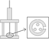 | Figure 1Measurement assembly for measuring biaxial flexural strength. The assembly had a specimen support and a loading steel rod, which was used in a universal testing machine. For the support of the test specimen, three steel balls with a diameter of 3.0 mm, positioned 120° apart on a support circle with a diameter of 10 mm, was provided. The sample was placed concentrically on these supports and the load was applied with a flat punch with a diameter of 1.5 mm at the center of the specimen.
|
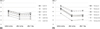 | Figure 2Changes in CIE L* values of each shade of (a) Premisa and (b) Denfil. Both products showed a relatively great decrease in the value (lightness) of each shade after curing. However, until 7 days, the values were a little increased.
|
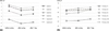 | Figure 3Changes in CIE a* values of each shade of Premisa and Denfil. (a) The products of Denfil showed a relatively great decrease in the redness of each shade after curing. However, until 7 days, the values were a little increased. (b) The products of Premisa showed a gradual increase in the redness of each shade after curing.
|
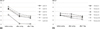 | Figure 4Changes in CIE b* values of each shade of (a) Denfil and (b) Premisa. Both products showed a gradual decrease in the yellowness of each shade after curing.
|
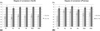 | Figure 5Changes in degree of conversion (%) of each shade of (a) Denfil and (b) Premisa. Both products showed a gradual increase in the degree of conversion of each shade from 2 hours after curing to 7 days.
|
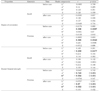
Table 2
Degree of conversion (DC) of two commercial dental composite resins measured at 2 hours and on 7 days after light-curing according to the shades (DC, mean ± SD; No., 10; unit, %)

![]()

References
1. Shortall AC, Wilson HJ, Harrington E. Depth of cure of radiation-activated composite restoratives-influence of shade and opacity. J Oral Rehabil. 1995. 22:337–342.

2. Ruyter IE, Oysaed H. Composites for use in posterior composites: composition and conversion. J Biomed Mater Res. 1987. 21:11–23.
3. Ferracane JL. Current trends in dental composites. Crit Rev Oral Biol Med. 1995. 6:302–318.
4. de Gee AJ, ten Harkel-Hagenaar E, Davidson CL. Color dye for identification of incompletely cured composite resins. J Prosthet Dent. 1984. 52:626–631.
5. Sideridou I, Tserki V, Papanastasiou G. Study of water sorption, solubility and modulus of elasticity of light-cured dimethacrylate-based dental resins. Biomaterials. 2003. 24:655–665.
6. Carmichael AJ, Gibson JJ, Walls AW. Allergic contact dermatitis to bisphenol-A-glycidyldimethacrylate (Bis-GMA) dental resin associated with sensitivity to epoxy resin. Br Dent J. 1997. 183:297–298.
7. Hansel C, Leyhausen G, Mai UE, Geurtsen W. Effetcs of various resin composite (co) monomers and extracts on two caries-associated micro-organisms in vitro. J Dent Res. 1998. 77:60–67.
8. Lovell LG, Stansbury JW, Syrpes DC, Bowman CN. Effects of composition and reactivity on the reaction kinetics of dimethacrylate/dimethacrylate copolymerizations. Macromolecules. 1999. 32:3913–3921.
9. Ruyter IE, Oysaed H. Conversion in different depths of ultraviolet and visible light activated composite materials. Acta Odontol Scand. 1982. 40:179–192.
10. Swartz ML, Phillips RW, Rhodes B. Visible light activated resins - depth of cure. J Am Dent Assoc. 1983. 106:634–637.
11. Ferracane JL, Aday P, Matsumoto H, Marker VA. Relationship between shade and depth of cure for light activated dental composite resins. Dent Mater. 1986. 2:80–84.
12. Davidson-Kaban SS, Davidson CL, Feilzer AJ, de Gee AJ, Erdilek N. The effect of curing light variationson bulk curing and wall-to-wall quality of two types and various shades of resin composites. Dent Mater. 1997. 13:344–352.
13. Yearn JA. Factors affecting cure of visible light activated composites. Int Dent J. 1985. 35:218–225.
14. Rueggeberg FA, Caughman WF, Curtis JW Jr. Effect of light intensity and exposure duration on cure of resin composite. Oper Dent. 1994. 19:26–32.
15. Unterbrink GL, Muessner R. Influence of light intensity on two restorative systems. J Dent. 1995. 23:183–189.
16. Sakaguchi RL, Berge HX. Reduced light energy density decreases post-gel contraction while maintaining degree of conversion in composites. J Dent. 1998. 26:695–700.
17. Pires JA, Cvitko E, Denehy GE, Swift EJ Jr. Effects of curing tip distance on light intensity and composite resin microhardness. Quintessence Int. 1993. 24:517–521.
18. Hansen EK, Asmussen E. Visible-light curing units: correlation between depth of cure and distance between exit window and resin surface. Acta Odontol Scand. 1997. 55:162–166.
19. Leloup G, Holvoet PE, Bebelman S, Devaux J. Raman scattering determination of the depth of cure of light-activated composites: influence of different clinically relevant parameters. J Oral Rehabil. 2002. 29:510–515.
20. Aravamudhan K, Floyd CJ, Rakowski D, Flaim G, Dickens SH, Eichmiller FC, Fan PL. Light-emitting diode curing light irradiance and polymerization of resin-based composite. J Am Dent Assoc. 2006. 137:213–223.
21. Omaima MS, Hamza A, Taheya A, Moussa . Influence of shade and time on the degree of conversion and fracture strength of light-cured composite resin. J Egyptian Dent Assoc. 1999. 45:4487.
22. Chen YC, Ferracane JL, Prahl SA. A pilot study of a simple photon migration model for predicting depth of cure in dental composite. Dent Mater. 2005. 21:1075–1086.
23. Stansbury JW, Dickens SH. Determination of double bond conversion in dental resins by near infrared spectroscopy. Dent Mater. 2001. 17:71–79.
24. Wei W, Sadeghipour K, Boberick K, Baran G. Predictive modeling of elastic properties of particulate-reinforced composites. Mater Sci Eng A. 2002. 332:362–370.
25. Braem M, Van Doren VE, Lambrechts P, Vanherle G. Determination of Young's modulus of dental composites: a phenomenological model. J Mater Sci. 1987. 22:2037–2042.
26. Chung SM, Yap AU, Koh WK, Tsai KT, Lim CT. Measurement of Poisson's ratio of dental composite restorative materials. Biomaterials. 2004. 25:2455–2460.
27. Cho YG, Kim MC. Color changes in composites according to various light curing sources. J Korean Acad Conserv Dent. 2002. 27:87–94.
28. Rueggeberg FA, Caughman WF, Curtis JW Jr, Davis HC. Factors affecting cure at depths within light-activated resin composites. Am J Dent. 1993. 6:91–95.
29. Park SJ, Noh EY, Cho HG, Hwang YC, Oh WM, Hwang IN. Color difference of the dental composites measured by different color measuring instruments. J Korean Acad Conserv Dent. 2009. 34:199–207.
30. Makinson OF. Colour changes on curing light-activated anterior restorative resins. Aust Dent J. 1989. 34:154–159.
31. Taira M, Okazaki M, Takahashi J. Studies on optical properties of two commercial visible-light-cured composite resins by diffuse reflectance measurements. J Oral Rehabil. 1999. 26:329–337.
32. Yu B, Lee YK. Influence of color parameters of resin composites on their translucency. Dent Mater. 2008. 24:1236–1242.
33. Kim IJ, Lee YK. Changes in color and color parameters of dental resin composites after polymerization. J Biomed Mater Res B Appl Biomater. 2007. 80:541–546.
34. Kawaguchi M, Fukushima T, Miyazaki K. The relationship between cure depth and transmission coefficient of visible light-activated resin composites. J Dent Res. 1994. 73:516–521.
35. Watts DC, Cash AJ. Analysis of optical transmission by 400-500 nm visible light into aesthetic dental biomaterials. J Dent. 1994. 22:112–117.
36. Seghi RR, Gritz MD, Kim J. Colorimetric changes in composites resulting from visible-light-initiated polymerization. Dent Mater. 1990. 6:133–137.
37. Uchida H, Vaidyanathan J, Viswanadhan T, Vaidyanathan TK. Color stability of dental composites as a function of shade. J Prosthet Dent. 1998. 79:372–377.
38. Cho YG, Seo JI, Kim SM, Jeong JH, Lee YG. Color changes in composite resins exposed to xenon lamp. J Korean Acad Conserv Dent. 2003. 28:195–202.
39. Bouschlicher MR, Rueggeberg FA, Wilson BM. Correlation of bottom-to-top surface microhardness and conversion ratios for a variety of resin composite compositions. Oper Dent. 2004. 29:698–704.
40. Rodrigues SA Jr, Scherrer SS, Ferracane JL, Della Bona A. Microstructural characterization and fracture behavior of a microhybrid and a nanofill composite. Dent Mater. 2008. 24:1281–1288.
41. Spanoudakis J, Young RJ. Crack propagation in a glass particle-filled epoxi resin. J Mater Sci. 1984. 19:473–486.
42. Ferracane JL, Berge HX, Condon JR. In vitro aging of dental composites in water-Effect of degree of conversion, filler volume, and filler/matrix coupling. J Biomed Mater Res. 1998. 42:465–472.
43. Kim KH, Ong JL, Okuno O. The effect of filler loading and morphology on the mechanical properties of contemporary composites. J Prosthet Dent. 2002. 87:642–649.
44. Chung SM, Yap AU, Chandra SP, Lim CT. Flexural strength of dental composite restoratives: comparison of biaxial and three-point bending test. J Biomed Mater Res B Appl Biomater. 2004. 71:278–283.
45. Cesar PF, Miranda WG Jr, Braga RR. Influence of shade and storage time on the flexural strength, flexural modulus, and hardness of composites used for indirect restorations. J Prosthet Dent. 2001. 86:289–296.
46. Lin-Gibson S, Sung L, Forster AM, Hu H, Cheng Y, Lin NJ. Effects of filler type and content on mechanical properties of photopolymerizable composites measured across two-dimensional combinatorial arrays. Acta Biomater. 2009. 5:2084–2094.
 XML Download