

 PDF
PDF ePub
ePub Citation
 Print
Print


Abstract
Objectives
The aim of this study was to evaluate the various NiTi rotary instruments regarding their ability to provide a circular apical preparation.
Materials and Methods
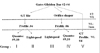
50 single canal roots were selected, cut at the cementodentinal junction and the coronal 1/3 of the canals was flared using Gates Glidden burs. Samples were randomly divided into 5 experimental groups of 10 each. In group I, GT files, Profile 04 and Quantec #9 and #10 files were used. In Group II Lightspeed was used instead of Quantec. In Group III, Orifice shaper, Profile .06 series and Lightspeed were used. In Group IV, Quantec #9 and #10 files were used instead of Lightspeed. In Group V, the GT file and the Profile .04 series were used to prepare the entire canal length. All tooth samples were cut at 1 mm, 3 mm and 5 mm from the apex and were examined under the microscope.
Results
Groups II and III (Lightspeed) showed a more circular preparation in the apical 1mm samples than the groups that used Quantec (Group I & IV) or GT files and Profile .04 series.(Group V)(p < 0.05) There was no significant difference statistically among the apical 3, 5 mm samples. In 5 mm samples, most of the samples showed complete circularity and none of them showed irregular shape.
Conclusions
Lightspeed showed circular preparation at apical 1 mm more frequently than other instruments used in this study. However only 35% of samples showed circularity even in the Lightspeed Group which were enlarged 3 ISO size from the initial apical binding file (IAF) size. So it must be considered that enlarging 3 ISO size isn't enough to make round preparation.
Figures and Tables
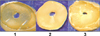 | Figure 2Photos from cross-section specimens. These specimens were sectioned 1, 3, 5 mm from the apex using Buehler saw (IsoMet 1000 Precision Saw, buehler, Lake Bluff, IL, USA) and examined under the microscope (×35). Circularity was scored. Score 1 was a complete circle, 2 was an incomplete circle, and 3 was an irregular shape.
|
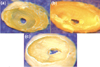 | Figure 3Photos from cross-section specimens at apical 1 mm level. (a) was a complete circle, (b) was an incomplete circle, and (c) was an irregular shape.
|
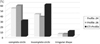
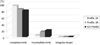
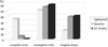 | Figure 4Comparison of circularity at apical 1 mm level for instrumentation modalities of 3 experimental groups.
|
References
1. Schilder H. Cleaning and shaping the root canal. Dent Clin North Am. 1974. 18:269–296.
2. Mizrahi SJ, Tucker JW, Seltzer S. A scanning electron microscopic study of the efficacy of various endodontic instruments. J Endod. 1975. 1:324–333.

3. Weine FS, Kelly RF, Lio PJ. The effect of preparation procedures on original canal shape and on apical foramen shape. J Endod. 1975. 1:255–262.
4. Walia HM, Brantley WA, Gerstein H. An initial investigation of the bending and torsional properties of Nitinol root canal files. J Endod. 1988. 14:346–351.
5. Kielt LW, Montgomery S. The effect of endosonic instrumentation in simulated curved root canals. J Endod. 1987. 13:215–219.
6. Seidler B. Root canal fillings: an evaluation ans method. J Am Dent Assoc. 1956. 53:567–576.
7. Schneider SW. A comparison of canal preparations in straight and curved root canals. Oral Surg Oral Med Oral Pathol. 1971. 32:271–275.
8. Hwang HK, Jou YT, Kim S. Sealing ability of isthmuses by different obturation techniques. J Endod. 1998. 24:283. Abstract.
9. Walton RE. Histologic evaluation of different methods of enlarging the pulp canal space. J Endod. 1976. 2:304–311.
10. Campos JM, del Rio C. Comparison of mechanical and standard hand instrumentation techniques in curved root canals. J Endod. 1990. 16:230–234.
11. Ehrlich AD, Boyer TJ, Hicks ML, Pelleu GB Jr. Effects of sonic instrumentation on the apical preparation of curved canals. J Endod. 1989. 15:200–203.
12. Burstone CJ, Qin B, Morton JY. Chinese NiTi wire-a new orthodontic alloy. Am J Orthod. 1985. 87:445–452.
13. Tharuni SL, Parameswaran A, Sukumaran VG. A comparison of canal preparation using the K-file and Lightspeed in resin blocks. J Endod. 1996. 22:474–476.
14. Portenier I, Lutz F, Barbakow F. Preparation of the apical part of the root canal by the Lightspeed and step-back techniques. Int Endod J. 1998. 31:103–111.
15. Chen JL, Messer HH. A comparison of stainless steel hand and rotary nickel-titanium instrumentation using a silicone impression technique. Aust Dent J. 2002. 47:12–20.
16. Paqué F, Ganahl D, Peters OA. Effects of root canal preparation on apical geometry assessed by micro-computed tomography. J Endod. 2009. 35:1056–1059.
17. Peters OA, Laib A, Goöhring TN, Barbakow F. Changes in root canal geometry after preparation assessed by high-resolution computed tomography. J Endod. 2001. 27:1–6.
18. Setzer FC, Kwon TK, Karabucak B. Comparison of apical transportation between two rotary file systems and two hybrid rotary instrumentation sequences. J Endod. 2010. 36:1226–1229.
 XML Download