

 PDF
PDF ePub
ePub Citation
 Print
Print


Introduction
Dental pulp is a one of the most important tissues among all of the organs in human body, and inflammatory process in it is complex anatomically and histologically, which is thought to be closely related together in neurogenic, inflammatory, osteoblastic/osteoclastic processes in dental pulp. Recently, SP has been introduced to induce inflammatory cytokines in human dental pulp cells, and several cytokines have been detected in dental pulp during inflammation including interleukin and tumor necrosis factor (TNF)-α.1,2 In addition, cytokines and growth factors were revealed to induce the expressions Matrix Metalloproteinses (MMPs) from human dental pulp tissues or cultured human dental pulp fibroblast.3,4 Moreover, three kinds of tissues cells (pulp, gingival, and periodontal ligament) may be expected to induce MMP and TIMP with stimulation of neuropeptides or proinflammatory cytokine because those three different kinds of tissues is originated from different parts of tooth germ seperately.
MMPs are a group of enzyme that together can degrade practically all extracellular matrix proteins, including native collagen. They participate in normal tissue formation and remodeling, and the expression of different MMPs may be upregulated during various pathological conditions, e.g, inflammation, tumor invasion, and metastasis.
Recent findings also indicate that human odontoblasts may express several other members of MMP-family. Even though the physiological roles of MMPs expressed by odontoblasts are currently unknown, it may be speculated that they could participate in dentin matrix organization prior to mineralization, regulation of matrix mineralization, intratubular dentin formation, or remodeling of the pulp connective tissue.
MMPs are usually divided into five groups: collagenase, gelatinases, stromelysins, membrane-type MMPs and others. Of the MMPs family, interstitial collagenase (MMP-1) is the key enzyme responsible for degrading type I and type II collagen since the initial breakdown of these fibrillar collagen network is mediated primarily by MMP-15,6 While MMP-8 preferentially degrades type I and MMP-13 type II collagen.
The biological activity of the MMPs can be regulated post-transcriptionally, or by interaction with specific tissue inhibitors of metalloproteinase (TIMPs).7 TIMP-1 to TIMP-4 have been recently identified. TIMP-1, a glycoprotein with a molecular mass of 28 kDa, can form a stoichiometric 1 : 1 complex with and inactive MMP-1.5,8 Because the same cells that produce MMP-1 are also capable of synthesizing TIMP-1, the overall molar ratio between MMP-1 and TIMP-1 therefore plays a significant role in connective tissue remodeling.9 TIMPs expression is regulated by growth factors and cytokines at the transcriptional level. TIMP-3, which is capable of binding to compartments of the ExtraCelluar Matrix (ECM) may also induce cell apoptosis. The TIMPs may also regulate the growth of several different cell types.
MMPs activity is regulated at least three levels: transcription, activation, and inhibition. In healthy adult tissues, the basal expression is usually low, but various external stimuli (cytokines, growth factors, changes in cell-cell and cell-ECM interactions) can induce the expression. MMP expression increases in pathological condition such as inflammation, tumor invasion, and metastasis.10 Most MMPs are secreted in latent proforms (zymogen) and activated at the cell surface or extracellularly. The activation requires the exposure of the catalystic site, which may be achieved by proteolytic enzymes (including other MMPs), or in vitro by chaotropic agents (such as organomercurials) and pH changes. Also autoactivation is possible, at least in vitro.11
Proinflammatory cytokines, such as interleukin-1 and tumor necrosis factor-α (TNF-α) will initiate and augment subsequent inflammatory cascades leading to tissue destruction. Recently large amount of interleukin-1 and tumor necrosis factor-α have been demostrated to induce rat pulpitis.12 These findings highlighted the significance of proinflammatory cytokines in pulpal injury. It is well established that interleukin-1 and tumor necrosis factor-α can mediate inflammatory tissue destruction by stimulating MMP-1 and TIMP-1 production in several cell types.13,14
Substance P (SP) and calcitonin gene related peptide (CGRP) are two important sensory neuropeptides expressed in the dental pulp.3 Neurogenic inflammatory reaction in the dental pulp describes the local release of neuropeptides (SP or CGRP) from afferent neurons and might play a role in the pathogenesis of pulpal disease. Although the primary function of these two neuropeptides, upon release, is to induce vasodilation and pulpal blood flow,15 they may play a direct role in initiating the local inflammatory cell infiltration.16 In addition, SP levels expressed from pulpal fibroblast cultured from carious teeth were higher compared with sound teeth,17 and Substance P remarkably enhanced MMP-2 activity in pulp cells via intracellular reactive oxygen species (ROS) pathway.18
Because three kinds of soft tissues (pulp, periodontal, and gingival tissues) are connected together without any obvious junctions between them, it may be meaningful to investigate if MMPs or TIMPs are induced from the cultured cells (PC, GC, and PDLC) from those tissues when they are stimulated with neuropeptides (SP or CGRP).
In the present study, it was investigated that MMPs and TIMPs are expressed in cultured cells (PC, GC, and PDLC) from those tissues and that the expression is regulated by neuropeptides (substance P and CGRP) or proinflammatory cytokine, TNF-α.
Materials and Methods
Sample collection and cell culture
Freshly extracted, intact, caries-free third molars (n=10) were obtained from the patients (15-25 years old) in the Department of Oromaxillofacial Surgery at Kyung-Hee Medical Center. Immediately after extraction, teeth were stored in phosphate buffered saline (PBS) and transferred to the laboratory. Gingival tissues were cut around the cementoenamel junction area and the periodontal ligaments were obtained by scratching the root surface. Each tooth was grooved longitudinally with a fissure bur at high speed. The tooth was then split with the driver, and the entire pulp (coronal and radicular) was elevated with cotton pliers to obtain maximum pulp tissue.
Repeated washing with PBS, the tissues obtained from the dental pulp, gingiva, and periodontal ligament were placed in a 48 well plate culture dish containing Dulbecco's Modified Eagle Medium (DMEM; Life Technologies/GIBCO BRL, Gaithersburg, MD, USA) supplemented with 10% fetal bovine serum (FBS).
The cells from pulp tissue (PF) and gingival tissue (GF), and periodontal ligaments (PDLF) were grown to confluence and passed at 1 : 2 ratio until used for experiments. Cell culture media were supplemented with 100 units/mL penicillin-G, 100 µg/mL streptomycin, and 0.25 µg/mL fungizone (Gemini Bio-Products, Inc., Woodland, CA, USA).
Once the cells obtained were confluent at passage 5-8, they were pooled in 100 mm culture dish and cultured until it became confluent for the stimulation with Substance P (SP), Calcitonin gene related peptides (CGRP) and TNF-α for the RNase Protection Assay. In addition, samples were seeded on the 6 well plate, and grown into confluent for the Enzyme-Linked Immunosorbent Assay after the stimulation with TNF-α.
Stimulation of the cells
Synthetic human SP, CGRP (Sigma, St. Louis, MO, USA), and TNF-α (R&D system, Mineapolis, MN, USA) were prepared in sterile water with 0.1% low-endotoxin bovine serum albumin (BSA, Sigma, St. Louis, MO, USA). Cells were seeded in 100mm culture dish and 6-well plates and grown to confluence before used for experiment. Alpha minimal essential medium containing L-glutamine (Life Technologies/GIBCO BRL, Gaithersburg, MD, USA) without FBS was used 24 hours before cells were stimulated with neuropeptiedes and TNF-α. Cells (PF, GF and PDLF) seeded in 100 mm culture dish were stimulated with SP (10-5, 10-8 M) or only with medium (Mock stimulation) for 4 hours and for 24 hours for RNase Protection Assay, and they were stimulated with CGRP (10-5M) and TNF-α (2 ng/mL) for 24 hours and with various concentraion of TNF-α (2, 10, and 100 ng/mL) for RNase Protection Assay as well.
In addition, cells (PF, GF and PDLF) were stimulated with Mock and various concentraion of TNF-α (2, 10, and 100 ng/mL) for 24 hours and with TNF-α (10 ng/mL) for 48 hours, and the supernatents from the cells were collected for Enzyme Linked Immunosor-bent Assay (ELISA) for MMP-1 and MMP-13. At this time, stimulation of the cells (PF, GF and PDLF) was 2 times longer (48 hours) with low concentration of TNF-α (10 ng/mL) to detect enough proteins from the supernatents out of the cells by using ELISA.
Extraction of total RNA
After the cells were stimulated, the supernatents were stored at -80℃. All kinds of cells stimulated were collected scraping with cell scraper (Fisher Scientific Korea, Seoul, Korea).
5 to 10-µg of total RNA was extracted from all kinds of cells stimulated using the RNeasy Mini kit following the manufacturer's instructions (Qiagen Korea Ltd. Seoul, Korea).
RNase protection assay
The total RNA extracted were subjected to an RNase protection assay as specified by the manufacturer, using the human MMP-1 probe set (Becton Dickinson Korea Ltd. Seoul, Korea) and BD RiboQuant Ribonuclease Protection Assay Systems (Becton Dickinson Korea Ltd. Seoul, Korea). The resulting protected RNAs were resolved on 5% denaturing polyacrylamide gels and exposed to X-ray films.
Enzyme linked immunosorbent assay
To detect the MMP-1 and MMP-13, Quantikine proMMP-1 and Quantikine MMP-13 (R&D system, Mineapolis, MN, USA) were purchased and the secretion of MMP-1 and MMP-13 were measured from the supernatant of the cells (PF, GF, PDLF) after the stimulation with TNF-α.
Briefly, after 100 µL of Assay Diluent from the kit was added to each well and 100 µL of standard and sample per well, the plate was incubated for 2 hours at room temperature on a horizontal orbital microplate shaker (0.12" orbit) set at 500 ± 50 rpm. Each well aspirated and washed, repeating the process three times for a total of four washes. The well washed with Wash Buffer (400 µL) multi-channel pipette. Complete removal of liquid at each step is essential to good performance. After the last wash, any remaining Wash Buffer removed by aspirating or decanting. Invert the plate and blot it against clean paper towels. 200 µL of Pro-MMP-1 Conjugate and pro-MMP-13. Conjugate were added to each well and incubated for 2 hours at room temperature on shaker. Repeating wash as in above, 200 µL of Substrate solution was added to each well. The plate were protected from light during the incubation for 20 minutes at room temperature on the benchtop. after 20 minutes, 50 µL of Stop Solution were added to each well. If color change does not appear uniform, gently tap the plate to ensure thorough mixing. Determine the optical density of each well within 30 minutes, using a microplate reader set to 570 nm.
Results
RNase protection assay
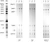
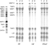
After stimulation of the cells for 4 and 24 hours in medium with SP (10-8 M, 10-5 M) or without SP (Mock), RNase Protection Assay demostrated that there were no differences between gene expression of MMPs and TIMPs. The effects of SP (10-8 M, 10-5 M) concentration on expression of MMPs and TIMPs showed no dose dependency when cells were incubated in the presence of SP (10-8 M, 10-5 M) as shown in Figures 1 and 2.
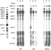
After stimulation of the cells for 24 hours in medium with CGRP (10-5 M) or TNF-α (2 ng/mL), RNase Protection Assay demostrated that the expression of MMP-12 in PF, GF, PDLF after stimulation with TNF-α were upregulated, compared with Mock especially in PDLF. The expression of MMP-1 in PF, GF, PDLF after stimulation with TNF-α were upregulated. The expression of MMP-13 in GF after stimulation with TNF-α were upregulated compared with Mock. The expression of TIMP-3, that is TNF-α converting enzyme inhibitor abundant in PF, GF, PDLF were downregulated in Mock compared with TNF-α (2 ng/mL) as shown in Figure 3.
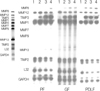
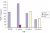
After stimulation of PF, GF, PDLF for 24 hours in medium with different dose of TNF-α (2 ng/mL, 10 ng/mL, 100 ng/mL), RNase Protection Assay demostrated that the expression of MMP-1 and MMP-12 in PF after stimulation with TNF-α (2 ng/mL, 10 ng/mL, 100 ng/mL) compared to Mock were upregulated. The effects of TNF-α (2 ng/mL, 10 ng/mL, 100 ng/mL) concentration on expression of MMP-1 and MMP-12 showed dose dependency when PF were incubated in the presence of TNF-α(2 ng/mL, 10 ng/mL, 100 ng/mL) for 24 hours as shown in Figure 4.
Enzyme linked immunosorbent assay
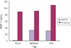
A signigicant induction of MMP-1 in the all kinds of cells studied was observed 24 hours and 48 hours after TNF-α stimulation as shown in Figures 5 and 6. Induction of MMP-1 by TNF-α was increased dose dependently in all kinds of cells studied. MMP-1 secretion from the PF after stimulation with 2 ng/mL and 10 ng/mL of TNF-α showed similar level, whereas 100 ng/mL of TNF-α showed significant higher level of MMP-1 at 24 hours. MMP-1 secretion from the GF was as same as that of PF for 24 hrs. Both 2 ng/mL and 10 ng/mL of TNF-α induced similar level of MMP-1, whereas 100 ng/mL of TNF-α showed higher level of MMP-1 than 2 ng/mL stimulation for 24 hours. An 11-fold increase of MMP-1 from PDLF after TNF-α (2 ng/mL) was when compared with mock stimulation for 24 hours, whereas 10 ng/mL and 100 ng/mL of TNF-α showed similar level of MMP-1. MMP-1 secretion from all kinds of cells studied with TNF-α (10 ng/mL) was significantly higher than that of mock stimulation (Figure 6).
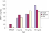
The secretion of MMP-13 was detected from PF after stimulation with TNF-α (2 ng/mL, 10 ng/mL, and 100 ng/mL) and Mock stimulation for 24 hours, nevertheless the secretion level was not dose-dependent shown as shown in Figure 7. The secretion from GF was detected only after 2 ng/mL of TNF-α stimulation for 24 hours and that from PDLF was after 10 ng/mL and 100 ng/mL of TNF-α stimulation for 24 hours (Figure 7). MMP-13 secretion from all kinds of cell studied 48 hours after stimulation with TNF-α (10 ng/mL) was significantly increased compared with Mock stimulation (Figure 8).
Discussion
Abnormal soft tissue degradation in tooth is one of the characteristic features of dental pulp and tissues surrounding tooth (periodontal ligaments and gingivae). Collagens type I and III are the primary components of the connective tissue of the dental pulp. These fibrillar collagens are resistant to most proteolytic enzymes. Among the members of an extensive family of MMPs, only a limited numbers of these can cleave the highly structured fibrillar collagens. These include MMP-1 (collagenase-1), MMP-8 (collagenase-2), MMP-13 (collagenase-3), and MMP-2, 9 (gelatinase). Therefore, this study demonstrated that cells cultured from pulps, periodontal ligaments, have the ability to degrade type Icollagen, which suggests that these cells play a major role in remodeling the extracellular matrix in pulp, gingiva, and periodontal ligament.
In dental pulp, the extracellular matrix (ECM) is composed mainly of type I and III collagens. MMP-1 is the only enzyme that can degrade the triple helix of type I and III collagens and render the sensitive to further digestion by other proteinases.5,6
Most of the MMPs characterized proved to be more abundant in the odontoblasts, MMP-1, -2, -9, -10, -11, -13, -14, -15, -16, -17, -19, -20, and -23 were expressed by mature human odontoblasts and pulp tissue.6,19 On the contrary, MMP-1, -7, -12, TIMP-1, and -2 were expressed in all of cells (PF, GF, and PDLF) studied in this experiment without any stimulation.
Bone Morphogenic Protein-2 (BMP-2) significantly up-regulated MMP-9 mRNA in human odontoblast,20 suppressed MMP-1 in bone cells.21 However, no expression of MMP-13 could be detected in osteoclasts, but expression of MMP-13 mRNA was detected in osteoblasts and fibroblasts primarily on the inner side of calvarial bone of skull.22 MMP-9 mRNA was rarely expressed from all kinds of cells studied, and the expression of MMP-9 mRNA was not increased after the stimulation with neuropeptide and proinflammatory cytokine.
IL-1 and TNF-α regulated the expression of MMP-1 and TIMP-1 mRNA synthesis from pulp derived cells.23-25 MMP-1 mRNA expression was detected in all of cells studied, and secretion of MMP-1 was increased from all kinds of cell studied in dose dependent manner 24 hours after stimulation with TNF-α.
The constitutive MMP-1 serves as remodeling element of connective tissue under physiological condition. Collagenase expression can be stimulated with cytokines. Providing further evidence for the suggestion that MMPs may participate to the regulation of defensive responses against external irrigation in the human dentin-pulp complex.
The expression of mMMP-8 was down-regulated by Transforming growth factor (TGF)-β1 in human odontoblasts and dental pulp cells,3 and MMP-8 had a role in pulpal and periapical inflammation.26 MMP-8 was expressed after TNF-α stimulation from all kinds of cells studied, neither Mock and neuropeptide stimulation. Nevertheless, the expression of MMP-12 (Metalloelastase) from all of cells studied were down-regulated after TNF-α stimulation.
TIMPs may have the ability to antagonize either the activity or zymogen form of different MMPs.27 TIMPs seem to be the major local inhibitor of MMPs.17 The concominant production of MMP-1 and TIMP-1 mRNA after TNF-α stimulation suggested a natural protective mechanism against dental pulp ECM degradation. Eventhough TIMP-1 mRNA intensity induced by TNF-α was lower than MMP-1. MMP-1 and TIMP-1 from pulp cells was increased compared with Mock stimulation.28 The expression of TIMP-3 from all of cells studied (especially in dose dependent manner from PF) was incresed 24 hours after TNF-α stimulation.
The expression of MMP-1 and MMP-13 has also been documented in mineralized tissue-producing cells other than odontoblasts, for example, human odontoblasts express both MMP-1 and MMP-13.21,22 MMP-1 was equally expressed by both odontoblasts and pulp tissues, whereas MMP-13 expression was five times more abundant in pulp tissue compared with odontoblasts. The MMP-1 expression level in healthy dentin-pulp complex cells was very low but inducible.20 The constitutive MMP-1 serves as remodeling element of connective tissue under physiological condition. Collagenase expression can be stimulated with cytokines. Providing further evidence for the suggestion that MMPs may participate to the regulation of defensive responses against external irrigation in the human dentin-pulp complex.
TIMPs may have the ability to antagonize either the activity or zymogen form of different MMPs. TIMPs seem to be the major local inhibitor of MMPs. The concomitant production of MMP-1 and TIMP-1 mRNA after TNF-α stimulation suggested a natural protective mechanism against dental pulp ECM degradation. However, the amount of MMP-1 gene synthesized was considerably higher than TIMP-1 after proinflammatory cytokine stimulation.23 Resident dental pulp fibroblasts might contribute to the pathogenesis of pulpitis in that they could produce a significant amount of mediators related to inflammatory tissue destruction.
The current study demonstrated that 3 kinds of cells in tooth (dental pulp, gingiva, and periodontal ligament) mediated type I collagen degradation can be enhanced by treatment with TNF-α, however, they were not regulated to induce MMPs by Substance P. These results further demonstrate that cytokines can influence the secretion of MMPs from pulp, gingiva, and periodontal ligament, which may lead to dental pulp and periodontal tissue degradation.
Conclusions
According to this study, the results were as follows:
The expression of MMPs in PF, GF, PDLF after stimulation with SP and CGRP were not changed compared with Mock stimulation for 4 hours and 24 hours.
The expression of MMP-1 in PF, GF, PDLF 24 hours after stimulation with TNF-α were upregulated.
The expression of MMP-12 in PF, GF, PDLF after stimulation with TNF-α were upregulated, especially in PDLF.
The expression of MMP-13 in all kinds of cells studied 24 hours after stimulation with TNF-α were upregulated.
The expression of TIMP-3 in PF, GF, PDLF after stimulation with TNF-α were downregulated.
TNF-α (2 ng/mL, 10 ng/mL, 100 ng/mL) increased MMP-1 and MMP-12 expression in PF dose dependently for 24 hours.
MMP-1 secretion in PF, GF, and PDLF can potentially be up regulated by TNF-α leading to type I/III collagens degradation. MMP-12 (metalloelastase) is induced in PDLF, although the tissue contains elastin-free fibers. MMP-13 (against type I/II collagen) was produced only in GF responding to TNF-α. Reduced TIMPs may enhance the action of MMPs. Taken together, our data suggest that TNF-α in the area of inflammation may play an important role in regulating the remodeling of dentin, cementum, and alveolar bone.

 XML Download