

 PDF
PDF ePub
ePub Citation
 Print
Print


Abstract
Objectives
The purpose of this study was to determine the optimal master apical file size with minimal transportation and optimal efficiency in removing infected dentin. We evaluated the transportation of the canal center and the change in untouched areas after sequential preparation with a #25 to #40 file using 3 different instruments: stainless steel K-type (SS K-file) hand file, ProFile and LightSpeed using microcomputed tomography (MCT).
Materials and Methods
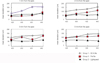
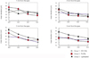
Thirty extracted human mandibular molars with separated orifices and apical foramens on mesial canals were used. Teeth were randomly divided into three groups: SS K-file, Profile, LightSpeed and the root canals were instrumented using corresponding instruments from #20 to #40. All teeth were scanned with MCT before and after instrumentation. Cross section images were used to evaluate canal transportation and untouched area at 1- , 2- , 3- , and 5- mm level from the apex. Data were statistically analyzed according to' repeated nested design'and Mann-Whitney test (p = 0.05).
Results
In SS K-file group, canal transportation was significantly increased over #30 instrument. In the ProFile group, canal transportation was significantly increased after preparation with the #40 instrument at the 1- and 2- mm levels. LightSpeed group showed better centering ability than ProFile group after preparation with the #40 instrument at the 1 and 2 mm levels.
Figures and Tables
Figure 1
Measuring the distance and the angle between original center and deviated center. a: original center of the canal. b: deviated center after preparation. Canal transportation (by distance) : Distance between a and b. Canal transportation (by degree) : A positive value indicates a movement of the canal center in the outer root surface (purple arrow), while a negative value signifies a shift in the opposite direction (green arrow).
* Original canal wall was drawn by red line and canal wall after preparation by black line.
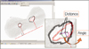
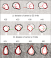
Figure 2
Cross sectional images acquired by µCT illustrate the sequential canal deviation by each group of instrument. Profile and Lightspeed group show more round and less deviated canal shapes.
References
1. Kerekes K, Tronstad L. Morphometric observations on the root canals of human molars. J Endod. 1977. 3:114–118.

2. Gani O, Visvisian C. Apical canal diameter in the first upper molar at various ages. J Endod. 1999. 25:689–691.
3. Short JA, Morgan LA, Baumgartner JC. A comparison of canal centering ability of four instrumentation techniques. J Endod. 1997. 23:503–507.
4. Albrecht LJ, Baumgartner JC, Marshall JG. Evaluation of apical debris removal using various sizes and tapers of ProFile GT files. J Endod. 2004. 30:425–428.
5. Rhodes JS, Ford TR, Lynch JA, Liepins PJ, Curtis RV. Micro-computed tomography: a new tool for experimental endodontology. Int Endod J. 1999. 32:165–170.
6. Rhodes JS, Ford TR, Lynch JA, Liepins PJ, Curtis RV. A comparison of two nickel-titanium instrumentation techniques in teeth using microcomputed tomography. Int Endod J. 2000. 33:279–285.
7. Bergmans L, Van Cleynenbreugel J, Wevers M, Lambrechts P. A methodology for quantitative evaluation of root canal instrumentation using microcomputed tomography. Int Endod J. 2001. 34:390–398.
8. Schneider SW. A comparison of canal preparations in straight and curved root canals. Oral Surg Oral Med Oral Pathol. 1971. 32:271–275.
9. Lee JH, Kim MJ, Seok CI. Evaluation of canal preparation with Ni-Ti rotary files by micro computed tomography. J Korean Acad Conserv Dent. 2004. 29:378–385.
10. Bramante CM, Berbert A, Borges RP. A methodology for evaluation of root canal instrumentation. J Endod. 1987. 13:243–245.
11. Ko HJ, Baek SH. A study of histomorphological change of curved root canal preparation using GT rotary file, profile and stainless steel K-file. J Korean Acad Conserv Dent. 2002. 27:612–621.
12. Gluskin AH, Brown DC, Buchanan LS. A reconstructed computerized tomographic comparison of Ni-Ti rotary GT files versus traditional instruments in canals shaped by novice operators. Int Endod J. 2001. 34:476–484.
13. Wu MK, Fan B, Wesselink PR. Leakage along apical root fillings in curved root canals. Part I: effects of apical transportation on seal of root fillings. J Endod. 2000. 26:210–216.
14. Deplazes P, Peters O, Barbakow F. Comparing apical preparations of root canals shaped by nickel-titanium rotary instruments and nickel-titanium hand instruments. J Endod. 2001. 27:196–202.
15. Thompson SA, Dummer PM. Shaping ability of Lightspeed rotary nickel-titanium instruments in simulated root canals. Part 2. J Endod. 1997. 23:742–747.
16. Versümer J, Hülsmann M, Schäfers F. A comparative study of root canal preparation using Profile .04 and Lightspeed rotary Ni-Ti instruments. Int Endod J. 2002. 35:37–46.
17. Iqbal MK, Maggiore F, Suh B, Edwards KR, Kang J, Kim S. Comparison of apical transportation in four Ni-Ti rotary instrumentation techniques. J Endod. 2003. 29:587–591.
18. Ruddle CJ. Cohen S, Burns RC, editors. Cleaning and shaping the root canal system. Pathways of the pulp. 2002. 8th ed. St. Louis: C.V Mosby;231–245.
19. Griffiths IT, Chassot AL, Nascimento MF, Bryant ST, Dummer PM. Canal shapes produced sequentially during instrumentation with Quantec SC rotary nickel-titanium instruments: a study in simulated canals. Int Endod J. 2001. 34:107–112.
20. Peters OA, Laib A, Göhring TN, Barbakow F. Changes in root canal geometry after preparation assessed by high-resolution computed tomography. J Endod. 2001. 27:1–6.
21. Kosa DA, Marshall G, Baumgartner JC. An analysis of canal centering using mechanical instrumentation techniques. J Endod. 1999. 25:441–445.
22. Peters OA, Schönenberger K, Laib A. Effects of four Ni-Ti preparation techniques on root canal geometry assessed by micro computed tomography. Int Endod J. 2001. 34:221–230.
23. Wu MK, Wesselink PR. A primary observation on the preparation and obturation of oval canals. Int Endod J. 2001. 34:137–141.
24. Paqué F, Ganahl D, Peters OA. Effects of root canal preparation on apical geometry assessed by micro-computed tomography. J Endod. 2009. 35:1056–1059.
 XML Download