

 PDF
PDF ePub
ePub Citation
 Print
Print


Ji-Hyun Bae1, Young-Gyun Kim2, Pil-Young Yoon2, Byeong-Hoon Cho3 , Yong-Hoon Choi1
, Yong-Hoon Choi1
Abstract
The purpose of this study was to evaluate the pulp tissue reaction to direct pulp capping of mechanically exposed beagle dogs'pulp with several capping materials. A total of 36 teeth of 2 healthy beagle dongs were used. The mechanically exposed pulps were capped with one of the followings: (1) Mineral Trioxide Aggregate (MTA: ProRoot® MTA, Dentsply, Tulsa, USA), (2) Clearfil SE Bond (Dentin adhesive system: Kuraray, Osaka, Japan), (3) Ultra-Blend (Photo-polymerized Calcium hydroxide: Ultradent, South Jordan, USA), (4) Dycal (Quick setting Calcium hydroxide: LD Caulk Co., Milford, USA) at 7, 30, and 90 days before sacrificing. The cavities were restored with Z350 flowable composite resin (3M ESPE, St. Paul. MN, USA). After the beagle dogs were sacrificed, the extracted teeth were fixed, decalcified, prepared for histological examination and stained with HE stain. The pulpal tissue responses to direct pulp capping materials were assessed.
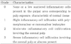
In MTA, calcium hydroxide, and photo-polymerized calcium hydroxide groups, initial mild inflammatory cell infiltration, newly formed odontoblast-like cell layer and hard tissue bridge formation were observed. Compared with dentin adhesive system, these materials were biocompatible and good for pulp tissue regeneration.
In dentin adhesive system group, severe inflammatory cell infiltration, pulp tissue degeneration and pulp tissue necrosis were observed. It seemed evident that application of dentin adhesive system in direct pulp capping of beagle dog teeth cannot lead to acceptable repair of the pulp tissue with dentine bridge formation.
Figures and Tables
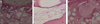
 | Figure 1a. Pulp capping with MTA at 7 days. Inflammatory cells infiltrated around the capping material under odontoblast-like cell. b. Pulp capping with MTA at 30 days. Newly formed dentinal bridge was observed. c. Pulp capping with MTA at 90 days. The thickness of hard tissue increased, compared with that of 30 days.
|
 | Figure 2a. Pulp capping with SE Bond at 7 days. Inflammatory cells infiltrated accompanied by soft tissue disturbances. b. Pulp capping with SE Bond at 30 days. Coronal pulp tissue necrosis beneath the exposure site was observed. c. Pulp capping with SE Bond at 90 days. Severe pulp tissue necrosis beneath the exposure site was seen.
|
 | Figure 3a. Pulp capping with Ultra-blend at 7 days. Inflammatory cell infiltration and hemorrhage was observed. b. Pulp capping with Ultra-blend at 30 days. Odontoblast-like cells arranged beneath exposure site. Pulp degeneration and partial pulp necrosis was seen. There was no hard tissue formation. c. Pulp capping with Ultra-blend at 90 days. Complete dentinal bridge protected the exposure site.
|
 | Figure 4a. Pulp capping with Dycal at 7 days. Pulp tissue degeneration was seen. b. Pulp capping with Dycal at 30 days. Partial pulp tissue degeneration and hard tissue formation were observed. c. Pulp capping with Dycal at 90 days. Complete hard tissue formation and normal soft tissue organization beneath the exposure site.
|

References
1. Tziafas D, Smith AJ, Lesot H. Designing new treatment strategies in vital pulp therapy. J Dent. 2000. 28:77–92.

2. Farhad A, Esfahan ZM. Calcium hydroxide: a review. Int dent J. 2005. 55:293–301.
3. Cox CF. Re-evaluating pulp protection: calcium hydroxide liners vs cohesive hybridization. J Am Dent Assoc. 1994. 125:823–831.
4. Cox CF, Bergenholtz G, Heys DR, Syed SA, Fitzgerald M, Heys RJ. Pulp-capping of the dental pulp mechanically exposed to oral microflora: a 1-2 year observation of wound healing in the monkey. J Oral Pathol. 1985. 14:156–168.
5. Lee TH. The physical properties of light-activated pulp capping material, substituted for Ca(OH)2 pastes. 2007. Seoul National Univ..
6. Brännström M, Nyborg H. Pulpal reaction to composite resin restorations. J Prosthet Dent. 1972. 27:181–189.
7. Stanley HR, Bowen RL, Cobb EN. Pulp responses to a dentin and enamel adhesive bonding procedure. Oper Dent. 1988. 13:107–113.
8. Hebling J, Giro EMA, Costa CAS. Biocompatibility of an adhesive system applied to exposed human dental pulp. J endod. 1999. 25:676–682.
9. de Souza Costa CAS, Nascimento ABL, Teixeira HM, Fontana UF. Response of human pulps capped with a self-etching adhesive system. Dent Mater. 2001. 17:230–240.
10. Koliniotou-Koumpia E, Tziafas D. Pulpal responses following direct pulp capping of healthy dog teeth with dentine adhesive systems. J Dent. 2005. 33:639–647.
11. Stanley HR, Swerdlow H, Buonocore MG. Pulp reactions to anterior restorative material. J Am Dent Assoc. 1967. 75:132–141.
12. Torabinejad M, Pitt Ford TR. Root-end filling materials: a review. Endod Dent Traumatol. 1996. 12:161–178.
13. Ford TR, Torabinejad M, Abedi HR, Bakland LK, Kariyawasam SP. Using mineral trioxide aggregate as a pulp-capping material. J Am Dent Assoc. 1996. 127:1491–1494.
14. Faraco IM Jr, Holland R. Response of the pulp of dogs to capping with mineral trioxide aggregate or a calcium hydroxide cement. Dent Traumatol. 2001. 17:163–166.
15. Holland R, de Souza V, Nery MJ, Otoboni Filho JA, Bernabe PF, Dezan Jr E. Reaction of rat connective tissue to implanted dentin tubules filled with mineral trioxide aggregate or calcium hydroxide. J Endod. 1999. 25:161–166.
16. Asgary S, Mohammad JE, Masoud P, Farzin G, Hessam R. A comparative study of histologic response to different pulp capping materials and a novel endodontic cement. Oral Surg Oral Med Oral Pathol Oral Radiol Endod. 2008. 106:609–614.
17. Torabinejad M, Hong CU, MacDonald F, Pitt Ford TR. Physical and chemical properties of a new root-end filling material. J Endod. 1995. 21:349–353.
18. Kakehashi S, Stanley HR, Fitzgerald RJ. The effects of surgical exposures of dental pulps in germ-free and conventional laboratory rats. Oral Surg Oral Med Oral Pathol. 1965. 20:340–349.
19. Rehman K, Saunders WP, Foye RH, Sharkey SW. Calcium ion diffusion from calcium hydroxide-containing materials in endodontically-treated teeth ; an in vitro study. Int Endod J. 1996. 29:271–279.
20. Hayashi Y. Ultrastructure of initial calcification in wound healing following pulpotomy. J Oral Pathol. 1982. 11:174–180.
21. Yamamura T. Differentiation of pulpal cells and inductive influences of various matrices with reference to pulpal wound healing. J Dent Res. 1985. 64 Spec No:530–540.
22. Goldberg F, Massone EJ, Spielberg C. Evaluation of the dentinal bridge after pulpotomy and calcium hydroxide dressing. J endod. 1984. 10:318–320.
23. Schröder U. Effects of calcium hydroxide-containing pulp-capping agents on pulp cell migration, proliferation and differentiation. J Dent Res. 1985. 64 Spec No:541–548.
24. Holland R, Pinheiro CE, de Mello W, Nery MJ, de Souza V. Histochemical analysis of the dogs dental pulp after pulp capping with calcium, barium, and strontium hydroxides. J Endod. 1982. 8:444–447.
25. Stanley HR, Pameijer CH. Pulp capping with a new visible-light-curing calcium hydroxide composition (Prisma VLC Dycal). Oper Dent. 1985. 10:156–163.
26. Kim JH, Hong JB, Lim BS, Cho BH. Histological evaluation of direct pulp capping with DSP-derived synthetic peptide in beagle dog. J Korean Acad Conserv Dent. 2009. 34:120–129.
27. Yun YR, Yang IS, Hwang YC, Hwang IN, Choi HR, Yoon SJ, Kim SH, Oh WM. Pulp response of mineral trioxide aggregate, calcium sulfate or calcium hydroxide. J Korean Acad Conserv Dent. 2007. 32:95–101.
28. Goldberg M, Six N, Decup F, Buch D, Soheili Majd E, Lasfargues JJ, Salih E, Stanislawski L. Application of bioactive molecules in pulp-capping situations. Adv Dent Res. 2001. 15:91–95.
 XML Download