

 PDF
PDF ePub
ePub Citation
 Print
Print


Introduction
Pre-treatment of the tooth surface with antibacterial agents is useful for eliminating the harmful effects caused by either the residual bacteria or bacterial microleakage.1 Chlorhexidine is one of the most widely used broad spectrum antibacterial or antiseptic agents in dentistry.2 The efficacy of chlorhexidine in caries prevention had been established in clinical trials.3
The latest development in dental adhesion is based on simplification and a reduced application time. The first self-etch adhesives were composed of two solutions, an acidic primer and bonding resin. Recently, many clinicians have shifted to one-step self-etch systems (all-in-one adhesives), in which the manufacturers have attempted to incorporate all the primary components of an adhesive system (etchant, primer, and bonding resin) into a single solution.
One potential problem with the use of chlorhexidine with dentin bonding agents is the possibility of adverse effects on the bond strength of the resin composites. Previous studies4-10 were carried out using etch-and-rinse and self-etch adhesives with conflicting results being reported. However, few studies have examined the effect of chlorhexidine application on the microtensile bond strength of resin composites to dentin using one-step self-etch adhesives. As the use of one-step self-etch adhesives has become popular at clinical practice recently, a study about the effect of chlorhexidine on resin composite restored with one-step self-etch adhesive is necessary.
Therefore, this study examined the effect of chlorhexidine on the microtensile bond strength (µTBS) of direct composite restorations in vitro using a one-step self-etch adhesive on human dentin.
Materials and Methods
1. Tooth preparation
Twenty-four extracted noncarious human permanent molars were washed and stored in distilled water. A plastic mold was filled with an autopolymerizing resin (Tokuso curefast; Tokuyama, Tokyo, Japan) and the root surface was embedded in acrylic resin, leaving the clinical crown exposed. After removing the plastic mold, the teeth were sectioned horizontally at mid-coronal level using a diamond-saw (Accutom-50; Struers, R.dovre, Denmark) under continuous water cooling. The flat dentin surface was exposed using 600 grit silicon carbide paper under running water.
2. Dentin bonding and resin composite buildups
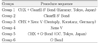
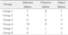
The teeth were then assigned randomly to six groups according to the adhesive system and application of chlorhexidine. Tables 1 and 2 listed the materials and groups used in this study, respectively. In the chlorhexidine applied groups, 1 mL of a 2% chlorhexidine solution was applied to the exposed dentin surface for 10 seconds and dried for 10 seconds without rinsing. Each adhesive system was applied according to the manufacturer's instruction. After the bonding procedure, the light-cure composite resin (Premise; Kerr, Orange, CA, USA) was bonded to the treated dentin surface in 1 mm increments. Each 1 mm increment was polymerized for 20 seconds using a light-emitting diode (LED) visible light-polymerizing unit (Bluephase; Ivoclar Vivadent, Schaan, Liechtenstein), and the height of the total resin build up was approximately 5 mm. The restored teeth were then stored in distilled water at room temperature for 24 hours.
3. µTBS testing
The restored teeth were sectioned longitudinally to make approximately 1×1 mm thick and 10 mm long specimens using a diamond-saw under copious amounts of water. Each group contained 10 specimens. Each specimen was glued to the jig of the microtensile testing machine (BISCO Inc; Bisco, Schaumburg, IL, USA) using cyanoacrylate cement (Zapit; Dental Ventures of America, Corona, CA, USA). A tensile load was applied at a 1.0 mm/min cross-head speed until bonding failure occurred and the maximum load at failure was recorded.
4. Failure mode investigation
The fractured test specimens were examined using an operating microscope (OPMI pico; Carl Zeiss, Obercohen, Germany) under×25 magnification, and the failure mode was classified as follows: adhesive, if the composite resin cone fractured at the adhesive-tooth adhesivetooth interface; cohesive, if the composite resin cone fractured inside the composite resin or dentin; or mixed, a combination of adhesive and cohesive failure.
5. Statistical analysis
Statistical analysis of the µTBS between chlorhexidine application groups and non-application groups were performed using a Student's t-test. For a comparison of the dentin adhesives, one-way ANOVA was performed and a Scheffé's test was used for a post-hoc multiple comparison. The level of significance was set to p < 0.05.
Results
Table 3 lists the µTBS of the chlorhexidine application and non-application groups of each dentin bonding agent. In each group, the chlorhexidine non-application group showed a higher µTBS than the application group. However there was no significant difference between application and non-application groups (p > 0.05). In the chlorhexidine application groups, Clearfil S3 Bond (group 1) showed the highest µTBS, followed by a G-Bond (group 5) and Xeno V (group 3). The µTBS values of the three adhesives were significant different (p < 0.05). Similarly, in the chlorhexidine non-application groups, Clearfil S3 Bond (group 2) showed the highest µTBS, followed by G-Bond (group 6) and Xeno V (group 4). The µTBS of the Clearfil S3 Bond was significant higher than that of the G-Bond (p < 0.05). However, there was no significant difference between the G-Bond and Xeno V (p > 0.05).
Table 4 shows the failure mode. Adhesive failure was the main failure mode observed in all groups. All specimens in group 4 showed adhesive failure.
Discussion
In many cases, an antiseptic can be useful for eliminating the effects caused by bacteria and provide a better prognosis for the minimal restorative treatment of dental caries. Chlorhexidine is a broad spectrum antiseptic with pronounced antimicrobial effects,11 and has been shown to be effective in reducing cariogenic bacteria.12
In addition to its antibacterial effect, chlorhexidine also functions as a potent matrix metalloproteinase (MMP) inhibitor.13 Despite the remarkable advances that have increased the dentin-resin bond strength, the premature loss of bond strength is a major problems with adhesive restorations. The loss of bond strength was attributed mainly to the degradation of the hybrid layer at the dentin-adhesive interface. Mild acids can activate MMPs.14,15 Both the etch-and-rinse adhesives and self-etching adhesives have mild acidity, and can release and activate endogenous MMPs during dentin bonding.16-18 These activated MMPs can slowly hydrolyze any unprotected collagen fibrils of the dentin hybrid layers, which are believed to be responsible for the thinning and disappearance of collagen fibrils from the incompletely infiltrated hybrid layers in aged, bonded dentin. Auto-degradation of the collagen matrix can be suppressed by synthetic protease inhibitors, such as chlorhexidine, indicating that MMP inhibition can be beneficial for preserving hybrid layers.19-21
According to these results, the application of chlorhexidine did not affect the µTBS value significantly. However, the difference in µTBS between the bonding agents was significant as follows: Clearfil S3 Bond showed the highest µTBS, followed by G-Bond and Xeno V. Xeno V had the lowest pH (pH < 2), followed by G-Bond (pH = 2.3), and Clearfil S3 Bond (pH = 2.4). Low-pH self-etch adhesives have rather low bond strengths, particularly to dentin.22 This may be why Clearfil S3 Bond showed the highest µTBS, followed by G-Bond and Xeno V.
Previous studies4,5,7,10 reported the association of chlorhexidine application and bond strength to be a contentious issue. There are studies that insist that the application of chlorhexidine affects the bond strength. Gurgan et al reported that the use of 2% chlorhexidine before or after acid etching may decrease the bond strength by altering the ability of the hydrophilic resin to seal dentin.7 Meiers and Shook indicated that bonding with Tenure (two-step etch-and-rinse adhesive) was unaffected by the application of chlorhexidine, whereas bonding with Syntac (two-step self-etch adhesive) was affected.10
However, Perdigão et al stated that chlorhexidine has strong affinity to the tooth surfaces, which is increased by acid-etching.5 Chlorhexidine also increases the surface free energy of the enamel and can have a similar effect on dentin. These properties might improve the adhesion of primers. However, the loss of calcium from hydroxyapatite and these effects are negated by the presence of chlorhexidine residue. According to their results, chlorhexidine had no significant effect on the shear bond strengths of the composite to dentin using the All-Bond 2 adhesive system. Say et al found that chlorhexidine applied before the acid etching of dentin caused changes in the smear layer, but an acid treatment of the surface nullified any effect of chlorhexidine on the smear layer.4 In addition, in the present study, there were no differences between the two groups, regardless of chlorhexidine application.
In this study, adhesive failure was the main failure mode in all groups. Adhesive failure may be due to the lower bond strength or a weak stability of the adhesive layer.
In conclusion, within the limits of this study, the application of 2% chlorhexidine did not affect the µTBS of the resin composite to the dentin using a one-step self-etch adhesive. Because chlorhexidine has antimicrobial properties and does not significantly affect the bond strength to dentin, the use of a chlorhexidine solution with a one-step self-etch adhesive might have the added potential of preventing collagen fibril degradation in the dentin hybrid layers. Further studies will be needed to determine the effects of chlorhexidine on the bond durability.

 XML Download