

 PDF
PDF ePub
ePub Citation
 Print
Print


Introduction
A range of filling materials are used for perforation repair and root end-fillings in clinical endodontics, such as reinforced zinc oxide eugenol cement, intermediate restorative material (IRM), super ethoxybenzoic acid (EBA) cement, calcium hydorixde, composite resins ,glass ionomer cements and mineral trioxide aggregate (MTA , Dentsply, Tulsa Dental, Tulsa, OK, USA). Among these, MTA, which is also called ProRoot MTA, has been used successfully in endodonctics for the past 10 years.1-3 More recently, new products similar to MTA have been introduced into the market including MTA angelus4-6 and MTA-bio7 (Angelus Solucoes Odontologicas, Londrina, Brazil) and Bioaggregate8 (Innovative Bioceramix, Vancouver, Canada). Bioaggregate is sold exclusively in Canada. Its recommended applications are the same as those of MTA.
This new material has received less research attention than other materials.9-12 Park et al. examined the chemical composition of BioAggregate and reported that it contained a significant amount of tantalum oxide instead of bismuth oxide.9 The major components were calcium silicate oxide and calcium silicate. Zhang et al. reported that MTA and Bioaggregate powder have a similar bactericidal effects on E. faecalis.8 More recently, Yuan et al. reported that Bioaggregate was nontoxic to mouse MC3T3-E1 osteoblast cells.12
Bioaggregate appears to be a modified or synthetic version of original MTA. Since the clinical indications of Bioaggregate are the same as those of MTA, it might be helpful to compare this new material with MTA, which has already been studied and proven to be effective and biocompatible over the past few years.13-15
This study examined the biocompatibility of BioAggregate compared to white ProRoot MTA on the cells derived from human dental pulp and periodontial tissue.
Materials and Methods
Cell Culture
Human premolars planned to be extracted for orthodontic treatment were used. The institutional Review Board of Gangnam Severance Hospital, Yensei University approved both the protocol and informed consent. Written informed consent was obtained from each patient (No 3-2009-0069). After extraction, the teeth were stored in 50 ml polyethylene tubes containing 10 ml alpha minimum essential medium (α-MEM)(Gibco/BRL, Grand Island, NY, USA) supplemented with 10% fetal bovine serum (FBS; Hyclone, Loga, UT, USA) and 1% penicillin. After washing 3 times with α-MEM (3% antibiotics/antimycotics), the tooth was held using sterilized gauze. The soft tissue on the middle 1/3 of the root was obtained by curettage with a No.15 blade. The tissue collected was kept in a 100 mm2 culture dish containing α-MEM and the culture plate was washed three times with the same culture media. A new culture dish was used to apply the tissue, which was kept in the incubator at 37℃ in a humidified atmosphere containing 5% CO2 in air until the plate became full with growing cells. The remaining tooth was split after making bucco-linugal grooves. The pulp tissue was removed using a sterilized spoon excavator and cultured in a similar manner as periodontal tissue.
Preparation of a cement-coated tissue culture plate
White MTA (ProRoot) and Bioaggregate were mixed according to the manufacturer's instructions. A 6-well tissue culture dish was coated with the MTA paste prepared with sterile water (10 mg cement/ml of H2O per well). PDL and pulp cells were seeded on culture dishes coated with the freshly mixed cements to determine if the initial toxicity of the freshly mixed material can affect cell attachment and growth.
Optical Microscopy
The cells were plated on either an MTA coated or plastic surface (5.0×10⁵ cells/well in a 6-well plate), and cultured for 1, 6, 48 and 72 hours before being examined by phase microscopy (Nikon TMS, Nikon Inc. Melville, NY, USA). Cells grown on cement coated dishes could not be visualized by this method since both cements used do not transmit light. Thus, the cells grown on plastic surface around cements were examined under the microscope and photos were taken.
Cell Viability test
The cells were grown for 3 days on a 12-well culture plate coated with each type of cement. After 1×10⁵ cells were seeded, the cells grown on a regular plastic plate were used as the control. An EZ-Cytox cell viability assay kit (DAEILL LAB Service Co., Seoul, Korea), which measures the cell dehydrogenase activity was used. This kit is similar to a MTT assay measuring the mitochondrial activity in viable cells. After adding 100 µL of the assay reagent, cell culture plate was kept in a 37℃ incubator for 1 hour. Subsequently, 100 µL of supernatant was taken and added to a 96-well plate. The color density at 450 nm was read using an ELISA reader (Molecular Devices Inc/E-max, Washington, DC, USA). The data is presented as a percentage when the density of the control was considered to be 100. The average of the triplicates was used for data analysis.
Results
Cell attachment
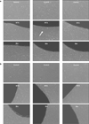
The effects of Bioaggregate cement on human pulp and PDL cell growth were examined by observing the cells grown on this cement using a phase microscope (Figure 1). The effects of MTA and Bioaggregate on cell growth were determined by examining the cells around the materials because cells grown on top of the material could not be visualized. Both types of cells on Bioaggregate showed no inhibition zones or gaps around the material at any time intervals. However, although cells were fully grown around MTA, an inhibition zone was detected in the human pulp and PDL cell culture grown with MTA.
Cell viability test
Before performing the viability tests, each cell culture plate was observed by optical microscopy to determine if the cell were growing healthily without dead floating cells.
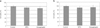
There were no significant differences in the human pulp and PDL cell cultures between control and experimental groups (Figure 2). There were 6.3 ± 8.02% and 9.25 ± 9.74% less viable cells in human PDL cells when cultured with MTA and Bioaggregate. The similar result was shown in pulp cells.
Discussion
In this study, the biocompatibility of Bioaggregate was compared with white MTA since its chemical composition was different from that of MTA from our previous study.9
Cell attachment and growth can be used as criteria to evaluate the biocompatibility or cytotoxicity of a material in a cell culture. MTA may not provide a favorable surface environment for cells while it sets because of the high pH generated during setting. Many clinical reports and animal studies have suggested that this harmful effect is transient.16,17 The lack of an inhibition gap in the cell culture grown with Bioaggregate indirectly demonstrated its favorable environment.
A cell viability test was performed using EZ-Cytox. Compared to the MTT assay, this method employs a soluble terazolium salt, which is unnecessary for dissolving formazan and removing the supernatant of the cell culture. There was no significant difference in the viable cell numbers between the MTA and Bioaggregate groups, which demonstrated both cements to be equally nontoxic to human pulp and PDL cells.
Our results are consistent with other previous studies, which demonstrated both cements were equally nontoxic to human pulp and PDL cells. De-Deus et al. showed in vitro biocompatibility compared to MTA when they tested this material using human mesenchymal cells.18
Also, the potentials that Bioaggregate can affect cell differentiation and/or mineralization have been shown in recent studies. Yan et al. found the increased level of alkaline phosphatase when cells were grown with Bioaggregate on 7 days.11 They concluded that Bioaggregate may enhance PDL cell differentiation. Yuan et al. also found the increased mineralization when osteoblasts were grown with Bioaggregate.12
MTA has been proved as biocompatible and nontoxic material in many previous studies. Recently, several new bioactive cements similar to MTA have been introduced. MTA angelus, MTA bio, and Bioaggreagte are the examples. These materials have been studied recently, and they are used in certain countries. In this study, white MTA was used as one of the experimental groups. By having MTA group as a reference, it was concluded that Bioaggregate might be compatible with MTA in terms with cell cytotoxicity and initial cell growth.
It is expected to have more new materials like MTA in the near future. At the same time, meticulous research should be needed before clinical applications.
 XML Download