

 PDF
PDF ePub
ePub Citation
 Print
Print


Introduction
Among several factors, success or failure of endodontic therapy depends on the quality of canal preparation.1 This is essential because preparation determines the efficacy of all subsequent procedures and includes mechanical debridement, creation of space for medicament delivery, and optimized canal geometries for adequate obturation. Unfortunately, canal preparation is adversely influenced by the highly variable root canal anatomy.2-4 To overcome this, a variety of endodontic instruments and canal preparation techniques have been introduced into the field of root canal treatment.
Canal preparation using stainless steel hand instruments is generally time-consuming and difficult in curved canals especially, because of its stiffness.5,6 Stainless steel instrument is likely to cause canal aberrations such as zips, elbow, ledges and perforations.7,8
During the late 1980s and 1990s, various types of endodontic instruments were developed with nickel-titanium alloys.9 The introduction of nickel-titanium alloy for the mechanically driven endodontic instruments has greatly simplified shaping of the root canal systems.9,10 Reports have shown that nickel-titanium (Ni-Ti) instruments are two or three times more flexible than conventional stainless steel files and that they have more resistance torsional fracture.11 Thus, these instruments have been found to be better than stainless steel instruments in maintaining the original anatomy and the shape and position of the apical foramen.6,12
However, despite the distinct advantages of the new technique, Ni-Ti rotary instruments have several unexpected disadvantages. One of these is the tendency to screw-in to the canal.13 This tendency is particularly accentuated during root canal preparation, especially when continuously rotating nickel-titanium instruments are used. Lack of tactile sense during the rotary instrumentation makes it more difficult to control the working length than during the hand instrumentation. For this reason, the screw-in effect during rotary instrumentation may cause overinstrumentation beyond the apical foramen.14 Undoubtedly, instrumentation beyond the apical foramen reduces the success rate of the endodontic treatment.9,15-19
Cross-sectional geometry, helical angles, pitch length and taper of files are the factors which have been investigated to be involved in the screw-in effect of Ni-Ti rotary files. Diemer and Calas evaluated the effect of pitch length on the torsional stress and tendency to screw-in using two instruments with same cross-section (triple helix; 0.6% taper).20 Their results showed that increasing the pitch length decreased torsional load sharing and the tendency to screw in. Schräder and Peter compared torques and forces developed by two sequences of nickel-titanium rotary instruments, using only .04 taper instruments or a combination of 04. and .06 tapers, respectively.21 Their conclusions were an instrumentation sequence encompassing various tapers seems to be safer in torsional and fatigue failure compared to sequence that used only one single taper. Ha et al. compared the screw-in effect among several Ni-Ti rotary file systems.13 The results showed that the design of the blade might be the cause of the significant differences of screw-in effect among the tested Ni-Ti rotary instruments. From the result of the study by Sung, rotary files with more taper produced more screw-in effect.22
Human dentition always contains root curvatures, and these curvatures may differ from mild to severe. In fact, if the root curvature is severe, then endodontic treatment of the associated pulp canal system becomes difficult. Objectives of the endodontic therapy include debriding the root canal system, continuously tapering in a conical form, and maintaining the original shape and position of the apical foramen.23 However, ledge formation, transportation of the apical foraman, and nontapered hourglass-shaped preparation are problems frequently observed after the instrumentation on curved root canals.24,25
From this point of view, it is necessary to evaluate the effect of the root canal curvature as a factor which is involved in the screw-in effect of Ni-Ti rotary files. However, there has been no report that demonstrate the relation between the screw-in effect and the root canal curvatures.
Therefore, the purpose of this study was to evaluate the screw-in effect on several root canal curvatures by using Ni-Ti rotary files.
Materials and Methods
1. Specimen preparation
A total of 80 simulated root canals in clear resin blocks (Root Canal Model, Item number: S6-U1, S4-U1, S4-U4, S4-U6; Nissin Dental Prod. Inc., Kyoto, Japan) were used in this study. Specimens were divided into the following groups according to root canal curvatures: 0 (S6-U1), 10 (S4-U1), 20 (S4-U4) and 30 (S4-U6) degree. Twenty specimens were used in each group to obtain the results.
Preflaring of resin blocks were done by following procedures.
The shaping procedure commenced with ProTaper instruments (Dentsply-Maillefer, Ballaigues, Switzerland) SX, S1, and S2. The coronal and middle third of the root canal was shaped if passive penetration was possible.
A ProFile (Dentsply-Maillefer) size 25 .06 taper instrument was inserted and used until 1.0-2.0 mm beyond the initial point of root curvature. This preflaring allowed instruments to reach the canal curvature without any interference. Therefore, it was possible to evaluate the effect of difference in the degree of root canal curvature.
This preflaring allowed instruments to reach the canal curvature without any interference. Therefore, it was possible to evaluate the effect of difference in the degree of root canal curvature.
2. Measurement of screw-in force
ProFile.06 was used in the experiment. The tip diameter of the instrument was the same as that of ISO size #30.
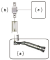
All canals were prepared by using a custom-made device (Figure 1) with a 16 : 1 reduction hand-piece powered by an electric motor (SurgiMotor II, Aseptico Corp., Woodinville, WA, USA). The contra-angled hand-piece with the rotary file was mounted on the custom-made device and the files were automatically inserted into the canal of resin block. The position of resin block was marked when the file bound the canal at the first time. The resin block was moved 1.0 mm toward the rotary file. 1.0 mm single pecking motion was generated with screw-in of clock-wise direction. New instrument was used for each canal with saline irrigation.
When the instruments were withdrawn at the end of the pecking movement, a force that resist the withdrawal of the instrument from the root canal was generated. This axial stress caused pulling of the resin blocks. The resistance force was considered as the screw-in force. The dynamometer (K1368-10N, Lorenz Messtechnik Gmbh, Alfdorf, Germany) in the device recorded the transmitted resistance force in mA. The generated signals (mA) were amplified with a sensor-interface with USB (LCV-USB, Lorenz Messtechnik Gmbh, Alfdorf, Germany) and transferred to the computer software. The transferred signals were recorded with a software (LCV-USE-VS, Lorenz Messtechnik Gmbh, Alfdorf, Germany).
Results
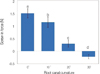
The screw-in forces at different canal curvatures are shown in Figure 2. Larger degree of canal curvature generated significantly less screw-in forces in all groups (p< 0.001).
Discussion
One aspect of canal configuration that has been shown to have an important influence on instrumentation is canal curvature. The amount of canal curvature may affect on access for instrumentation and also on the risk of instrument separation. Enlargement of curved canals may be difficult due to the potential complications of ledging, zipping, stripping, and apical debris compaction.24 However, despite the importance of canal curvature in endodontic treatment, there was no research investigated on the outcomes regarding the screw-in effect of Ni-Ti rotary files on the severity of the root curvatures. Thus, in the present study, we evaluated the screw-in effect of Ni-Ti rotary files at various root curvature.
The fact that roots are curved was appreciated by stating the angle of the curve and then categorizing roots as mildly (< 5°), moderately (10° to 20°) or severely (> 20°) curved.26 Thus, in the present study, specimens were divided into four groups according to the canal curvatures; 0, 10, 20 and 30 degree.
Determination of the real anatomical diameter is difficult when no preflaring is performed. This accuracy may be enhanced when anatomical diameter determination is performed after flaring.27-29 Thus, in the present study, ProTaper Shaping Files (SX, S1, S2) and ProFile size 25 .06 taper instrument were used for preflaring in order to eliminate possible factors which may influence the result.
In the present study, simulated canals in resin blocks were used to eliminate variations of canal anatomy without using lubricants which may limit the number of variables and avoid stress measurement because of axial or transversal irrigation force.30 However, although simulated canals in resin blocks allow comparisons between root canal curvatures under identical conditions, i.e, the standardization of shape, taper, and curvature of canal, there are certain disadvantages as their surface texture, abrasion behavior and hardness as well as cross-sections differ from those in natural teeth.
The results of present study showed that larger degree of canal curvature generated significantly lesser screw-in forces in all groups. It may be considered that following two reasons are the cause of this outcome.
As an instrument rotates in the canal, it binds against tooth structure, which places friction on the instrument called torque. The amount of torque generated while rotating in the canal is positively related to the mass of the instrument. Larger sized and greater taper files, although being stronger and having better torque resistance, will create more torque value on contact with the canal wall.31,32 Canal curvature is the important factor in the determining the torque value. If the canal curvature is larger, it results in higher torque on file. As a result, it may be speculated that lesser screw-in force occurred in severely curved groups on this experiment.
In other point of view, the other reason may be that file with increased taper are likely to bind along the shaft. When the rotary instruments are continuously rotating, the stresses generated in the instruments are increased. Thus, more tapered instruments screw into dentin with more forces.22 Moreover, most root canals are curved, whereas endodontic instruments are manufactured from straight metal blanks. This results in uneven force distribution in certain contact areas3,34 and a tendency of the instrument to straighten itself inside the root canal.35 If the degree of canal curvature decreases, files are likely to bind more along the shaft. As a result, it is assumed that the stresses generated in the instruments increase. Therefore, greater screw-in force to dentin occur.
In conclusion, within the condition of the present study, larger degree of canal curvature generated lesser screw-in forces. More attention should be paid when using rotary instruments in canals with less curvatures than canals with more curvatures to prevent or reduce the screw-in forces. Further research is needed to evaluate the relationship between the screw-in effect and root canal curvatures on natural teeth and to find out the techniques to control this screw-in effect during rotary instrumentation.
 XML Download