

 PDF
PDF ePub
ePub Citation
 Print
Print


Abstract
Objectives
This study investigated the effect of infection control barrier thickness on power density, wavelength, and light diffusion of light curing units.
Materials and Methods
Infection control barrier (Cleanwrap) in one-fold, two-fold, four-fold, and eight-fold, and a halogen light curing unit (Optilux 360) and a light emitting diode (LED) light curing unit (Elipar FreeLight 2) were used in this study. Power density of light curing units with infection control barriers covering the fiberoptic bundle was measured with a hand held dental radiometer (Cure Rite). Wavelength of light curing units fixed on a custom made optical breadboard was measured with a portable spectroradiometer (CS-1000). Light diffusion of light curing units was photographed with DSLR (Nikon D70s) as above.
Results
Power density decreased significantly as the layer thickness of the infection control barrier increased, except the one-fold and two-fold in halogen light curing unit. Especially, when the barrier was four-fold and more in the halogen light curing unit, the decrease of power density was more prominent. The wavelength of light curing units was not affected by the barriers and almost no change was detected in the peak wavelength. Light diffusion of LED light curing unit was not affected by barriers, however, halogen light curing unit showed decrease in light diffusion angle when the barrier was four-fold and statistically different decrease when the barrier was eight-fold (p < 0.05).
Figures and Tables
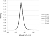 | Figure 1Wavelength (nm) of LED light curing unit with infection control barriers covering the filberoptic bundle.
|
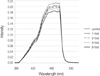 | Figure 2Wavelength (nm) of halogen light curing unit with infection control barriers covering the filberoptic bundle.
|
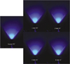 | Figure 3Light diffusion from LED light curing unit with infection control barriers covering the fiberoptic bundle.
|
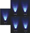 | Figure 4Light diffusion from halogen light curing unit with infection control barriers covering the fiberoptic bundle.
|


References
1. Caughman GB, Caughman WF, Napier N, Schuster GS. Disinfection of visible-light-curing devices. Oper Dent. 1989. 14(1):2–7.
2. Kakaboura A, Tzoutzas J, Pitsinigos D, Vougiouklakis G. The effect of sterilization methods on the light transmission characteristics and structure of light-curing tips. J Oral Rehabil. 2004. 31:918–923.

3. Rueggeberg FA, Caughman WF, Comer RW. The effect of autoclaving on energy transmission through light-curing tips. J Am Dent Assoc. 1996. 127:1183–1187.
4. Nelson SK, Caughman WF, Rueggeberg FA, Lockwood PE. Effect of glutaraldehyde cold sterilants on light transmission of curing tips. Quintessence Int. 1997. 28:725–730.
5. Chong SL, Lam YK, Lee FKF, Ramalingam L, Yeo ACP, Lim CC. Effect of various infection-control methods for light-cure units on the cure of composite resins. Oper Dent. 1998. 23:150–154.
6. Pollington S, Kahakachchi N, van Noort R. The Influence of Plastic Light Cure Sheaths on the Hardness of Resin Composite. Oper Dent. 2009. 34:741–745.
7. Scott BA, Felix CA, Price RB. Effect of disposable infection control barriers on light output from dental curing lights. J Can Dent Assoc. 2004. 70:105–110.
8. Fan PL, Schumacher RM, Azzolin K, Geary R, Eichmiller FC. Curing-light intensity and depth of cure of resin-based composites tested according to international standards. J Am Dent Assoc. 2002. 133:429–434.
9. Park SM, Shin DH. Microhardness and microleakage of composite resin according to the change of curing light intensity. J Korean Acad Conserv Dent. 2001. 26:363–371.
10. Park JG, Cho BH, Lee IB, Kwon HC, Um CM. The effects of various light intensity on the polymerization of resin composites. J Korean Acad Conserv Dent. 2001. 26:86–94.
11. Shortall AC, Harrington E, Wilson HJ. Light curing unit effectiveness assessed by dental radiometers. J Dent. 1995. 23:227–232.
12. Rueggeberg FA. Precision of hand-held dental radiometers. Quintessence Int. 1993. 24:391–396.
13. Peutzfeldt A. Correlation between recordings obtained with a light-intensity tester and degree of conversion of a light-curing resin. Scand J Dent Res. 1994. 102:73–75.
14. Kim SY, Lee IB, Cho BH, Son HH, Kim MJ, Seok CI, et al. Dentin bond strength of bonding agents cured with Light Emitting Diode. J Korean Acad Conserv Dent. 2004. 29:504–514.
15. Price RB, Derand T, Sedarous M, Andreou P, Loney RW. Effect of distance on the power density from two light guides. J Esthet Dent. 2000. 12:320–327.
16. Corciolani G, Vichi A, Davidson CL, Ferrari M. The influence of tip geometry and distance on light-curing efficacy. Oper Dent. 2008. 33:325–331.
 XML Download