

 PDF
PDF ePub
ePub Citation
 Print
Print


Introduction
One of the objectives of successful endodontic treatment is to prevent infection or reinfection by complete obturation of root canal system.1 However, no available material and/or technique produce a complete seal of the entire root canal system.2
Ingle et al.3 suggested up to 59% of endodontic failures are attributable to apical leakage. Failure of the apical seal allows undebrided irritants to escape from the canal into the periapcial tissue to initiate an inflammatory response.4
The most commonly used core filling material is gutta percha but these materials inherently leave discrepancies between the obturator and the canal wall and possess no natural adhesion to tooth structure.5 Therefore, root canal sealers are used with these materials to provide the required adhesion to dentin and to fill the irregularities and minor discrepancies between the core filling material and the canal walls. The sealer also acts as a lubricant during the obturation procedure. Hence, the sealing quality of a root canal filling depends on the sealing ability of the sealer.6
In some cases, the practitioner has difficulty to dry the root canal space completely before obturation which is potentially caused by poor isolation, ingress of periapical exudate or incomplete apical extension, extremely small or tortuous for using paper point. Thus, clinically these various environmental conditions of the root canal system may be related to some physical properties of sealer include setting time, solubility or water absorption, adhesion, penetration and sealing ability of a sealer.7,8
Previous endodontic textbooks recommend that the root canal space should be completely dry prior to obturation in order to increase the adhesion of sealers to the dentinal wall and the filling material.9,10 But, with recent advances in bonding system, hydrophilic methacrylate resin based sealers were introduced in dental material. The manufacturers recommended that dentinal walls are kept moist, not dehydrated to take maximum advantage of the hydrophilic properties of the sealers. Therefore, the effect of residual moisture on the apical seal should be determined.
This study compared the sealing abilities of four different types of sealer which were used in the different moisture conditions of root canal.
Materials and Methods
Ninety recently extracted single rooted human teeth were selected which have single straight root canal with fully developed apices and they are free of cracks, caries or fractures. The root was rid of attached tissues and calculus and the tooth then immersed in 0.02% sodium azide, NaN3 (DUKSAN PURE CHEMICAL CO., LTD., Ansan city, Korea) at 37℃ until use.
Instrumentation and obturation of root canals
The coronal portion of all teeth was removed with high speed handpiece and diamond bur. (Shofu INC., Kyoto, Japan) so that each specimen was 13 mm long Straight-line entries were made and the apical patency was determined by passing a size 15 K-file. Canal working lengths were established 1.0 mm short of the apical foramina. The root canals were prepared using K3 NiTi rotary instruments (SybronEndo, Glendora, Ca, USA) at 300 rpm. All canals were enlarged with a 0.06 taper, size 45 instrument. Irrigation between each filing with 0.5 mL of 2.5% NaOCl and final flush with 3 mL of 17% EDTA (2 minute) were applied to remove smear layer.11 To remove all of the residual chemicals, final rinse with 3 mL of sterile water were applied.
All teeth were stored in distilled water of 37℃ until used. The prepared roots were randomly divided into 12 experimental groups of 7 roots, and 2 control groups of 2 roots each. Specimens were then subjected to 3 different moisture conditions.
Moisture condition I (DRY)
Excess water was removed with paper points followed by dehydration with 90% ethanol and 200℃ probe of System B (Analytic Technology, Orange, CA, USA) was inserted into the root canals, twice for 5 seconds dwell time.
Moisture condition II (PAPER POINT DRY)
Blot-dried with paper point until the last one appearing dry.
Group 1-12
Applying the 3 moisture conditions above, the teeth were filled with the warm vertical compaction technique by using one of the 4 types of sealer. Group 1-3 were filled with a calcium hydroxide based sealer, Sealapex (Kerr, SybronEndo, Orange, CA, USA). Group 4-6 were filled with a epoxy resin based sealer, AH plus (Dentsply-Maillefer, Tulsa, OK, German). Group 7-9 were filled with a zinc oxide-eugenol-based sealer, Tubuli-seal (Kerr, SybronEndo, Orange, CA, USA). Group 7-9 were filled with a methacrylate resin-based sealer, EndoRez (Ultradent Products Inc, South Joran, UT, USA)(Table 1).
The sealers were used according to the manufacturer's recommendations.
The 3 positive control teeth were filled in a same manner but without sealer and 3 negative control teeth were filled with GP and AH Plus. The filled roots were stored in a moist gauze at 37℃ for 7 days to allow for complete setting.
Glucose leakage model
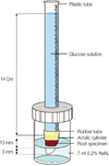
The specimens of the negative control group were completely coated with nail varnish. The specimens of the positive control and experimental groups were coated except for the area 3 mm of the apical end. The teeth were placed in a device designed to measure glucose leakage (Figure 1).12
The resin block around the coronal 4 mm of each root was inserted to a rubber tube which was itself connected to a 15 cm long plastic tube. This assembly was then placed in a 20 mL sterile glass bottle with a screw cap and sealed with Zapit cyanoacrylate (Dental Bentures of America, Inc., Corona, CA, USA). 7 mL of 0.2% NaN3 solution was inserted into the glass bottle so that the 3 mm of root apical end was immersed in the solution. NaN3 was used to inhibit the growth of microorganisms that might influence the glucose readings.12 1mol/L of glucose solution (pH 7.0) containing 0.2% NaN3 was injected into the plastic tube until the top of the solution was 14 cm higher than the top of gutta-percha in the canal which created a hydrostatic pressure of 1.5 kPa (15 cm H2O).13 This apparatus was then transferred into an incubator providing 100% humidity at 37℃. To examine the evaporation of the solution in the glass bottle, another glass bottle containing 7 ml of 0.2% NaN3, was also placed in the incubator and used to keep the volume of NaN3 solution same in the apparatus from evaporation. The weight of the supplemental solution was measured every day to figure out the amount of evaporation during test time.
Measurement of microleakage
A 10 µL aliquot of solution was drawn by using a micro-pipette from the glass bottle at 1, 3, 7, 14, 21, and 30 days. After drawing the sample, 10 µL of fresh 0.2 % NaN3 was added to the glass bottle reservoir to maintain a constant volume. To determine the concentration of glucose, the phenol-sulfuric acid method was used. Each 100 µL of resulted product was quantified with spectrophotometer (PowerWave; D. I. Biotech Ltd, Korea) at 490 nm wavelength. The lowest glucose level for which the current procedure is believed to be accurate is 0.75 mmol/L. The values below this were considered unreliable and recorded as o (no leakage).14
Results
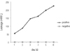
The negative control group showed no leakage of glucose. The positive control group had substantial leakage of glucose from the first day which increased rapidly over time until 30 days (Figure 2) and it showed highly significant amount of penetration compared to the experimental groups (p < 0.05).
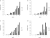
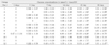
The mean glucose leakage values for each moisture condition and for each material are shown in Table 2 and Figures 3a-d.
There was a tendency of increase in leakage in all experimental groups during experimental period. There was no significant differences were found among the dry and paper point dry groups (Group 1 and Group 2 or Group 4 and Group 5), within dry and paper point dry groups (Group 1 and Group 2 or Group 4 and Group 5), within the Sealapex and AH plus groups (p > 0.05). but significantly more leakage was observed on the wet group (Group 3 and Group 6)(p < 0.05). Within the Tubli-seal groups, significantly less leakage was observed on the paper point dry group (Group 8) (p < 0.05) when compared with dry and wet groups. (Group 7, 9) Within the EndoRez groups, only significant differences were found between the dry (Group 10) and paper point dry groups (Group 11) (p < 0.05)).
Throughout the experimental period Tubuli-seal/wet (Group 9) showed the highest mean cumulative glucose penetration (17.38.75 mmol/L), whereas AH plus/dry (Group 4) had the least (2.000.78 mmol/L).
Discussion
To evaluate the sealing ability of root canal filling materials, various test methods have been applied; dye penetration, radioactive isotopes test, bacterial or bacterial metabolites leakage test, and fluid filtration. But these linear tracer penetration could not provide information that is penetrated volume of tracer and often yielded a large variation of the result.
In present study, new glucose penetration model was used. This model technique indeed, seems to have many advantages over the previous technique because of possibility of quantitative measurements, reproducibility, and sensitivity.15 Glucose is hydrophilic, has a low molecular weight (180 Da), and is an effective tracer to detect endodontic microleakage.16 Moreover, the glucose test might be more sensitive because the detected threshold measurement by eye is higher than that of the spectrophotometer. 15
In this study, phenol and sulfuric acid method, that was the easiest and most well known colorimetric assay, has been used for measurement of glucose concentration.
The fundamental principles of this assay, neutral sugars adding phenol is sulfuric acid catalyzed dehydration and condensation of the products with different developers to form characteristic chromogen.17
The results of this study demonstrated that the moisture condition of root canals at the time of obturation and the type of sealer that was used had a significant effect on microleakage.
The manufacturer of the calcium hydroxide sealer, Sealapex, states in the product insertion instructions that the canal walls must be dry before obturation.
The results of this study demonstrated that Sealapex has shown a significant more leakages when the moisture content in the canals is high. A possible reason for the results could be related to the decrease in setting time. Horning and Kessler,18 demonstrated that the setting reaction of calcium hydroxide sealers is effectively accelerated by the presence of moisture and it prevents complete wetting of the gutta-percha and the dentin of the canal walls by the sealer. This would result in poor adaptation to both surfaces as well as the formation of voids that would permit glucose penetration and leakage.
Leakage of sealer is mainly related to their solubility. Tagger et al.19 reported that Ca(OH)2 dissociated into Ca++ and OH-. The solid consequently loses content and calcium hydroxide content may dissolve leaving obturation voids. Hence this could be the cause of the poor ability of sealing.
AH plus is a epoxy resin based sealer. Some studies report the apical sealing ability of resin based sealers to be superior to that of other sealers.5,20
Under the conditions of the present study, AH plus used in dry canals showed the significantly better apical sealing ability and these data are in accordance with the results of previous reports. In the study about solubility of root-canal sealer in water and artificial saliva, AH plus presented the least leakage when compared with other sealers. This is because of its low solubility that might be related to the characteristics of its resinous matrix, which is more resistant to the solubility.21
Also AH plus showed some expansion during setting procedure, so this property compensates for the polymerization stress that are created during shrinkage.
But, in present study, significantly more leakages occur when AH plus group were used in moist and/or wet canal. Roggendorf et al.22 has described similar result. It is believed that the AH plus sealer did not adapt well to dentin walls because of the hydrophobic properties of epoxy resins.23
And a study reported that all the specimens filled with AH plus sealer leaked within 13 days. This could be because of shrinkage of this epoxy type sealer during setting.24 Thus moisture may be the relevant factor for these adverse effects.
Tubuli-seal is a ZnO-eugenol based sealer. In present study, Tubuli-seal showed the higher mean cumulative glucose penetration compared to the other groups in all moisture condition. Especially Tubuli-seal/wet (Group 9) group presented the highest leakage values and these results are in accordance with ZnO-eugenol based sealers that showed poor sealing and adhesive properties to dentin in some other investigations.25,26
In an aqueous environment, ZOE based sealers exhibited continuous solubility. This response was interpreted as water hydrolyzed the zinc-eugenolate into ZnO or Zn(OH)2 and eugenol that were detected as a slow, continuous loss of mass.27 Therefore, Tubuli-seal have shown a significant decrease in their physical properties when contaminated with moisture because of their high solubility,28 especially in the early stage of the setting reaction.29 Also as the manufacturer's instructions, the conditions of canal should be thoroughly dried prior to application because humidity will shorten the setting time of the mixed components.
But, Kuhre and Kessler,29 determined that no significant difference in the quality of the apical seal produced by zinc oxide eugenol sealer and lateral condensation of gutta percha when moisture was present in root during obturation. It is thought that this difference is due to the sealing and leakage test method.
But, for the Tubli-seal significantly less leakage was observed on the paper point dry group (Group 8). The controlled amount of moisture may work as a lubricant for these sealers that allows a better attachment to the root canal wall.22
EndoRez has been recently introduced in the market that the main component of this methacrylate based sealer is a urethane dimethacrylate (UDMA), which is an inherently hydrophobic monomer. For the purpose of making this sealer easier to insert and adapt, phosphate esters were added which render the material hydrophilic. It has been reported by the manufacturer that EndoREZ may be used on slightly moist canal because of the hydrophilicity of this sealer.
In present study, within the EndoRez groups, only the paper point dry group (Group 10) presented significantly lower leakage. So, this paper point dry condition maybe was more desirable than a totally dry or wet canal. The hydrophilicity allows penetration of resin tags and the formation of hybrid layer, resulting in micromechanical interlocking and increasing quality of sealing ability.30,31 As it is not possible to obtain completely dry surface throughout canal surface, this characteristic may be advantageous for the sealer.
But, in spite of hydrophilicity of this sealer, water can apparently not completely be displaced. Zmener et al.32 demonstrated that water permeation during the polymerization process might result in the entrapment of water droplets within the sealer dentin interface and this might result in bond disruption and further increased leakage. Wong and Spencer,33 also reported that excess water can inhibit polymerization of methacrylate based resin.
But, further long-term studies are warranted before these data can be recommended for clinical application.
Conclusions
The results of this study demonstrated that the moisture condition of root canals at the time of obturation and the type of sealer that was used had a significant effect on leakage and sealing ability. Thus drying procedure according to sealer types is a critical step and should not be missed in endodontic treatment.
The glucose model represents a useful method for performing quantitative leakage assessments.

 XML Download