

 PDF
PDF ePub
ePub Citation
 Print
Print


Abstract
This study investigated the changes in gene expression when mineral trioxide aggregate (MTA) was applied in vitro to human dental pulp cells (HDPCs). MTA in a teflon tube (diameter 10 mm, height 2 mm) was applied to HDPCs. Empty tube-applied HDPCs were used as negative control. For microarray analysis, total RNA was extracted at 6, 24, and 72 hrs after MTA application. The results were confirmed selectively by performing reverse transcriptase polymerase chain reaction for genes that showed changes of more than two-fold or less than half. Of the 24,546 genes, 109 genes were up-regulated greater than two-fold (e.g., FOSB, THBS1, BHLHB2, EDN1, IL11, FN1, COL10A1, and TUFT1) and 69 genes were down-regulated below 50% (e.g., SMAD6 and DCN). These results suggest that MTA, rather than being a bio-inert material, may have potential to affect the proliferation and differentiation of pulp cells in various ways.
Figures and Tables
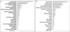
Figure 1
Differentially expressed genes (178 genes) up- or down-regulated by at least two-fold following a cellular treatment with MTA at every time point. (a) The biological processes of up-regulated 109 genes, (b) The biological processes of down-regulated 69 genes.

References
1. American Association of Endodontists. Glossary of endodontic terms. 2003. 7th ed. 40.
2. Cox CF, Subay RK, Ostro E, Suzuki S, Suzuki SH. Tunnel defects in dentin bridges: their formation following direct pulp capping. Oper Dent. 1996. 21:4–11.
3. Cox CF, Hafez AA, Akimoto N, Otsuki M, Suzuki S, Tarim B. Biocompatibility of primer, adhesive and resin composite systems on non-exposed and exposed pulps of non-human primate teeth. Am J Dent. 1998. 11:s55–s63.
4. Cox CF, Tarim B, Kopel H, Gürel G, Hafez A. Technique sensitivity: biological factors contributing to clinical success with various restorative materials. Adv Dent Res. 2001. 15:85–90.

5. Torabinejad M, Chivian N. Clinical applications of mineral trioxide aggregate. J Endod. 1999. 25:197–205.
6. Torabinejad M, Hong CU, Pitt-Ford TR, Kettering JD. Cytotoxicity of four root end filling materials. J Endod. 1995. 21:489–492.
7. Dominguez MS, Witherspoon DE, Gutmann JL, Opperman LA. Histological and scanning electron microscopy assessment of various vital pulp-therapy materials. J Endod. 2003. 29:324–333.
8. Ford TR, Torabinejad M, Abedi HR, Bakland LK, Kariyawasam SP. Using mineral trioxide aggregate as a pulp-capping material. J Am Dent Assoc. 1996. 127:1491–1494.
9. Yun YR, Yang IS, Hwang YC, Hwang IN, Choi HR, Yoon SJ, Kim SH, Oh WM. Pulp response of mineral trioxide aggregate, calcium sulfate or calcium hydroxide. J Korean Acad Conserv Dent. 2007. 32:95–101.
10. Bae JH, Kim YG, Yoon PY, Cho BH, Choi YH. Histological study. J Korean Acad Conserv Dent. 2010. 35:5–12.
11. Andelin W, Shabahang S, Wright K, Torabinejad M. Identification of hard tissue after experimental pulp capping using dentin sialoprotein (DSP) as a marker. J Endod. 2003. 29:646–650.
12. Kuratate M, Yoshiba K, Shigetani Y, Yoshiba N, Ohshima H, Okiji T. Immunohistochemical analysis of nestin, osteopontin, and proliferating cells in the reparative process of exposed dental pulp capped with mineral trioxide aggregate. J Endod. 2008. 34:970–974.
13. Min KS, Yang SH, Kim EC. The Combined Effect of Mineral Trioxide Aggregate and Enamel Matrix Derivative on Odontoblastic Differentiation in Human Dental Pulp Cells. J Endod. 2009. 35:847–851.
14. Brown PO, Botstein D. Exploring the new world of the genome with DNA microarrays. Nat Genet. 1999. 21:33–37.
15. Shi S, Robey PG, Gronthos S. Comparison of human dental pulp and bone marrow stromal stem cells by cDNA microarray analysis. Bone. 2001. 29:532–539.
16. McLachlan JL, Smith AJ, Bujalska IJ, Cooper PR. Gene expression profiling of pulpal tissue reveals the molecular complexity of dental caries. Biochim Biophys Acta. 2005. 1741:271–281.
17. Syudo M, Yamada S, Yanagiguchi K, Matsunaga T, Hayashi Y. Early gene expression analyzed by a genome microarray and real-time PCR in osteoblasts cultured with a 4-META/MMA-TBB adhesive resin sealer. Oral Surg Oral Med Oral Pathol Oral Radiol Endod. 2009. 107:e77–e81.
18. So H, Park SH, Choi GW. The comparison of gene expression from human dental pulp cells and periodontal ligament cells. J Korean Acad Conserv Dent. 2009. 34:430–441.
19. Yokose S, Kadokura H, Tajima Y. Establishment and characterization of a culture system for enzymatically released rat dental pulp cells. Calcif Tissue Int. 2000. 66:139–144.
20. Goldberg M, Farges JC, Lacerda-Pinheiro S. Inflammatory and immunological aspects of dental pulp repair. Pharmacol Res. 2008. 58:137–147.
21. Matsui S, Takeuchi H, Tsujimoto Y, Matsushima K. Effects of Smads and BMPs induced by Ga-Al-As laser irradiation on calcification ability of human dental pulp cells. J Oral Sci. 2008. 50:75–81.
22. Imamura T, Takase M, Nishihara A, Oeda E, Hanai J, Kawabata M, Miyazono K. Smad6 inhibits signaling by the TGF-β superfamily. Nature. 1997. 389:622–626.
23. Keklikoglu N. The localization of Fos B, a member of transcription factor AP-1 family, in rat odontoblasts and pulpal undifferentiated ectomesenchymal cells. Folia Histochem Cytobiol. 2004. 42:191–193.
24. Sabatakos G, Sims NA, Chen J, Aoki K, Kelz MB, Amling M, Bouali Y, Mukhopadhyay K, Ford K, Nestler EJ, Baron R. Overexpression of DeltaFosB transcription factor(s) increases bone formation and inhibits adipogenesis. Nat Med. 2000. 6:985–990.
25. Shen M, Yoshida E, Yan W, Kawamoto T, Suardita K, Koyano Y, Fujimoto K, Noshiro M, Kato Y. Basic helix-loop-helix protein DEC1 promotes chondrocyte differentiation at the early and terminal stages. J Biol Chem. 2002. 277:50112–50120.
26. Iwata T, Kawamoto T, Sasabe E, Miyazaki K, Fujimoto K, Noshiro M, Kurihara H, Kato Y. Effects of overexpression of basic helix-loop-helix transcription factor Dec1 on osteogenic and adipogenic differentiation of mesenchymal stem cells. Eur J Cell Biol. 2006. 85:423–431.
27. Lawler J. The functions of thrombospondin-1 and-2. Curr Opin Cell Biol. 2000. 12:634–640.
28. Murphy-Ullrich JE, Schultz-Cherry S, Höök M. Transforming growth factor-beta complexes with thrombospondin. Mol Biol Cell. 1992. 3:181–188.
29. Ueno A, Yamashita K, Nagata T, Tsurumi C, Miwa Y, Kitamura S, Inoue H. cDNA cloning of bovine thrombospondin 1 and its expression in odontoblasts and predentin. Biochim Biophys Acta. 1998. 1382:17–22.
30. Casasco A, Calligaro A, Casasco M, Springall DR, Tenti P, Marchetti C. Immunohistochemical localization of endothelin-like immunoreactivity in human tooth germ and mature dental pulp. Anat Embryol (Berl). 1991. 183:515–520.
31. Neuhaus SJ, Byers MR. Endothelin receptors and endothelin-1 in developing rat teeth. Arch Oral Biol. 2007. 52:655–662.
32. Guidry C, Hook M. Endothelins produced by endothelial cells promote collagen gel contraction by fibroblasts. J Cell Biol. 1991. 115:873–880.
33. Marini M, Carpi S, Bellini A, Patalano F, Mattoli S. Endothelin-1 induces increased fibronectin expression in human bronchial epithelial cells. Biochem Biophys Res Commun. 1996. 220:896–899.
34. Khan ZA, Farhangkhoee H, Mahon JL, Bere L, Gonder JR, Chan BM. Endothelins: regulators of extracellular matrix protein production in diabetes. Exp Biol Med (Maywood). 2006. 231:1022–1029.
35. Yan Y, Liu Z, Zhang WG. In vitro study of the effects of endothelin-1 on human dental pulp cells. Chin J Dent Res. 1999. 2:5–13.
36. Kido S, Kuriwaka-Kido R, Imamura T, Ito Y, Inoue D, Matsumoto T. Mechanical stress induces Interleukin-11 expression to stimulate osteoblast differentiation. Bone. 2009. 45:1125–1132.
37. Suga K, Saitoh M, Fukushima S, Takahashi K, Nara H, Yasuda S. Interleukin-11 induces osteoblast differentiation and acts synergistically with bone morphogenetic protein-2 in C3H10T1/2 cells. J Interferon Cytokine Res. 2001. 21:695–707.
38. Takeuchi Y, Watanabe S, Ishii G, Takeda S, Nakayama K, Fukumoto S. Interleukin-11 as a stimulatory factor for bone formation prevents bone loss with advancing age in mice. J Biol Chem. 2002. 277:49011–49018.
39. Hynes R. Molecular biology of fibronectin. Ann Rev Cell Biol. 1985. 1:67–90.
40. Lesot H, Osman M, Ruch JV. Immunofluorescent localization of collagens, fibronectin and laminin during terminal differentiation of odontoblasts. Dev Biol. 1981. 82:371–381.
41. Yoshiba N, Yoshiba K, Twaku M, Nakamura H, Osawa H. A confocal laser scanning microscopic study of the immunofluorescent localization of FN in the odontoblast layer of human teeth. Arch Oral Biol. 1994. 39:395–400.
42. Wang P, Hao J, Shi J. Actions of bovine plasma fibronectin on cultured human dental pulp cells. Chin J Dent Res. 2000. 3:55–62.
43. Hoshi K, Kemmotsu S, Takeuchi Y, Amizuka N, Ozawa H. The primary calcification in bones follows removal of decorin and fusion of collagen fibrils. J Bone Miner Res. 1999. 14:273–280.
44. Mochida Y, Duarte WR, Tanzawa H, Paschalis EP, Yamauchi M. Decorin modulates matrix mineralization in vitro. Biochem Biophys Res Commun. 2003. 305:6–9.
45. Alini M, Marriott A, Chen T, Abe S, Poole AR. A novel angiogenic molecule produced at the time of chondrocyte hypertrophy during endochondral bone formation. Dev Biol. 1996. 176:124–132.
46. Felszeghy S, Hollo K, Modis L, Lammi MJ. Type X collagen in human enamel development: a possible role in mineralization. Acta Odontol Scand. 2000. 58:171–176.
47. Kwan KM, Pang MK, Zhou S, Cowan SK, Kong RY, Pfordte T. Abnormal compartmentalization of cartilage matrix components in mice lacking collagen X: Implications for function. J Cell Biol. 1997. 136:459–471.
48. Deutsch D, Palmon A, Fisher LW, Kolodny N, Termine JD, Young MF. Sequencing of bovine enamelin ("tuftelin") a novel acidic enamel protein. J Biol Chem. 1991. 266:16021–16028.
49. Paine CT, Paine ML, Luo W, Okamoto CT, Lyngstadaas SP, Snead ML. A tuftelin-interacting protein (TIP39) localizes to the apical secretory pole of mouse ameloblasts. J Biol Chem. 2000. 275:22284–22292.
 XML Download