

 PDF
PDF ePub
ePub Citation
 Print
Print


I. Introduction
For several decades, dentists have been using conventional mechanical cutting and drilling system to remove diseased dental hard tissues and to prepare cavities for restorations. But the current trend towards minimum-intervention dentistry has introduced alterative techniques for dental cavity preparation in order to replace the invasive approach using high-speed burs.1) The use of laser irradiation has been widely studied and applied for that purpose, due to its precise and effective ability to eliminate carious tissue while avoiding removal of sound tooth substrate, thus resulting in a more conservative cavity design. And their advantages have also drawn numerous researchers to investigate various applications of lasers in dentistry.2)
For instance, an additional therapeutic option is available for the treatment of dentinal hypersensitivity with the advent of laser technology and its growing utilization in dentistry.6) According to the consulted literature, lasers have been effective in the treatment of dentin hypersensitivity. Villa et al.3) demonstrated the laser photobiomodulating action in the dental pulp, showing evidence of a large quantity of tertiary dentin production, causing the physiological obliteration of the dentinal tubules. Moritz et al.4) and Schwarz et al.7) also demonstrated that the effectiveness of laser treatment is due to an occlusion or narrowing of dentinal tubules. And Walsh5) reported the laser-induced changes in neural transmission networks within the dental pulp (depressed nerve transmission) by affecting the depolarization of nerve ending. In addition, the immediate analgesic effect in the treatment of dentin hypersensitivity with diode was reported.3)
Recently, new laser system, the erbium, chromium: yttrium scandium gallium garnet (Er,Cr:YSGG) laser was introduced into the dentistry. It is the laser of 2780 nm wavelength using a pulsed-beam system and fiber delivery. The mechanism of the effect of this laser was generally accepted that water droplets produced violent microexpansion after efficiently absorbing the laser energy which subsequently formed hydrokinetic forces that could quickly ablate the dental hard tissue.8) It has been recommended for minimally invasive purposes, due to its precise ablation of dental structure without side-effects to the pulp and surrounding tissues.9) According to the manufacturers, Er,Cr:YSGG laser can be applied in various procedures including laser-etching, soft-tissue mucous membrane and cutaneous surgery and relief of dentin hypersensitivity, as well as cutting enamel, dentin and bone.
Although laser application provides many advantageous aspects and is deemed clinically viable in various dental procedures, there still has been concern that the laser irradiation may deleteriously affect the bond strength of adhesive restoration. Previous studies 10-13) on the effects of erbium laser irradiation on bonding procedures are somewhat controversial.
Some authors10,11) indicated no significant effect or slight improvement. Carrieri et al.10) reported that the use of Er:YAG laser irradiation of cavity preparation or pretreatment of dentin surface previously to adhesive procedures using two-step total-etching system yielded similar bonding quality to dental hard tissue compared to conventional high-speed drill. Visuri et al.11) also reported that the condition of dentin following Er:YAG laser ablation was sufficient to bond with three-step total-etching system and composite restorative materials, and persisted that an Er:YAG laser might eliminate the need for acidetching dentin as a pretreatment for composite bonding.
On the other hand, some authors12,13) indicated a significant deterioration of bond strength of composite restorative materials. Botta et al.12) reported that Er,Cr:YSGG laser conditioning on dentin significantly reduced bond strength of etch-and-rinse and one-step self-etch systems. Also, Armengol et al.13) reported that Er:YAG and Nd:YAP laser preparation did not improve bonding using three-step total-etching system to enamel and dentin because laser treatment probably modified the surface energy of enamel and dentin, providing surfaces less suitable for adhesive resin.
To overcome this problem, mechanical or chemical removing of laser-modified layer has been suggested 14-16). Gutknrcht et al.14) and Carvalho et al.15) suggested additional acid-etching of the laser-prepared cavity. Also, Obeidi et al.16) found that adjunctive use of phosphoric acid following water-rinsing appeared to have eliminated the laser-modified layer and recommended longer etching time to obtain higher bond strength.
Although the bond strengths of Er,Cr:YSGG laser-irradiated dentin have been investigated, earlier studies were conducted almost exclusively by applying the Er,Cr:YSGG laser of parameters for preparation or laser-etching. To the extent of knowledge of the authors of this study, there are currently no comparable studies using Er,Cr:YSGG with low power setting of the hypersensitivity mode. Therefore, the purpose of this in vitro study is to evaluate the effect of Er,Cr:YSGG irradiation in the hypersensitivity mode on microtensile bond strength of composite resin restoration.
II. Materials and Methods
1. Specimen preparation
Twenty extracted human permanent molars without caries were stored in distilled water. The crown portions were horizontally-sectioned at mid-coronal level using a diamond-saw (3200 rpm, Accutom-50; Struers, Rødovre, Denmark) under continuous water cooling. A 600-grit silicon carbide paper was used under running water to produce flat dentin surface and smear layer. The teeth were then randomly divided to six groups, according to laser irradiation and the adhesive system. The materials and groups used in this study are showed in Table 1 and Figure 1.
2. Laser irradiation
The specimens in group 1, 2 and 3 were irradiated with an Er,Cr:YSGG hydrokinetic system (Waterlaser; Biolase, Irvine, CA, USA) one by one to simulate the laser irradiation for reliving dentin hypersensitivity. A complete description of laser parameters is included in Table 2. The laser beam was aligned perpendicular to the surface and moved in a sweeping fashion by hand during the exposure period.
3. Dentin bonding and resin composite restoration procedures
Dentin surface was etched for 15 sec in group 1 and 4, and for 20 sec in group 2 and 5 with 37.5% phosphoric acid etchant. After that, the Optibond FL applied to the dentin surfaces. In group 3 and 6, self-etching primer and adhesive were applied. All procedures were according to the manufacturer's instruction (Table 2) except the longer etching time in group 2 and 5. After the bonding procedure, the teeth were restored with light-cure composite resin. The restored teeth were stored in distilled water at room temperature for 24 hr.
4. Microtensile bond strength (µTBS) test
The teeth were cut longitudinally to make the specimens approximately 1 × 1 mm thick and 10 mm long. Each group was consisted of 12 specimens. The specimens were glued to the jig of microtensile testing machine (BISCO Inc, Schaumburg, IL, USA) using cyanoacrylate cement (Zapit; Dental Ventures of America, Corona, CA, USA). Tensile load was applied at a cross-head speed of 1 mm/min until the failure of specimen.
5. Failure mode investigation
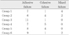
Failure mode was investigated using operating microscope (OPMI pico; Carl zeiss, Obercohen, Germany) under 25 × magnification and the results were classified as follows: adhesive, cohesive and mixed. If the composite resin restoration had fractured at the adhesive-tooth interface, it was recorded as adhesive failure. If the composite resin restoration had fractured inside the composite resin or dentin, it was recorded as cohesive failure. If a combination of adhesive and cohesive in dentin or in resin had occurred, it was recorded as mixed failure.
6. Statistical analysis
Statistical analysis was performed with using SPSS 12.0 software (SPSS, Chicago, IL, USA). Student t-test was used for comparison between µTBS according to the laser irradiation. And one-way ANOVA was used for comparison between µTBS according to the bonding agent and Scheffe's test was used for post-hoc multiple comparisons. The level of significance was set at p < 0.05.
7. Scanning electron microscopy
To compare the SEM analyses of the prepared dentin surface and irradiated dentin surface, the specimens were dehydrated in an ascending series of ethanol (50% for 2 hr, 70% for 2 hr, 95% for 2 hr, 100% for 2 hr) and dried. They were then mounted on aluminum stubs and sputter coated with gold/palladium. Examination of SEM (JSM-6480LV; JEOL, Tokyo, Japan) operating at 20 kV was performed.
III. Results
1. Comparison between µTBS
Comparison between µTBS according to the laser irradiation, dentin bonding agents and etching time was showed in Table 3.
The laser irradiation group showed lower bond strength than the no laser irradiation group (p < 0.05). In laser irradiation groups, Optibond FL of 3-step etching for 20 sec group showed the highest µTBS, followed by Optibond FL of 3-step etching for 15 sec, Clearfil SE bond of 2-step self-etch. And there were significant differences among groups. In no laser irradiation groups, Optibond FL of 3-step etching for 20 sec showed the highest µTBS, followed by Optibond FL of 3-step etching for 15 sec, Clearfil SE bond of 2-step self-etch. And there were significant differences among groups.
2. Failure mode
Failure mode was presented in Table 4. Adhesive failure was predominantly observed in all groups. In 2-step self-etch groups (group 3 and group 6), adhesive failure was more commonly observed than the other groups.
3. SEM evaluation
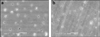
The laser-irradiated dentin samples revealed rough surfaces with opened dentinal tubules, an absence of a smear layer, and more prominent peritubular dentin than intertubular dentin. The use of the laser at low power level seemed not to create abundant craters or scale-like appearance which were observed in general laser irradiated dentin surface (Figure 2a). On the other hand, the micrographs of no laser-irradiated dentin surfaces showed the presence of a smear layer that occluded dentinal tubules (Figure 2b).
IV. Discussion
Since Goldman et al.17) used a ruby laser to attempt caries removal in 1964, lasers have been used for caries prevention, caries removal, endodontic treatment, treatment of dentin hypersensitivity, and as an alternative to conventional acid-etching in clinical applications.11,18,20) Recently the pulsed erbium laser, Er,Cr:YSGG laser has been advocated for hard and soft tissue ablation.12,19)
However, there has been a growing debate on the use of laser for cavity preparation and for conditioning enamel and dentin because of the conflicting effect on the bond strength of composite resin restoration. According to some studies,10,11,19-21) lasers may be used to facilitate adhesion between the tooth structure and the restorative material. Kataumi et al.21) showed that the irradiated dentin exhibited bond strengths comparable to those of cut-dentin. On the other hand, other studies12,13,22) demonstrated that laser treatment damaged the dentin bonding. Gurgan et al.22) showed that adhesion to dental hard tissues after etching with Er:YAG laser, the same erbium family as Er,Cr:YSGG, was inferior to that obtained after conventional acid etching.
The result of this study showed that the bond strength of laser-irradiated dentin was lower than that of no-laser irradiated dentin, supporting the latter findings. There are several simultaneous effects that can interfere with bonding during laser irradiation. In the dentin surface, the increase of temperature is produced by laser/dentin interaction.34) It results in a modified surface, in which the collagen network completely melted and vaporized. Thus the denatured collagen fibrils are fused together and poorly attached to the underlying unaffected dentin, preventing proper dentin hybridization.23) These altered microstructures would hamper the infiltration of primer and the hybrid layer would become more susceptible to hydrolysis.24)
Also, it has been demonstrated that the denatured organic matrix blocks the diffusion pathways of adhesive in dentin. The diffusion pathway blockage affects the porosity of the structures and consequently impairs penetration of the adhesive components.9)
Moreover, after laser irradiation, the dental surfaces are chemically modified. Irradiation with erbium lasers promotes loss of carbonate, formation of new hydroxyapatite-like crystals, and consequently more acid-resistant surfaces.25) Asli et al.26) found that during laser irradiation, the evaporation of organic components may well lead to an increase in Ca, P, Mg contents in the laser-modified dentin, resulting in acquired acid resistance of dentin surface, in turn, affecting the adhesion of dental materials to dental hard tissue. Hossain et al.27) also asserted that acid resistance may be promoted by Er,Cr:YSGG laser irradiation.
The analysis of failure mode also supports the result of the microtensile bond strength test. Adhesive failures between resin and dentin dominated in the laser-irradiated group, while mixed failures dominated in the no laser-irradiated group. And after acid etching of laser-irradiate dentin, adhesive failures decreased. From this result, it might be thought that acid etching could reinforce the hybrid layer by eliminating the laser-modified layer.
With respect to the micromorphological changes seen in the laser-irradiated dentin surface, the SEM examinations showed characteristic features. It revealed opened dentinal tubules, absence of a smear layer and more prominent peritubular dentin than intertubular dentin.28) Since intertubular dentin contains more water and has a lower mineral content than does peritubular dentin, it is selectively more ablated than the peritubular dentin, leaving protruding dentinal tubules with a cuff-like appearance and resulting in more irregular dentin surface.
As the efficacy of dentin surface treatment is related to the power setting chosen for Er,Cr:YSGG laser ablation, the morphology of dentin surface irradiated with the hypersensitivity mode could be different from that irradiated for laser etching or preparation. The abundant crater or scale-like appearance, microcrack and rough surface usually found in the dentin surface irradiated with high laser power output was not shown in this study.
Considering the result of SEM evaluation in relation to the result of µTBS test, the formation of an inter-diffusion zone of laser-irradiated dentin which is not similar to that described for acid-etched would cause the deterioration of bond strength.16) Cardoso et al.23) suggested that the irregularities on the laserirradiated dentin were so prominent that they may reduce the bond strength by preventing uniform stress distribution at the adhesive-dentin interface. Moreover, because of these irregularities, the thickness of the adhesive layer was not uniform on the dentin surface, thus resulting in diminished bonding effectiveness.
Based on the results of current study, an increase of phosphoric acid etching time could be considered as a way to overcome the acid resistance and offset irregularities of the dentin. Some studies16,29,30) evaluating the influence of acid etching time on bond strength in dentin irradiated with Er,Cr:YSGG lasers used the etching time of 15 sec, the conventional etching time of dentin and 30 sec with 37% phosphoric acid. And the laser output of 3 W or 4 W was used in those studies, which was much higher than used in current study (0.25 W). Since the laser output that is used in this study was lower than those of other studies, the etching time of 20 sec was selected as a longer etching time instead of 30 sec to compare the effect of acid etching time. The results of this study showed that the bond strength of laserirradiated dentin after 20 sec acid-etching time is comparable to that of no laser-irradiated dentin (15 sec etching time) and indicated that the longer acid etching could have eliminated the laser-modified layer.
In addition to the etching time, caution should be taken in terms of selection of the bonding agents. Lately, the remarkable evolution of contemporary dentin adhesive systems was aimed at the simplification of clinical step. Factors such as time and simplification of the clinical steps have led to an increased use of the simplified-step adhesives in association with resin materials. Self-etching systems are required to completely dissolve and incorporate the smear layer in order to interact with the underlying mineralized dentin.31,32) However, in the presence of this more acid-resistant surface, the weak acids present in the self-etching system cannot sufficiently modify the surface to promote adhesive penetration. Since Clearfil SE bond used in current study is categorized in mild self-etching bonding system according to its acidity (pH = 1.8), the µTBS of laser-irradiated Clearfil SE bond group was the lowest of all tested groups. Other authors23,33) also support this finding that the Er,Cr:YSGG laser-irradiated dentin is poor substrate for bonding with the self-etching system. For example, Cardoso et al.23) assessed the bonding effectiveness of adhesives to Er,Cr:YSGG laser-irradiated dentin using irradiation settings specific for cavity preparation, and demonstrated that the bond strength of Optibind FL (three-step total-etch) was the highest, followed by Clearfil SE bond (two-step self-etch), Clearfil S3 bond and Adper Prompt L-Pop (one-step self-etch).
In summary, the results showed that the µTBS of Er,Cr:YSGG laser irradiated dentin in the hypersensitivity mode was adversely influenced in all adhesive system used in current study. Also, the bond strengths of three-step total-etch system were higher than those of two-step total-etch system irrespective of laser irradiation and the bond strength of etching for 20 sec group was higher than that of etching for 15 sec group in three-step total-etch groups. Therefore, three-step total-etching system seems preferable to mild self-etching system when applied on laser-irradiated dentin in the hypersensitivity mode and increase of etching time for three-step total-etching system would be beneficial.
V. Conclusions
Within the limitation of this study, it may be concluded that since Er,Cr:YSGG laser-irradiation in the hypersensitivity mode on dentin surface could adversely affect the bonding strength of composite resin restoration due to acquired acid resistance, total-etching system seems preferable and increase of acid etching time for total-etching system would be better.

 XML Download