

 PDF
PDF ePub
ePub Citation
 Print
Print


Abstract
The final preparation (MAF) size in infected root canals is still controversial. Nonetheless, recent studies demonstrated that larger apical preparation sizes produces a greater reduction in remaining bacteria and dentinal debris as compared to smaller apical preparation sizes. Therefore, clinicians should be practiced with treatment strategies guided by evidence-based information, especially in infected/failed root canals.
Figures and Tables
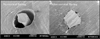
Figure 1
Influence of cervical preflaring on determination of apical file size in curved canal. Coronal flaring can help to select the suitable IAF that closely matches the size of apical foramen. Pictures were quoted from the journal of Baugh and Wallace (reference #12).
References
1. Kakehashi S, Stanley HR, Fitzgerald RJ. The effects of surgical exposure of dental pulps in germ-free and conventional laboratory rats. Oral Surg Oral Med Oral Pathol. 1965. 20:340–349.

2. Sundqvist G. Bacteriological studies of necrotic dental pulps. 1976. Umeå University Odontological Dissertations No.7.
3. Figdor D. Microbial etiology of endodontic treatment failure and pathologic properties of selected species. 2002. Umeå University Odontological Dissertations No.79.
4. Bystrom A, Sundqvist G. Bacteriological evaluation of the efficacy of mechanical instrumentation in endodontic therapy. Scand J Dent Res. 1981. 89:321–328.
5. Falk KW, Sedgley CM. The influence of preparation size on the mechanical efficacy of root canal irrigation in vitro. J Endod. 2005. 31:742–745.
6. Jung IY, Seo MA, Fouad AF, Spangberg LSW, Lee SJ, Kum KY. Apical anatomy of mesiobuccal and mesial root of permanent first molars. J Endod. 2005. 31:364–368.
7. Dummer PMH, McGinn JH, Rees DG. The position and topography of the apical canal constriction and apical foramen. Int Endod J. 1984. 17:192–198.
8. Peters OA, Schönenberger K, Laib A. Effects of four Ni-Ti preparation techniques on root canal geometry assessed by micro computed tomography. Int Endod J. 2001. 34:221–230.
9. Baugh D, Wallace J. The role of apical instrumentation in root canal treatment: A review of the literature. J Endod. 2005. 31:333–340.
10. Rollison S, Barnett F, Stevens R. Efficacy of bacterial removal from instrumented root canals in vitro related to instrumentation technique and size. Oral Surg Oral Med Oral Pathol Oral Radiol Endod. 2002. 94:366–371.
11. Grossman LI. Endodontic practice. 1970. 7th ed. Philadelphia: Lea & Febiger.
12. Barroso JM, Guerisoli DMZ, Capelli A, Saquy PC, Pěcora JM. Influence of cervical preflaring on determination of apical file size in maxillary premolars: SEM analysis. Braz Dent J. 2005. 16:30–34.
13. Silveira LFM, Martos J, Pintado LS, Teixeeira RA, Neto JBC. Early preflaring and crown-down shaping influences the first file binds to the canal apical third. Oral Surg Oral Med Oral Pathol Oral Radiol Endod. 2008. 106:e99–e101.
14. Kfir A, Rosenberg E, Fuss Z. Comparison in vivo of the first tapered and nontapered instruments that bind at the apical constriction. Oral Surg Oral Med Oral Pathol Oral Radiol Endod. 2006. 102:395–398.
15. Wu MK, Wesselink PR. Efficacy of three techniques in cleaning the apical portion of the curved root canals. Oral Surg Oral Med Oral Pathol Oral Radiol Endod. 1995. 79:492–496.
16. Shuping G, Orstavik D, Sigurdsson A, Trope M. Reduction of intracanal bacteria using nickel-titanium rotary instrumentation and various medications. J Endod. 2000. 26:751–755.
 XML Download