

 PDF
PDF ePub
ePub Citation
 Print
Print


I. Introduction
Endodontically treated teeth often lose substantial tooth structure from previous caries, pre-existing restorations, and/or endodontic procedures1). The amount of sound tooth structure that will remain following root canal therapy and any subsequent preparation is an important factor in planning the restoration of endodontically treated tooth2). Clinically, the necessity of placing a post arises when too insufficient crown structure is present to sustain a coronal restoration.
Cast posts and cores are commonly used for teeth with little remaining coronal structure or for uniradicular teeth with small coronal volume because of their superior physical properties3,4). Cast gold alloy is an inert material with elastic modulus and coefficient of thermal expansion similar to those of enamel, and yet it has good compressive strength that can withstand normal occlusal forces5). But their hardness might be a major disadvantage in adjustment and may predispose the tooth to root fracture. Another disadvantage of the cast post and core placement procedure is that it requires two visits and laboratory fabrication.
Prefabricated metal posts allow fast, cheap, easy and simple techniques, but they do not take into account the individual shape of the root canal and their adaptation is not always ideal6,7). Restoration of endodontically treated teeth can include a definitive restoration, core, post, cement, and the tooth structure with potential interfaces of component-to-component.
So stress concentrations at the interface between the metal post and resin core and the reconstructed crown may cause cracks and crown detachment because of the significant strain gradient caused by the large difference in stiffness between crown material, post and core7). As the number of interfaces increases, the potential for failure will also increased8).
Recently, fiber reinforced composite (FRC) posts can be used in high-demand cosmetic procedures, such as with all-ceramic and indirect resin restorations. The use of fiber posts appears to be a good restorative choice to rebuild endodontically treated teeth. Their elastic modulus is claimed to be close to that of dentin. Although this feature could reduce the risk of root fractures, their flexibility is generally greater than that of metal posts, and a flexible post may not be effective in stabilizing the composite core in teeth without their remaining crown9).
Compared with various post and core systems, the ideal post and core material should have physical properties-such as elastic modulus, compressive strength and coefficient of thermal expansion-that are similar to those of dentin. And the creation of a mono-block dentin-post-core system through the dentinal bonding would allow better distribution of forces along the root. Therefore if excessive loads are applied to the tooth, the post will be able to absorb stresses, reducing the possibility of root fracture.
A completely different approach recommends the use of so-called EndoCrown instead of posts and cores. An EndoCrown is a full or compact crown that extends a post into the pulp chamber or root canals as one unit and not a separate post10). The development of indirect resin with elastic modulus closer to dentin and luting agents with lower elastic moduli has been suggested to reduce the tension that can develop at the tooth/cement/restorative material interface11). Since any significant mismatch between stiffness of intraradicular devices and dental tissues will cause large stress concentration at the restorative materials and tooth interfaces, the best solution appears to be the use of customized post systems made of materials whose elastic properties are very close to those of the natural tooth. EndoCrown fabricated with indirect composite resins seem to be the most reliable restoration, because the EndoCrown configuration significantly reduces the number of interfaces and dentin like elastic modulus generates low amounts of stress concentration12).
Mechanical response of dental posts can be experimentally investigated by suitable tests under various loading conditions. On the other hand, more detailed information about the stress distribution surrounding endodontic posts can be evaluated by photoelastic analysis, strain gauge, speckle interferometry or by numerical simulations with finite element (FE) analysis, as proposed by a number of authors13-16) which also provides stress distribution inside the post. The FE method is based on a mathematical model which approximates the geometry and the loading conditions of the structure (i.e. a pulpless tooth reconstructed with an endodontic post) to be analyzed. Deformations and stresses in any point of the model can be evaluated and the most stressed areas can thus be evidenced.
The purpose of this study was to evaluate the influence of elastic modulus of restorative materials and the number of interfaces of post and core systems on the stress distribution of three differently restored endodontically treated maxillary second premolars using three-dimensional (3D) FE analysis.
II. Materials and Methods
1. Finite element models
To develop a 3D FE model, intact normal extracted human maxillary second premolars were used. The extracted premolars were scanned serially with micro-CT (SkyScan1072; SkyScan, Aartselaar, Belgium) to expose the tooth sections perpendicular to the long axis of the tooth (58 µm in thickness) and parallel to the occlusal plane. 3D-DOCTOR (Able Software Co., Lexington, MA, USA) image processing software was employed to detect the boundaries of enamel, dentin and pulp from the sectioned two dimensional images and to make 3D surface model. HyperMesh Ver. 6 (Altair Engineering, Inc., Troy, USA) and ANSYS Ver. 9 (Swanson Analysis Systems, Inc., Houston, USA) were used to mesh and analyze 3D FE model.
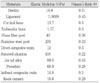
The physical properties of the tooth and supporting structures used in this study are given in Table 1. All the vital tissues were presumed linearly elastic, homogeneous and isotropic. The corresponding elastic properties such as Young's modulus and Poisson's ratio were determined according to literature survey10,15-18).
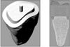
The periodontal ligament was assumed to be 0.3 mm wide, and the alveolar bone was also generated by growing the outer surface of the tooth from 2 mm below the cemento-enamel junction (CEJ). In all loading cases, the mesial, distal and base nodes of simulated alveolar bone were assumed fixed to prevent rigid body motion. Endodontically treated maxillary second premolar which has 2 mm ferrule above CEJ was used as a tooth models (Figure 1-A). Restored structure of models was shown in Figure 1-B.
2. Experimental models of different post and core systems and definitive restorations
The three experimental models were designed as follows and summarized at Table 2.
Model 1: It consisted of 426245 elements with 81976 nodes and was restored with a stainless steel post which has 0.9 mm of tip diameter and 8 mm of length from CEJ to post apex. A porcelain-fused to gold (PFG) crown was considered. The amount of tooth preparation and gold coping thickness is excerpted from the text of prosthodontics19).
Model 2: It consisted of 427563 elements with 82211 nodes and was restored with a glass fiber post which has 0.9 mm of tip diameter and 1.4 mm of coronal diameter. A sintered alumina crown was considered.
Model 3: It consisted of 427563 elements with 82211 nodes, was restored by EndoCrown which was composed of post, core and crown in all-in-one.
It was assumed that complete bonding between components was considered and the thickness of adhesive resin cement was 0.1 mm.
3. Loading condition and stress analysis
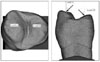
As shown in Figure 2, an oblique 500 N load of 45° direction to the long axis of tooth was applied on the buccal (Load A) and palatal (Load B) cusp15).
The von Mises stresses in the coronal and root structure of each model were analyzed using ANSYS.
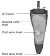
The stress distributions of coronal structure were evaluated at the shoulder margin (SM), lower margin of ferrule (LF), and upper margin of ferrule (UF) as shown in Figure 3.
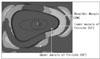
The stress distributions of root structure was evaluated vertically at the slightly mesiobuccal and distopalatal outer surface from CEJ to the root apex as shown in Figure 4.
III. Results
[Stress analysis of coronal structure]
1. Under the condition of Load A
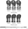
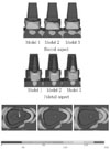
1-1) External view of stress distribution
The stresses were distributed along the buccal and palatal crown margins. High level of stress was concentrated at mid-palatal crown cervical margin of Model 2 (Figure 5).
1-2) Internal view of stress distribution
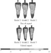
The stresses were distributed along the buccal and palatal shoulder margins. High stress was concentrated at mesio-buccal and mid-palatal shoulder margin of all models. In Model 1 and 2, low level of stress was distributed at ferrule and core. At ferrule and core of Model 3, the stress level was higher than Model 1 and 2 as shown in buccal, palatal and occlusal aspect (Figure 6).
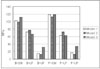
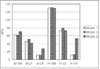
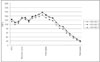
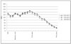
1-3) Peak stresses at SM, LF and UF
The von Mises stresses in SM, LF, and UF were presented at Figure 7. Along the SM, the peak stress of palatal aspect was higher than that of buccal aspect. Along the buccal SM, the peak stress of Model 3 was highest of all and along the palatal SM, the peak stress was highest in Model 1. Along the buccal and palatal LF, the peak stress was highest in Model 2. At UF, Model 3 had highest peak stress among all models.
2. Under the condition of Load B
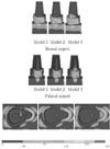
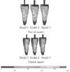
2-1) External view of stress distribution
The stresses were distributed along the buccal and palatal crown margins. High stress was concentrated at mid-palatal crown cervical margin of Model 2 (Figure 8).
2-2) Internal view of stress distribution
The stresses were distributed along the buccal and palatal shoulder margins. High stress was concentrated at mesio-buccal and mid-palatal shoulder margin of all models.
At ferrule and core of Model 3, the stresses level was higher than Model 1 and 2 as shown in buccal, palatal, occlusal aspect (Figure 9).
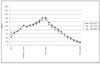
2-3) Peak stresses at SM, LF and UF
The von Mises stresses in SM, LF, and UF were presented at Figure 10. Peak stress of Model 3 was highest among the models at buccal SM and at buccal and palatal UF. At the buccal and palatal LF, the peak stress of Model 2 was highest of all.
[Stress analysis of root structure]
1. Under the condition of Load A
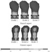
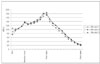
1-1) Stress distribution of root surface
Stress distributed along buccal and palatal root area of all models. Peak stress of all models concentrated at middle third area of the root at buccal and palatal aspect (Figure 11).
2. Under the condition of Load B
2-1) Stress distribution of root surface
Stress distributed along buccal and palatal area of all models. Peak stresses of all models concentrated at middle third area of the root in buccal and palatal aspect (Figure 14).
IV. Discussion
After root canal treatment, the dental practitioner is faced with the task of restoring the tooth to rehabilitate oral functions. The restoration must achieve adequate retention and circumvent damage to the remaining hard tissue through the prevention of bacterial microleakage and hard tissue fractures.
Many investigators20-25) reported that the design as well as the material of the post and core affect the resistance to fracture of endodontically treated teeth restored with post and core systems, while other studies reported that resistance to fracture of endodontically treated teeth covered by a crown with a 2 mm margin on healthy tooth structure was not affected by the post and core complex26,27).
For the successful rehabilitation of the endodontically treated tooth, it is imperative to understand the disparity and complexity of the relationship of interfaces with various restorative materials. When evaluating the interfaces of any system, their failures provide dentists with design principles that can be used with any post, core and crown retained system.
The purpose of this study was to evaluate the influence of elastic modulus of restorative materials and the number of interfaces of post and core systems on the stress distribution.
But the limitation of designing the FE models used in this study, interface characteristics in terms of adhesive bonding could not be simulated. However the different elastic modulus of components may play a role of interfaces. Any large mismatch between the physical properties of posts, cements, and residual tooth tissues may cause adverse stress concentrations at the different interfaces involved28).
In the present study, endodontically treated single root maxillary premolar was used, which present a restorative and an esthetic challenge because the occlusal forces on them are greater and their directions are different in the premolar area compared to the anterior teeth.
According to analysis of coronal structure distribution, the extent of stress distribution and magnitude of peak stress at the shoulder was highest in Model 3 and lowest in Model 2. But at the cervical margin of the crown, the peak stress of Model 2 was highest and lowest in Model 3. It would be explained that the rigid crown can resist deformation more effectively than the less rigid crown. Thus the stress was concentrated at the cervical margin of crown itself rather than at the shoulder margin. The study of Hunter and Flood29) suggested that their experiments have indicated that the type of post and core restoration may be less important than the placement of a well-fitting crown with margins beyond the core restoration and it was agreed with this study.
Pegoretti s.30) claimed that stresses at the cervical margins could be lowered using less stiff crown materials like composite resins, but their claim is not accordance with the result of this study. On the contrary, Stricker et al.31) suggested that all-ceramic crown with a high elastic modulus exhibited better and more stress-resistant marginal adaptation at the tooth-to-luting composite interface than EndoCrown. And this is in agreement with the result of present study.
The reason of highest stress distribution at UF of Model 3 is that less stiff EndoCrown was deformed easily compared to other models and this result in stress accumulation at the underlying upper ferrule. But the reason of highest stress distribution at LF of Model 2 could not be explained with the rigidity of the full veneer crown.
Although the buccal cusp was loaded under Load A, the peak stress of palatal SM was higher than that of buccal SM. Compared to buccal aspect, the palatal core was short and flat and this caused the high stress concentration at palatal cervical margin because of the low resistance to displacement.
Oyar et al.32) suggested that anatomical occlusal preparation designs were advantageous in stress distribution at buccal areas than palatal area because of geometry of prepared tooth structure. In addition, it could be speculated that the length of palatal cervical margin is shorter than buccal cervical margin and this lead to heavy stress concentration at palatal shoulder margin.
According to analysis of root structure stress distribution, peak stress was located at level of post apex in all models. The development of stress concentration in the region adjacent to the apex of the endodontic post is in agreement with previous FE analysis33,34)
Albuquerque et al.35) and Hayashi et al.36) reported that stiff post generated stress concentration at root surface. Rigid metal posts resisted lateral forces without distortion and this resulted in stress transfer to the less rigid dentin causing potential root cracking and fracture. But in this study, peak stress of Model 2 which was restored with fiber post was highest of all.
Others37,38) have suggested that a Young's modulus approaching that of dentin is more desirable, as stress transmitted to the root on loading of the post will decrease, thus reducing the risk of root fracture. The ideal root canal post must be sufficiently elastic to accompany the natural flexural movements of the structure of the tooth, something that a very rigid metal post cannot do. EndoCrown used in present study had low stress at outer root surface, which was coincided with the study of Zarone et al.10)
From the results of this study, stress concentration at the root surface of post apex level was determined by the elastic modulus of definitive restoration rather than the rigidity of the post itself. Thus, the higher the elastic modulus of definitive restoration leads to the more stress concentration at the root surface.
From the results of this study, stress concentration at the root surface of post apex level was determined by the elastic modulus of definitive restoration rather than the rigidity of the post itself. Thus, the higher the elastic modulus of definitive restoration leads to the more stress concentration at the root surface.
In general, when a system with component of different rigidity is loaded, the more rigid component is capable of resisting greater forces without distortion. The less rigid component fails and relieves stress.
The elastic modulus of definitive restorations governed stress distribution, the influence of the post on the stress distribution of restored teeth could not be observed. For the explanation of this result, 2.0 mm ferrule height, 0.1 mm thickness of adhesive cement layer, and also considerable thickness of resin core in the pulp chamber could be considered as other factors that influence the stress distribution. Thus, further study is needed to compare the influence of post and core system using the models restored with same full veneer crown but with different post and core systems and varying ferrule height.
V. Conclusions
Within the limits of this study, the following conclusions were drawn:
The elastic modulus of the definitive restorations rather than the type of post and core system was the primary factor that influenced the stress distribution of endodontically treated maxillary premolars.
The stress concentration at the coronal structure could be lowered through the use of definitive restoration of high elastic modulus.
The stress concentration at the root structure could be lowered through the use of definitive restoration of low elastic modulus.

 XML Download