

 PDF
PDF ePub
ePub Citation
 Print
Print


Abstract
The purpose of this study was to evaluate the effect of soft chelating irrigant on the sealing ability of root fillings by using a glucose leakage test.
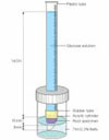
A total of 45 single-rooted teeth were selected for the study. The teeth were decoronated leaving a total length of 13mm. The root canals prepared using K3 NiTi rotary instruments to an apical dimension of size 45(0.06 taper). The specimens were then randomly divided into 3 experimental groups of 13 roots each and 2 control groups of 3 roots each. Specimen in each group were prepared with different irrigation protocols : group 1, 2.5% NaOCl; group 2, 2.5% NaOCl and 17% EDTA; group 3, 2.5% NaOCl and 15% HEBP. The root canals were filled with gutta-percha and AH Plus sealer using lateral condensation. After 7 days in 37℃, 100% humidity, the coronal-to-apical microleakage was evaluated quantitatively using a glucose leakage model. The leaked glucose concentration was measured with spectrophotometry at 1, 4, 7, 14, 21 and 28 days.
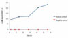
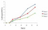
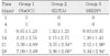
There was a tendency of increase in leakage in all experimental groups during experimental period. HEBP-treated dentin showed no significant difference with EDTA-treated dentin during experimental period. From the 21th day onward, HEBP-treated dentin showed significantly lower leakage than smear-covered dentin. HEBP-treated dentin displayed a similar sealing pattern to EDTA-treated dentin and a better sealing ability than smear-covered dentin. Consequently, a soft chelator(HEBP) could be considered as the possible alternative to EDTA.
Figures and Tables
References
1. Czonstkowsky M, Wilson EG, Holstein FA. The smear layer in endodontics. Dent Clin North Am. 1990. 34:13–25.
2. Pashley DH, Michelich V, Kehl TJ. Dentin permeability: effects of smear layer removal. J Prosthet Dent. 1981. 46:531–537.

3. Pashley DH. Smear layer: Physiological considerations. Oper Dent Suppl. 1984. 3:13–29.
4. Foster KH, Kulild JC, Weller RN. Effect of smear layer removal on the diffusion of calcium hydroxide through radicular dentin. J Endod. 1993. 19:136–140.
5. White RR, Goldman M, Lin PS. The influence of the smeared layer upon dentinal tubule penetration by plastic filling materials. J Endod. 1984. 10:558–562.
6. Okşan T, Aktener BO, Sen BH, Tezel H. The penetration of root canal sealers into dentinal tubules. Int Endod J. 1993. 26:301–305.
7. Economides N, Liolios E, Kolokuris I, Beltes P. Long-term evaluation of the influence of smear layer removal on the sealing ability of different sealers. J Endod. 1999. 25:123–125.
8. Lee JM, Park SH, Choi KW. The Effect of Smear Layer Treatment on the Microleakage. J Korean Acad Conserv Dent. 2006. 31:378–389.
9. Johnson WT, Gutmann JL. Cohen S, Hargreaves KM, editors. Obturation of the cleaned and shaped root canal system. Pathways of the Pulp. 2005. 9th edn. St Louis, MO: Mosby Elsevier;358–399.
10. Shahravan A, Haghdoost AA, Adl A, Rahimi H, Shadifar F. Effect of smear layer on sealing ability of canal obturation: a systematic review and meta-analysis. J Endod. 2007. 33:96–105.
11. Lester KS, Boyde A. Scanning electron microscopy of instrumented, irrigated and filled root canals. Br Dent J. 1977. 143:359–367.
12. Hulsmann N, Heckendorff M, Lennon A. Chelating agents in root canal treatment: mode of action and indications for their use. Int Endod J. 2003. 36:810–830.
13. Zehnder M, Schmidlin P, Sener B, Waltimo T. Chelation in root canal therapy reconsidered. J Endod. 2005. 31:817–820.
14. Grawehr M, Sener B, Waltimo T, Zehnder M. Interaction of ethylenediamine tetracetic acid with sodium hypochlorite in aqueous solutions. Int Endod J. 2003. 36:411–415.
15. Baumgartner JC, Marder CL. A scanning electron microscopic evaluation of four root canal irrigation regimens. J Endod. 1987. 13:147–157.
16. De-Deus G, Zehnder M, Reis C, Fidel S, Fidel RA, Galan J Jr, Paciornik S. Longitudinal co-site optical microscopy study on the chelating ability of etidronate and EDTA using a comparative single-tooth model. J Endod. 2008. 34:71–75.
17. Ari H, Erdemir A, Belli S. Evaluation of the effect of endodontic irrigation solution on the microhardness and the roughness of root canal dentin. J Endod. 2004. 30:792–795.
18. De-Deus G, Namen F, Galan J Jr, Zehnder M. Soft chelating irrigation protocol optimizes bonding quality of Resilon/Epiphany root fillings. J Endod. 2008. 34:703–705.
19. Russell RG, Gogers MJ. Bisphosphonates: from the laboratory to the clinic and back again. Bone. 1999. 25:97–106.
20. Xu Q, Fan MW, Fan B, Cheung GS, Hu HL. A new quantitative method using glucose for analysis of endodontic leakage. Oral Surg Oral Med Oral Pathol Oral Radiol Endod. 2005. 99:107–111.
21. Shemesh H, Wu MK, Wesselink PR. Leakage along apical root fillings with and without smear layer usting two different leakage models: a two-month longitudinal ex vivo study. Int Endod J. 2006. 39:968–976.
22. Wu MK, Wesselink PR. Endodontic leakage studies reconsidered. Part I. Methodology, Application and relevance. Int endod J. 1993. 26:37–43.
23. AliGhamdi A, Wennberg A. Testing of sealing ability of endodontic filling materials. Endod Dent Traumatol. 1994. 10:249–255.
24. Pommel L, Camps J. Effects of pressure and measurement time on the fluid filtration method in endodontics. J Endod. 2001. 27:256–258.
25. Rao P, Pattabiraman TN. Reevaluation of the phenol-sulfuric acid reaction for the estimation of hexoses and pentoses. Anal Biochem. 1989. 181:18–22.
26. Eldeniz AU, Erdemir A, Belli S. Shear bond strengths of three resin based sealers to dentin with and without the smear layer. J Endod. 2005. 31:293–296.
27. Behrend GD, Cutler CW, Gutmann JL. An in-vitro study of smear layer removal and microbial leakage along root-canal fiilings. Int Endod J. 1996. 29:99–107.
28. De-Deus G, Soares J, Leal F, Luna AS, Fidel S, Fidel RA. Similar glucose leakage pattern on smear-covered, EDTA-treated and BioPure MTAD-treated dentin. J Endod. 2008. 34:459–462.
29. Park DS. The effect of MTAD on the apical leakage of obturated root canals: an electrochemical study. J Korean Acad Conserv Dent. 2006. 31:119–124.
30. Shemesh H, Wu M-K, Wesselink PR. Leakage along apical root fillings with and without smear layer using two different leakage models:a two-month longitudinal ex vivo study. Int Endod J. 2006. 39:968–976.
31. Calt S, Serper A. Smear layer removal by EGTA. J Endod. 2000. 26:459–461.
32. Eldeniz AU, Erdemir A, Belli S. Effect of EDTA and citric acid solutions on the microhardness and the roughness of human root canal dentin. J Endod. 2005. 31:107–110.
33. Schwartz R. Adhesive dentistry and endodontics. Part 2: Bonding in the root canal system-The promise and the problems: A review. J Endod. 2006. 32:1125–1134.
34. Pashley DH, Tay FR, Yiu C, Hashimoto M, Breschi L, Carvalho RM, Ito S. Collagen degradation by host-derived enzymes during aging. J Dent Res. 2004. 83:216–221.
35. Eldeniz AU, Erdemir A, Belli S. Shear bond strength of three resin based sealers to dentin with and without the smear layer. J Endod. 2005. 31:293–296.
36. Mamootil K, Messer HH. Penetration of dentinal tubules by endodontic sealer cements in extracted teeth and in vivo. Int endod J. 2007. 40:873–881.
37. Tay FR, Hosoya Y, Loushine RJ, Pashley DH, Weller RN. Ultrastructure of intraradicular dentin after irrigation with BioPure MTAD. II. The consequence of obturation with an epoxy resin-based sealer. J Endod. 2006. 32:473–477.
 XML Download