

 PDF
PDF ePub
ePub Citation
 Print
Print


Abstract
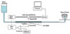
The aim of this study was to measure the dentinal tubular fluid flow (DFF) during and after amalgam and composite restorations. A newly designed fluid flow measurement instrument was made. A third molar cut at 3 mm apical from the CEJ was connected to the flow measuring device under a hydrostatic pressure of 15 cmH2O. Class I cavity was prepared and restored with either amalgam (Copalite varnish and Bestaloy) or composite (Z-250 with ScotchBond MultiPurpose: MP, Single Bond 2: SB, Clearfil SE Bond: CE and Easy Bond: EB as bonding systems). The DFF was measured from the intact tooth state through restoration procedures to 30 minutes after restoration, and re-measured at 3 and 7days after restoration.
Inward fluid flow (IF) during cavity preparation was followed by outward flow (OF) after preparation. In amalgam restoration, the OF changed to IF during amalgam filling and slight OF followed after finishing.
In composite restoration, application CE and EB showed a continuous OF and air-dry increased rapidly the OF until light-curing, whereas in MP and SB, rinse and dry caused IF and OF, respectively. Application of hydrophobic bonding resin in MP and CE caused a decrease in flow rate or even slight IF. Light-curing of adhesive and composite showed an abrupt IF. There was no statistically significant difference in the reduction of DFF among the materials at 30 min, 3 and 7 days after restoration (P > 0.05).
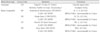
Figures and Tables
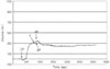 | Figure 2Consecutive DFF during amalgam restoration. Upward movement (+) vs time on graph indicates outward DFF, whereas downward movement (-) indicates inward DFF. CP: cavity preparation; V: varnish application; ga: gentle air stream; AF: amalgam filling.
|
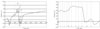 | Figure 3(a) Consecutive DFF during composite restoration with MP. (b) Magnified view of consecutive DFF during bonding procedure of MP. Upward movement (+) vs time on graph indicates outward DFF, whereas downward movement (-) indicates inward DFF. CP: cavity preparation; LC: light curing; C1: first layer filling of composite; C2: second layer filling of composite; E: acid-etching; r: rinse; d: blot-dry; P: primer application; ga: gentle air; A: adhesive application.
|
 | Figure 4(a) Consecutive DFF during composite restoration with SB. (b) Magnified view of consecutive DFF during bonding procedure of SB. Upward movement (+) vs time on graph indicates outward DFF, whereas downward movement (-) indicates inward DFF. CP: cavity preparation; LC: light curing; C1: first layer filling of composite; C2: second layer filling of composite; E: acid-etching; r: rinse; d: blot-dry; sb: Single Bond application; ga: gentle air.
|
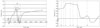 | Figure 5(a) Consecutive DFF during composite restoration with CE. (b) Magnified view of consecutive DFF during bonding procedure of CE. Upward movement (+) vs time on graph indicates outward DFF, whereas downward movement (-) indicates inward DFF. CP: cavity preparation; LC: light curing; C1: first layer filling of composite; C2: second layer filling of composite; SE: self-etching primer application; a: air-dry; A: adhesive application.
|
 | Figure 6(a) Consecutive DFF during composite restoration with EB. (b) Magnified view of consecutive DFF during bonding procedure of EB. Upward movement (+) vs time on graph indicates outward DFF, whereas downward movement (-) indicates inward DFF. CP: cavity preparation; LC: light curing; C1: first layer filling of composite; C2: second layer filling of composite; eb: Easy Bond application; a: air-dry.
|

References
1. Addy M, West N. Etiology, mechanisms, and management of dentine hypersensitivity. Curr Opin Periodontol. 1994. 71–77.
2. Walters PA. Dentinal hypersensitivity: a review. J Contemp Dent Pract. 2005. 6(2):107–117.

3. Johnson GH, Gordon GE, Bales DJ. Postoperative sensitivity associated with posterior composite and amalgam restorations. Oper Dent. 1988. 13(2):66–73.
4. Gordan VV, Mjör IA, Hucke RD, Smith GE. Effect of different liner treatments on postoperative sensitivity of amalgam restorations. Quintessence Int. 1999. 30(1):55–59.
5. Grover PS, Hollinger J, Lorton L. A review of the incidence of pain after an operative treatment visit: Part I. J Prosthet Dent. 1984. 51(2):224–225.
6. Brännstrom M, Lindén LA, Aström A. The hydrodynamics of the dental tubule and of pulp fluid. A discussion of its significance in relation to dentinal sensitivity. Caries Res. 1967. 1(4):310–317.
7. Brännstrom M, Lindén LA, Johnson G. Movement of dentinal & pulpal fluid caused by clinical procedures. J Dent Res. 1968. 47(5):679–682.
8. Brännstrom M. The hydrodynamic theory of dentinal pain: sensation in preparations, caries, and the dentinal crack syndrome. J Endod. 1986. 12(10):453–457.
9. Pashley DH, Matthews WG, Zhang Y, Johnson M. Fluid shifts across human dentin in vitro in response to hydrodynamic stimuli. Arch Oral Biol. 1996. 41(11):1065–1072.
10. Paphangkorakit J, Osborn JW. The effect of normal occlusal forces on fluid movement through human dentine in vitro. Arch Oral Biol. 2000. 45(12):1033–1041.
11. Hashimoto M, Ito S, Tay FR, Svizero NR, Sano H, Kaga M, Pashley DH. Fluid movement across the resin-dentin interface during and after bonding. J Dent Res. 2004. 83(11):843–848.
12. Ratih DN, Palamara JE, Messer HH. Dentinal fluid flow and cuspal displacement in response to resin composite restorative procedures. Dent Mater. 2007. 23(11):1405–1411.
13. Ciucchi B, Bouillaguet S, Holz J, Pashley D. Dentinal fluid dynamics in human teeth, In vivo. J Endod. 1995. 21(4):191–194.
14. Horiuchi H, Matthews B. In-vitro observations on fluid flow through human dentine caused by pain-producing stimuli. Arch Oral Biol. 1973. 18(2):275–294.
15. Benenson W, Harris JW, Stocker H, Lutz H. Handbook of physics. 2006. New York: Springer.
16. Bausch JR, de Lange K, Davidson CL, Peters A, de Gee AJ. Clinical significance of polymerization shrinkage of composite resins. J Prosthet Dent. 1982. 48(1):59–67.
17. Pashley DH. Dynamics of the pulpo-dentin complex. Crit Rev Oral Biol Med. 1996. 7(2):104–133.
18. Tay FR, Pashley DH, Suh BI, Carvalho RM, Itthagarun A. Single-step adhesives are permeable membranes. J Dent. 2002. 30(7-8):371–382.
19. Tay FR, Pashley DH. Water treeing-a potential mechanism for degradation of dentin adhesives. Am J Dent. 2003. 16(1):6–12.
20. Ozok AR, De Gee AJ, Wu MK, Wesselink PR. The influence of resin composite and bonded amalgam restorations on dentine permeability in Class II cavities in vitro. Dent Mater. 2001. 17(6):477–484.
21. Hersek N, Canay S, Akça K, Ciftçi Y. Comparison of microleakage properties of three different filling materials. An autoradiographic study. J Oral Rehabil. 2002. 29(12):1212–1217.
22. Murray PE, Hafez AA, Smith AJ, Cox CF. Bacterial microleakage and pulp inflammation associated with various restorative materials. Dent Mater. 2002. 18(6):470–478.
23. Chersoni S, Suppa P, Grandini S, Goracci C, Monticelli F, Yiu C, Huang C, Prati C, Breschi L, Ferrari M, Pashley DH, Tay FR. In vivo and in vitro permeability of one-step self-etch adhesives. J Dent Res. 2004. 83(6):459–464.
24. Grégoire G, Guignes P, Millas A. Effect of self-etching adhesives on dentin permeability in a fluid flow model. J Prosthet Dent. 2005. 93(1):56–63.
25. Lee IB, Kim MH, Kim SY, Jang JH, Cho BH, Son HH, Baek SH. Development of Nano-fluid Movement Measuring Device and its Application to Hydrodynamic Analysis of Dentinal Fluid. J Korean Acad Conserv Dent. 2008. 33(2):141–147.
 XML Download