

 PDF
PDF ePub
ePub Citation
 Print
Print


Abstract
In this study we evaluated the influence of both the thickness of residual enamel and the color of the composite resins applied to lingual surface on the labial surface color.
Background plates were made by randomly (A1, A2, A6D, B1, B2, B3, C1, C2, C6D) selected colors of Filtek Supreme (3M ESPE, St. Paul, U.S.A.) composite resin.
Crown portion of 9 maxillary central incisors were cut off and embedded with acrylic resin except labial surface. Samples of average thickness of 2.2 mm were obtained after cutting it in a thickness of 2.5 mm from the labial surface and sandpaper polish.
The shade of composite resin background was measured using Spectrophotometer (Spectrolino® GretagMacbeth, Regensdorf, Switzerland). And CIE L*a*b* value of 2.2 mm thickness tooth samples were measured on the 9 composite resin backgrounds. And then, the cutting side of tooth samples was ground to the extent of 1.9 mm, 1.6 mm, 1.3 mm, 1.0 mm and placed on composite resin backgrounds and measured L*a*b* values with the same method.
In all samples, L* value and b* value seemed to have a tendency of decreasing as thickness of tooth sample becomes thinner regardless of background colors (p < 0.05). But, a* value didn't show the significant differences depending on the thickness.
Figures and Tables
Figure 1
L* value of the TS1 (upper) and TS8 (lower) according to thickness of teeth specimen on various resin backgrounds.
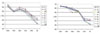
Figure 2
a* value of the TS1 (upper) and TS8 (lower) according to thickness of teeth specimen on various resin backgrounds.
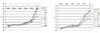
Figure 3
b* value of the TS1 (upper) and TS8 (lower) according to thickness of teeth specimen on various resin backgrounds.
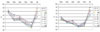
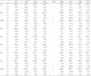
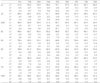
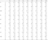
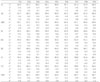

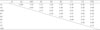
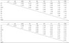
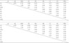
Table 7
Color difference (ΔE*) of the TS1 (upper) and TS8 (lower) tooth specimen among different background at 2.2 mm thickness
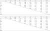
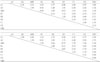
Table 8
Color difference (ΔE*) of the TS1 (upper) and TS8 (lower) tooth specimen among different background at 1.9 mm thickness
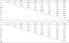
Table 9
Color difference (ΔE*) of the TS1 (upper) and TS8 (lower) tooth specimen among different background at 1.6 mm thickness
References
1. Kramer IRH, McLean JW. Alterations in the staining reaction of dentin resulting from a constituent of a self-polymerizing resin. Br Dent J. 1952. 93:150–153.
2. Buonocore M. A simple method of increasing the adhesion of acrylic filling materials to enamel surfaces. J Dent Res. 1955. 34:849–853.

3. Ward GT, Buonocore MG, Woolridge ED Jr. Preliminary report of a technique using Nuva-Seal in the treatment and repair of anterior fractures without pins. N Y State Dent J. 1972. 38:269–274.
4. Jinks GM. Fluoride impregnated cements and their effect on the activity of interproximal caries. J Dent Child. 1963. 30:87–92.
5. Knight GM. The use of adhesive materials in the conservative restoration of selected posterior teeth. Aust Dent J. 1984. 29:324–331.
6. Morand JM, Jonas P. Resin-modified glass ionomer cement restorations of posterior teeth with proximal carious lesions. Quintessence Int. 1995. 26:389–394.
7. Mount GJ. Minimal treatment of the carious lesion. Int Dent J. 1991. 41:55–59.
8. Magne P, Douglas W. Porcelain veneer : Dentin bonding optimization and biomimetic recovery at the crown. Int J Prosthodont. 1999. 12:111–121.
9. Magne P, Versilius A, Douglas WH. Rationalization of incisor shape : Experimental-numerical analysis. J Prosthet Dent. 1999. 81:345–355.
10. Clark EB. The color problem in dentistry. Dent Digest. 1931. 499–509.
11. Clark EB. An analysis of tooth color. J Am Dent Assoc. 1931. 18:2093–2103.
12. Clark EB. Tooth color selection. J Am Dent Assoc. 1933. 20:1065–1073.
13. Grajower R, Revah A, Sorin S. Reflectance spectra of natural and acrylic teeth. J Prosthet Dent. 1976. 36:570–579.
14. Hwang IN, Lee KW. Translucency of light cured composite resins depends on thickness & its influence on color of restorations. J Korean Acad Conserv Dent. 1999. 24:585–603.
15. Yeh CL, Miyagawa Y, Powers JM. Optical properties of composites of selected shades. J Dent Res. 1982. 61:797–801.
16. CERA. Tooth-colored restoratives principles and techniques. 2003. 9th ed. 196–200.
17. Kubelka P, Munk F. Ein beitrag zur optik der farbanstriche. Z Tech Physik. 1931. 12:593–601.
18. Cook WD, McAree DC. Optical properties of esthetic restorative materials and natural dentition. J Biomed Mater Res. 1985. 19:469–488.
19. Grajower R, Fuss Z, Hirschfeld Z. Reflectance spectra of composite resins on liners. J Prosthet Dent. 1979. 41:650–656.
20. Grajower R, Wozniak WT, Lindsay JM. Optical properties of composite resins. J Oral Rehabil. 1982. 9:389–399.
21. Johnston WM, Ma T, Kienle BH. Translucency parameter of colorants for maxillofacial prostheses. Int J Prosthodont. 1995. 8:79–86.
22. Miyagawa Y, Powers JM. Prediction of color of an esthetic restorative material. J Dent Res. 1983. 62:581–584.
23. Davis BK, Auilino SA, Lund PS, DiazArnold AM, Denehy GE. Colorimetric evaluation of the effect of porcelain opacity on the resultant color of porcelain veneers. Int J Prosthodont. 1992. 5:130–136.
24. Terada Y, Macyama S, Hirayasu R. The influence of different thicknesses of dentin porcelain on the color reflected from thin opaque porcelain fused to metal. Int J Prosthodont. 1989. 2:352–356.
25. Sproull RC. Color matching in dentistry. Part II: Practical applications of the organization of color. J Prosthet Dent. 1973. 29:556–566.
26. Goodkind RJ, Loupe MJ. Teaching of color in predoctoral and postdoctoral dental education in 1988. J Prosthet Dent. 1992. 67:713–717.
27. Culpepper WD. A comparative study of shade matching procedures. J Prosthet Dent. 1970. 24:166–173.
28. O'Neal SJ, Powell WD. A comparative study of shade matching procedures. J Prosthet Dent. 1984. 63:174.
29. Van der Burgt TP, ten Bosch JJ, Borsboom PC, Plasschaert AJ. A new method for matching tooth colors with color standard. J Dent Res. 1985. 64:837–841.
30. Jorgenson MW, Goodkind RJ. Spectrophotometric study of five porcelain shades relative to the dimensions of color, porcelain thickness, and repeated firings. J Prosthet Dent. 1979. 42:96–105.
31. Seghi RR, Johnston WM, O'Brien WJ. Spectrophotometric analysis of color differences between porcelain systems. J Prosthet Dent. 1986. 56:35–40.
32. Seghi RR, Hewlett ER, Kim J. Visual and instrumental colorimetric assessments of small color differences on translucent dental porcelain. J Dent Res. 1989. 68:1760–1764.
33. Seghi RR. Effects of instrument-measuring geometry on colorimetric assessments of dental porcelains. J Dent Res. 1990. 69:1180–1183.
34. Lund TW, Schwabacher WB, Goodkind RJ. Spectrophotometric study of the relationship between body porcelain color and applied metallic oxide pigments. J Prosthet Dent. 1985. 53:790–796.
35. Anusavice KJ, Zhang NZ, Moorhead JE. Influence of P2O5, AgNO3, and Fecl3 on color and translucency of lithiabased-ceramics. Dent Mater. 1994. 10:230–235.
36. Hwang IN, Oh WM. Colorimetric analysis of extracted human teeth and five shade guides. J Korean Acad Conserv Dent. 1997. 22:769–781.
37. Cho KM, Shin DH. Color analysis of the natural teeth with a modified intraoral spectrophotometer. J Korean Acad Conserv Dent. 1998. 23:223–235.
 XML Download