

 PDF
PDF ePub
ePub Citation
 Print
Print


Abstract
The purpose of this study is to observe and compare the dynamic change of artificially demineralized enamel by remineralization solutions of different degrees of saturation at pH 4.3.
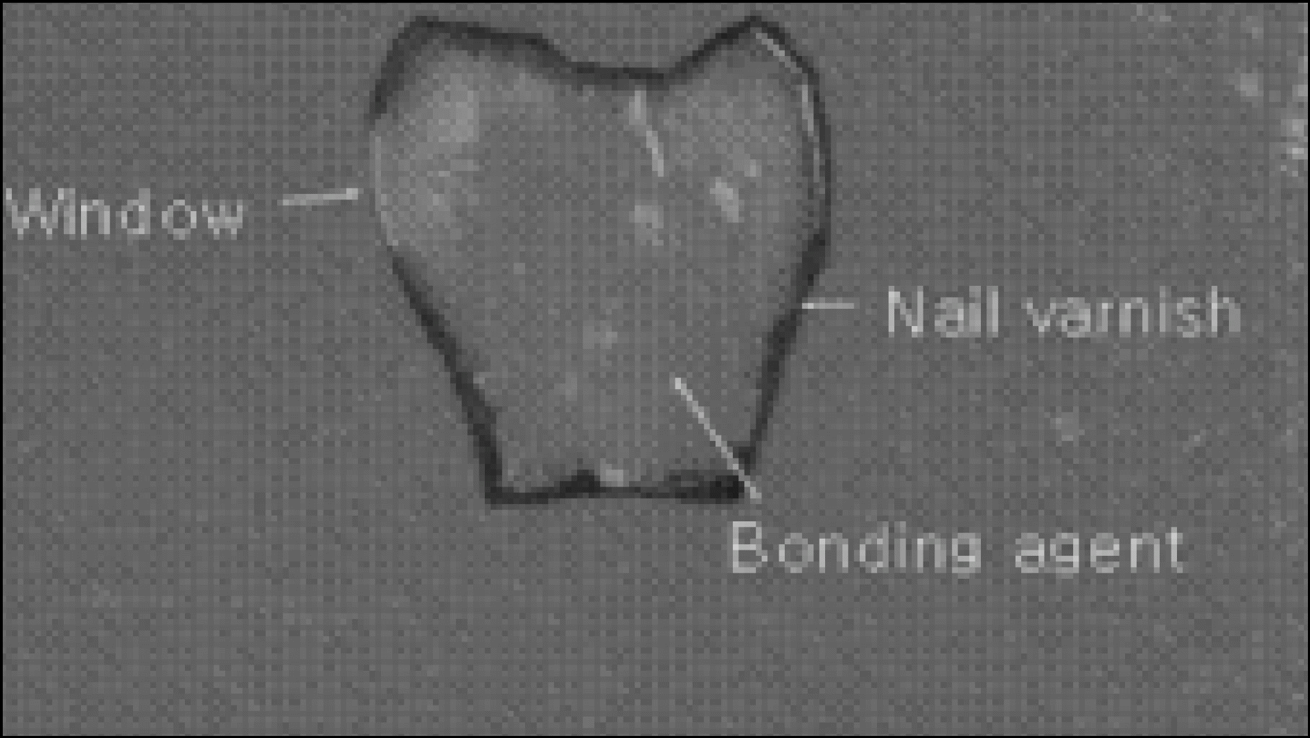
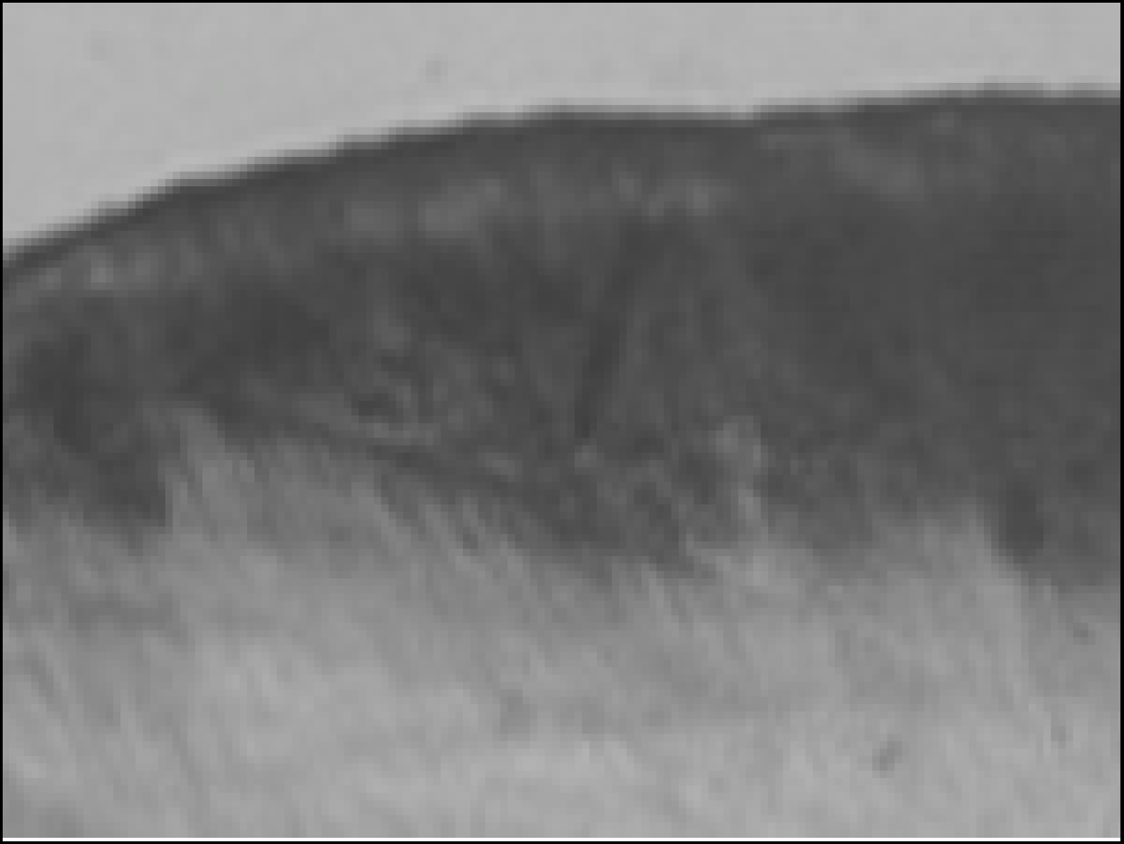
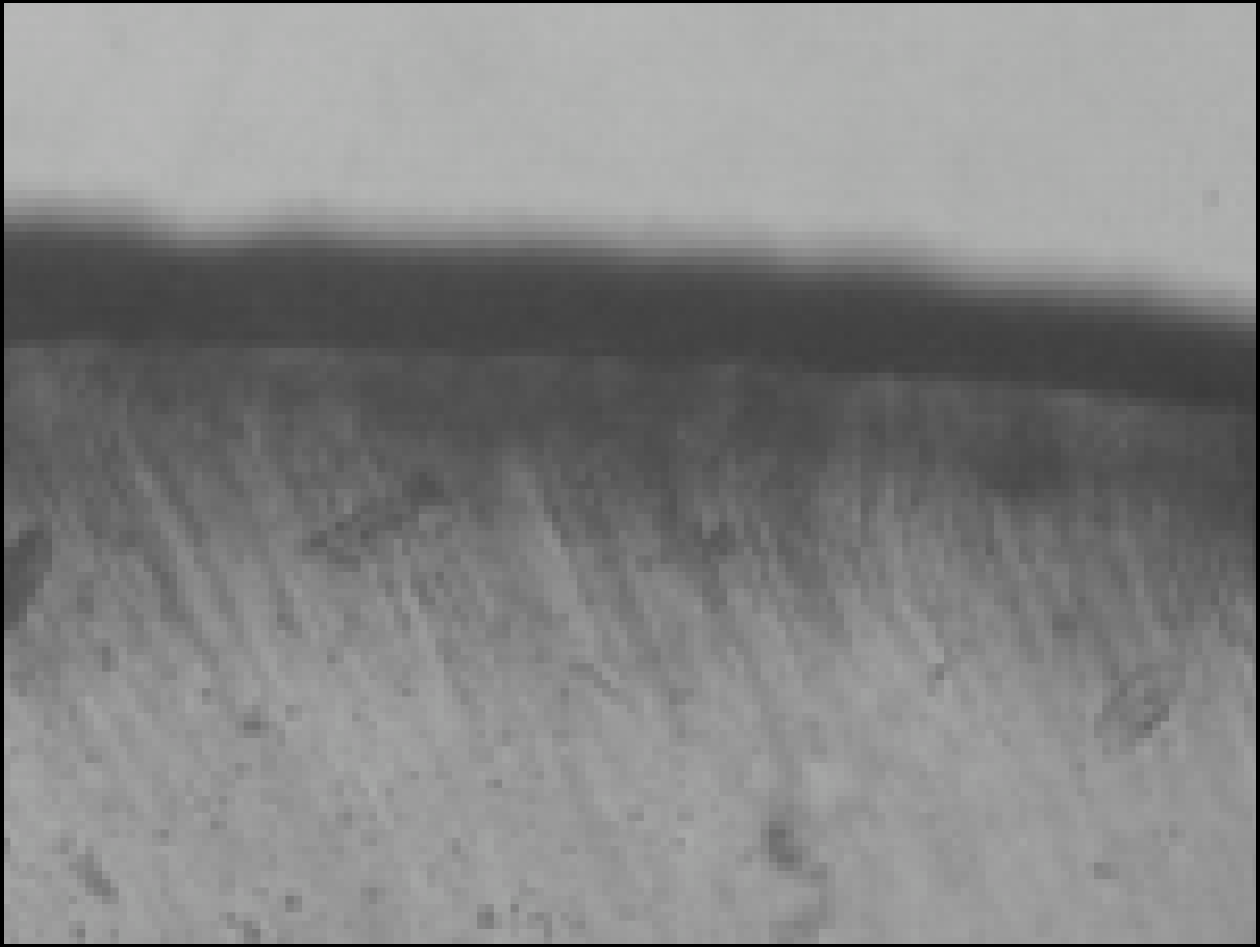
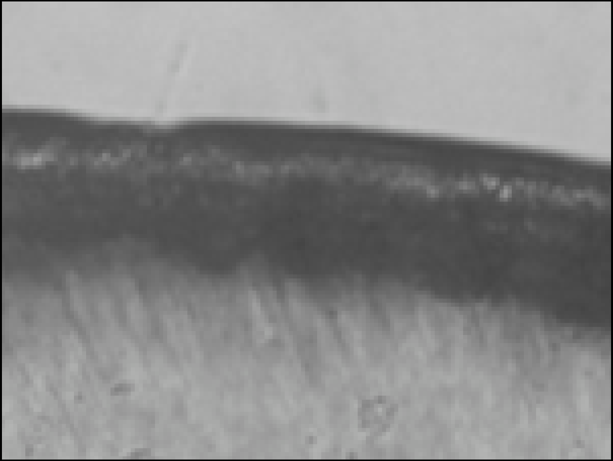
In this study, 30 enamel specimens were demineralized artificially by lactic acid buffered solution. Each of 10 specimens was immersed in pH 4.3 remineralization solution of three different degrees of saturation (0.22, 0.30, 0.35) for 10 days. After demineralization and remineralization, images were taken by a polarizing microscope (× 100). The density of lesion were determined from images taken after demineralization and remineralization.
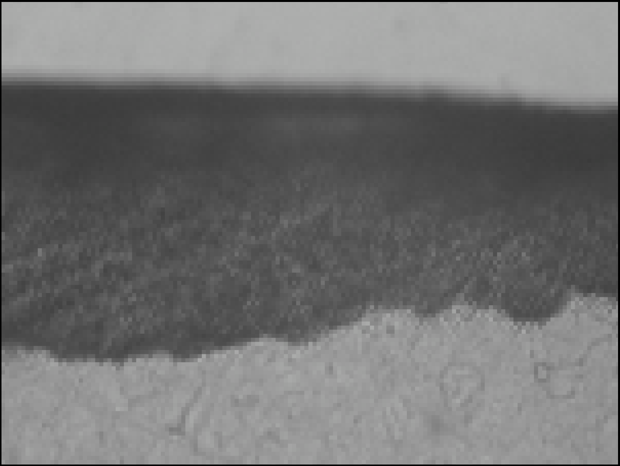
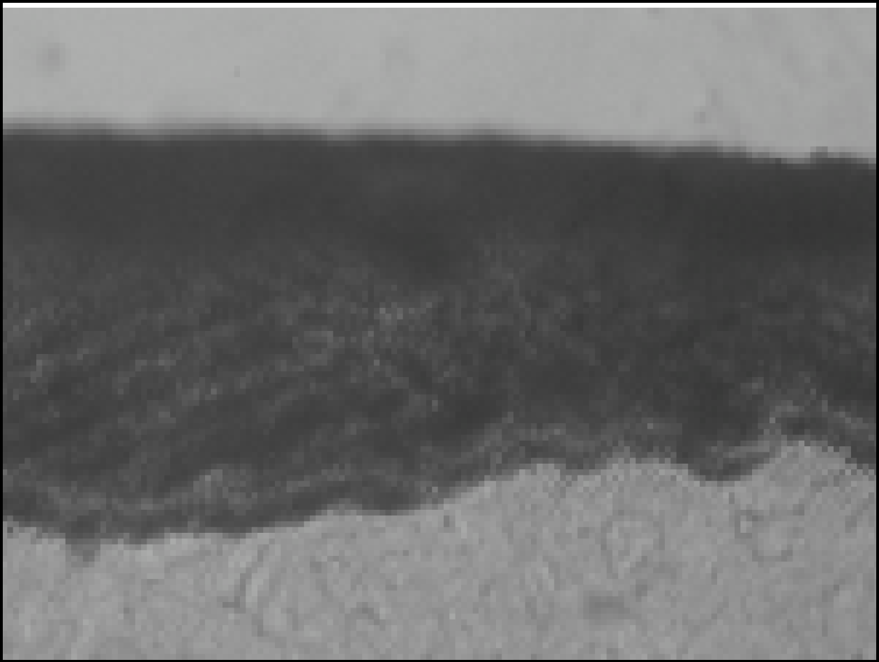
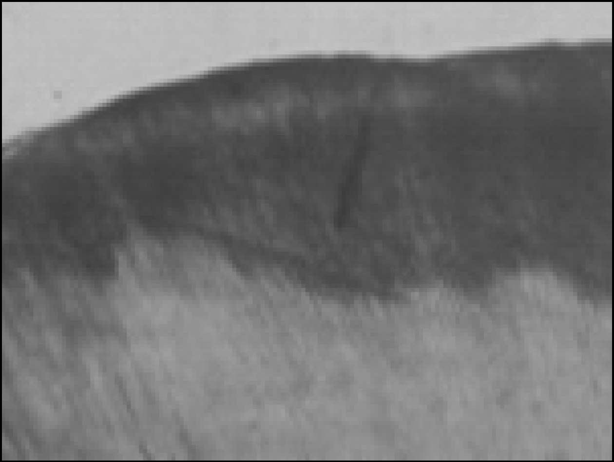
During remineralization process, mineral deposition and mineral loss occurred at the same time. After remineralization, total mineral amount and width of surface lesion increased in all groups. The higher degree of saturation was, the more mineral deposition occurred in surface lesion and the amount of mineral deposition was not much in subsurface lesion. Total demineralized depth increased in all groups.
Go to : 
References
1. Featherstone JDB, Duncan JF, Cutress TW. A Mechanism for dental caries based on chemical process and diffusion phenomena during in-vitro caries simulation on human tooth enamel. Arch Oral Biol. 24:101–112. 1979.
2. Margolis HC, Moreno EC. Physicochemical perspectives on the cariostatic mechanisms of systemic and topical fluorides. J Dent Res. 69:606–613. 1990.

3. Head JA. A study of saliva and its action on tooth enamel in reference to its hardening and softening. J Am Med Assoc. 59:2118–2122. 1912.
4. Anderson BG. Clinical study of arresting dental caries. J Dent Res. 17:443–452. 1938.
5. Backer , Dirks O. Posterupted changes in dental enamel. J Dent Res. 45:503–511. 1966.
6. ten Cate JM, Jongebloed WL, Arends J. Remineralization of artificial enamel lesions in vitro. IV. Influence of fluorides and diphophates on short and long term remineralization. Caries Res. 15:60–69. 1981.
7. Margolis HC, Murphy BJ, Moreno EC. Development of carious-like lesions in partially saturated lactate buffers. Caries Res. 19:36–45. 1985.
8. Margolis HC, Moreno EC. Kinetic and thermodynamic aspect of enamel demineralization. Caries Res. 19:22–35. 1985.
9. Moreno EC, Zahradnik RT. Chemistry of enamel subsurface demineralization in vitro. J Dent Res. 53:226–235. 1974.
10. 이찬 영. 산 완충 용액을 이용한 인공 치아 우식 형성. 연세치대 논문집. 7:34–41. 1992.
11. 박 정원, 허 복, 이 찬영. 유기산 완충 용액의 포화도가 법랑질 및 상아질의 재광화에 미치는 영향과 산화 인회석의 AFM 관찰. 대한치과보존학회지. 25:459–473. 2000.
12. Aoba T, Okazaki M, Takahashi J, Moriwaki Y. X-ray diffraction study on remineralization using systhetic hydroxyapatite pellets. Caries Res. 12:223–230. 1978.
13. Featherstone JDB, Mellerg JR. Relative rates of progress of artificial caries lesions in bovine, ovine and human enamel. Caries Res. 15(1):109–114. 1981.
14. Featherstone JDB, Rodgers BE, Smith MW. Physicochemical requirements for rapid remineralization of early carious lesions. Caries Res. 15:221–235. 1981.
15. Margolis HC, Moreno EC, Murphy BJ. Effect of low levels of fluoride in solution on enamel demineralization. J Dent Res. 65:23–29. 1986.
16. Theuns HM, van Dijk JWE, Driessens FCM, Groeneveld A. Effect of the pH of buffer solution on artificial carious lesion formation in human tooth enamel. Caries Res. 18:7–11. 1984.
17. Nikiforuk G. Formation, structure and metabolism of dental plaque. Understanding dental caries Vol. 1 Etiology and Mechanism. p. 119–157. Karger, Basel and New York;1985.
18. Nikiforuk G. The use of topical fluoride. Understanding dental caries Vol. 11. Prevention. p. 63–86. Karger, Basel and New York;1985.
19. Lammers PC. Borggreven JMPMm, Driessens FCM. Influence of fluorid on in vitro remineralization of artificial subsurface lesions determined with a sandwich technique. Caries Res. 24:81. 1990.
20. 박 성호, 이 찬영, 이 정석. 유산 완충액을 이용한 인공 치아 우식 의 형성에 미치는 산의 농도와 pH 에 관한 연구. 대한치과보존 학회지. 18:277–290. 1993.
21. 김 민경, 금 기연, 이 찬영. 법랑질 인공우식의 재광화에 미치는 pH의 영향에 관한 연구. 대한치과보존학회지. 22:193–208. 1997.
22. 권 중원, 이 찬영. 완충 용액의 유산 농도와 pH가 법랑질의 재광 화에 미치는 영향. 연세대학교 대학원 치의학과 박사학위논문. 2006.
23. Nancollas GH, Purdie N. The kinetics of crystal growth. Quart Rev. 18:1–20. 1964.
24. ten Cate JM, Arends J. Remineralization of artificial enamel lesions in vitro. Caries Res. 11:277. 1977.
25. Varughese , Moreno EC. Crystal growth of calcium apatites in dilute solutions containing fluorides. Calcified tissue Internationals. 33(4):431–439. 1981.
26. Amjad Z, Nancollas GH. Effect of fluoride on the growth of hydroxyapatite and human dental enamel. Caries Res. 13:250–258. 1979.
27. 한 원섭, 금 기연, 이 찬영. 인공 치아 우식의 재광화에 미치는 불 소의 영향. 대한치과보존학회지. 21:161–173. 1996.
28. Silverstone LM. Remineralization phenomena. Caries Res. 11:59–84. 1977.
29. Haikel Y, Frank RM, Voegel JC. Scanning electron microscopy of the human enamel surface layer of incipient caries lesions. Caries Res. 17(1):1–13. 1983.
30. Featherstone JDB. Comparison of artificial caries like lesions by quantitative microradiography and microhardness profiles. Caries Res. 17:385–391. 1983.
31. Groeneveld A, Jongebloed W, Arends J. The mineral content of decalcified surface enamel. A combined microprobe-quantitative microradiography study. Caries Res. 8(3):267–274. 1974.
32. Darling AI. Studies of the early lesion of enamel caries with transmitted light, polarized light and radiography. Brit Dent J. 6:289–341. 1956.
33. Darling AI. Studies of the early lesion of enamel caries its nature, mode of spread and points of entry. Brit Dent J. 8:119–135. 1958.
34. Silverstone LM, Wefel JS, Zimmerman BF, Clarkson BH, Featherstone MJ. Re-mineralization of natural and artificial lesions in human dental enamel in vitro effect of calcium concentration of the calcific fluid. Caries Res. 15:138–157. 1981.
35. White DJ, Chen WC, Nacollas GH. Kinetic and physical aspects of enamel remineralization-A constant composition study. Caries Res. 22:11–19. 1988.
Go to :
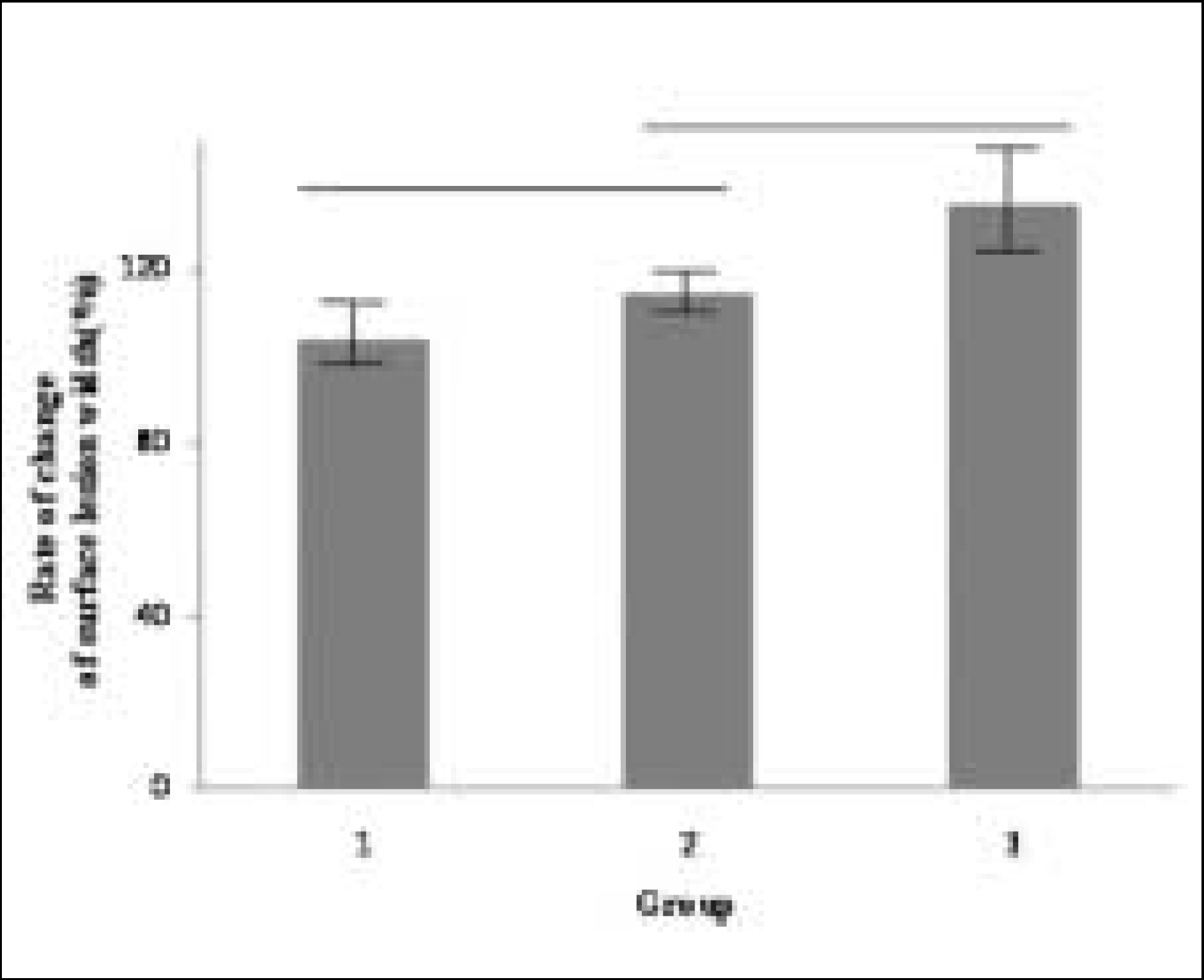 | Figure 8.Rate of change of surface lesion width at Group 1, 2, 3 ((width of surface lesion after remineralization / width of surface lesion before remineralization) × 100(%)). Horizontal bars represent no statistically significant differences (P > .05). |
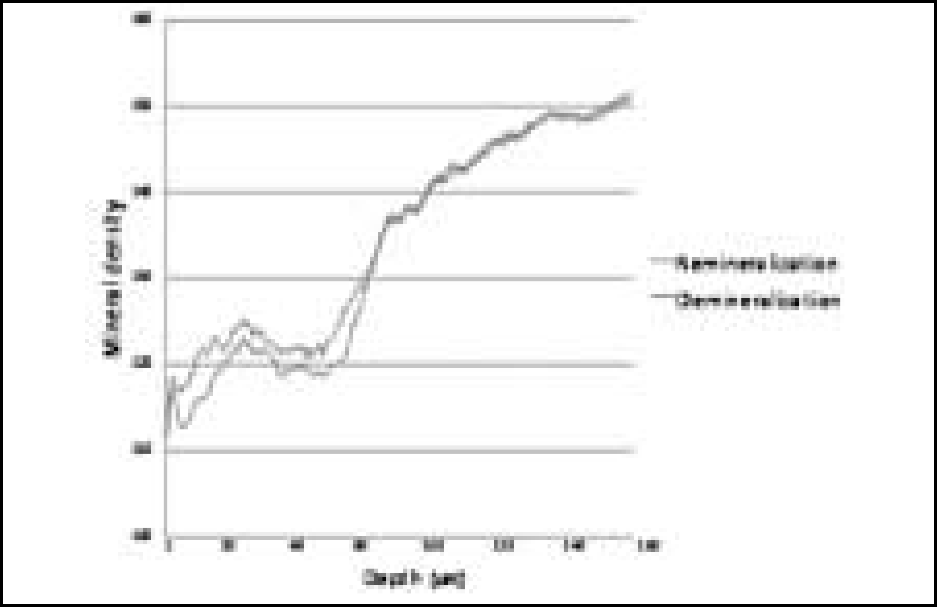 | Figure 9.Comparison of mineral density in enamel before and after remineralization (Group 1) |
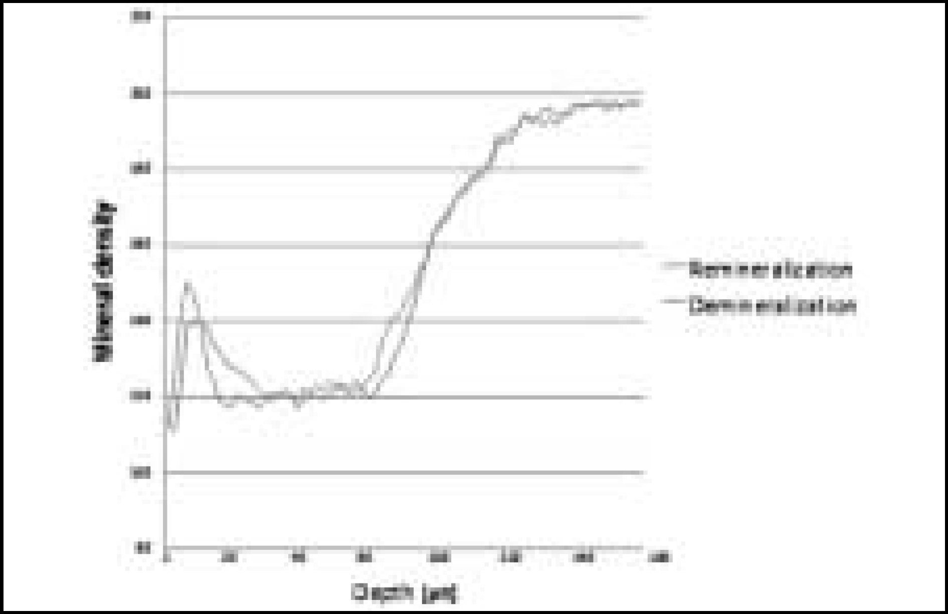 | Figure 10.Comparison of mineral density in enamel before and after remineralization (Group 2) |
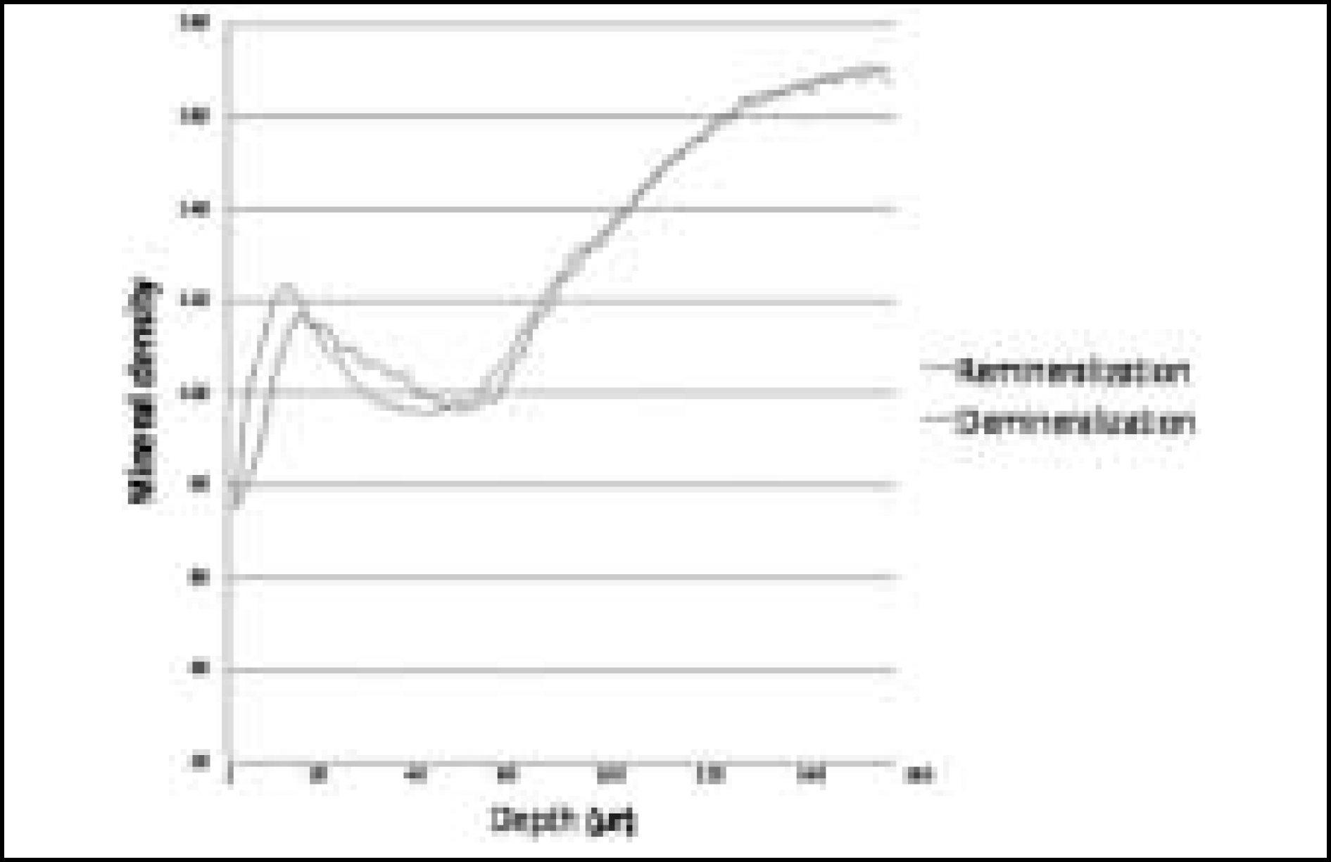 | Figure 11.Comparison of mineral density in enamel before and after remineralization (Group 3) |
Table 1.
Initial composition of demineralization solution
| Composition | Concentration |
|---|---|
| Lactic acid (mM) | 100 |
| Calcium (mM) | 15.5 |
| Phosphate (mM) | 8.5 |
| Sodium azide (mM) | 3.08 |
| pH | 4.3 |
Table 2.
Initial composition of remineralization solution
Table 3.
Rate of change of quantitative value during demineralization & remineralization at enamel
| Condition | Demineralized | Surface lesion | Mineral |
|---|---|---|---|
|
Depth (%) |
Width (%) |
Change (%) |
|
| Group | (Mean ±S.D.) | (Mean ±S.D.) ( | (Mean ±S.D.) |
| 1 | 115.1 ±11.4 % |
 * *
|
109.8 ±3.8 % |
| 2 | 112.6 ±7.9 % | 101.9 ±2.2 % | |
| 3 | 111.4 ±6.5 % | 102.7 ±4.7 % |
 XML Download