

 PDF
PDF ePub
ePub Citation
 Print
Print


Abstract
This study examined the influence of the apical sizes on the sealing ability of a root canal filling.
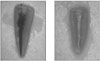
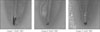
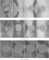
Thirty-six single rooted teeth with a single canal were divided into 3 groups (n = 12) and instrumented with either the Profile® or LightSpeed® system to achieve three different apical sizes (master apical file [MAF] of #25, #40, or #60). The teeth were filled with gutta percha using a modified continuous wave technique. The level of microleakage was determined by immersing ten teeth from each group into India ink for 1 week followed by clearing with nitric acid, ethyl-alcohol, and methylsalicylate. The microleakage was measured using vernier calipers. The data was analyzed statistically using Kruskal-Wallis one-way ANOVA and a Student-Newman-Keuls Method. Two teeth from each group were sectioned horizontally at 1, 2, 3 and 4 mm from the apex in order to observe a cross section.
The apical size was significantly (p < .05) influenced the level of microleakage. In the Student-Newman-Deuls Method, MAF sizes of #25 and #40; and MAF sizes of #25 and #60, respectively showed a statistically significant difference. There was no significant difference between #40 and #60. In most cross sections, oval-shaped canals were observed, and the irregularity of the internal surface increased with decreasing apical size. There was also an increase in the area of recess, which is the area where the canal space is not filled with either gutta-percha or sealer.
When the root canals are filled using a modified continuous wave technique, canal filling with more consistent and predictable outcome may be expected as the apical preparation size is increased.
Figures and Tables

References
1. Schilder H. Filling root canals in three dimensions. Dent Clin North Am. 1967. 723–744.

2. Sjögren U, Hăagglund B, Sundqvist G, Wing K. Factors affecting the long-term results of endodontic treatment. J Endod. 1990. 16:498–504.
3. Rud J, Andreasen JO. A study of failures after endodontic surgery by radiographic, histologic, and stereomicroscopic methods. Int J Oral Surg. 1972. 1:311–328.
4. Gutmann JL. Clinical, radiographic, and histologic perspectives on success and failure in endodontics. Dent Clin North Am. 1992. 36:379–392.
5. Buckley M, Spángberg L. The prevalence and technical quality of endodontic treatment in an American subpopulation. Oral Surg Oral Med Oral Pathol Oral Radiol Endod. 1995. 79:92–100.
6. Youngson CC, Nattress BR, Manogue M, Speirs AF. In vitro radiographic representation of the extent of voids within obturated root canals. Int Endod J. 1995. 28:77–81.
7. Ainley JE. Fluorometric assay of the apical seal of root canal fillings. Oral Surg Oral Med Oral Pathol. 1970. 29:753–762.
8. Kuttler J. Fundamentos de Endo-metaendodoncia Practica. 1980. 2nd ed. Mexico: Mendez Ostero;8.
9. Green D. A stereo-binocular microscopic study of the root apices and surrounding areas of 100 mandibular molars ; preliminary study. Oral Surg Oral Med Oral Pathol. 1955. 8:1298–1304.
10. Ponce EH, Vilar Fernandez JA. The cemento-dentinocanal junction, the apical foramen, and the apical constriction : evaluation by optical microscopy. J Endod. 2003. 29:214–219.
11. Wu MK, Roris A, Barkis D, Wesselink PR. Prevalence and extent of long oval canals in the apical third. Oral Surg Oral Med Oral Pathol Oral Radiol Endod. 2000. 89:739–743.
12. Buchanan LS. The standardized-taper root canal preparation : part1. Concepts for variably tapered shaping instruments. Int Endod J. 2000. 33:516–529.
13. Buchanan LS. The standardized-taper root canal preparation : part2. GT file selection and safe handpiece-driven file use. Int Endod J. 2001. 34:63–71.
14. Buchanan LS. The standardized-taper root canal preparation : part3. GT file technique in large root canals with small apical diameters. Int Endod J. 2001. 34:149–156.
15. Buchanan LS. The standardized-taper root canal preparation : part4. GT file technique in large root canals with large apical diameters. Int Endod J. 2001. 34:149–156.
16. Buchanan LS. The standardized-taper root canal preparation : part5. GT file technique in small root canals. Int Endod J. 2001. 34:244–249.
17. Buchanan LS. The standardized-taper root canal preparation : part6. GT file technique in abruptly curved canals. Int Endod J. 2001. 34:250–259.
18. Weine F. Endodontic therapy. 1972. St. Louis: C.V. Mosby;209–222.
19. Wu MK, Barkis D, Roris A, Wesselink PR. Does the first file to bind correspond to the diameter of the canal in the apical region? Int Endod J. 2002. 35:264–267.
20. Strindberg LZ. The dependence of results of pulp therapy on certain factors : an analytic study based on radiographic and clinical follow-up examination. Acta Odontol Scand. 1956. 14:Suppl. 1.
21. Hoskinson SE, Ng YL, Hoskinson AE, Moles DR, Gulabivala K. A retrospective comparison of outcome of root canal treatment using two different protocols. Oral Surg Oral Med Oral Pathol Oral Radiol Endod. 2002. 93:705–715.
22. Friedman S, Abitbol S, Lawrence HP. Treatment outcome in endodontics : the Toronto Study. Phase 1 : initial treatment. J Endod. 2003. 29:787–793.
23. Wu MK, Schwartz FB, Van des Sluis LW, Wesselink PR. The quality of root fillings remaining in mandibular incisors after root-end cavity preparation. Int Endod J. 2001. 34:613–619.
24. Wu MK, Wesselink PR. Efficacy of three techniques in cleaning the apical portion of the curved root canals. Oral Surg Oral Med Oral Pathol Oral Radiol Endod. 1995. 79:492–496.
25. Siqueira JE, Araujo MC, Garcia PF, Fraga RC, Dantas CJ. Histological evaluation of the effectiveness of five instrumentation techniques for cleaning the apical third of root canals. J Endod. 1997. 23:499–502.
26. Rollison S, Barnett F, Stevens RM. Efficacy of bacterial removal from instrumented root canals in vitro related to instrumentation technique and size. Oral Surg Oral Med Oral Pathol Oral Radiol Endod. 2002. 94:366–371.
27. Card SJ, Sigurdsson A, Orstavik D, Trope M. The effectiveness of increased apical enlargement in reducing intracanal bacteria. J Endod. 2002. 28:779–783.
28. Tan BT, Messer HH. The quality of apical canal preparation using hand and rotary instruments with specific criteria for enlargement based on initial apical file size. J Endod. 2002. 28:658–664.
29. Shuping GB, Ørstavik D, Sigurdsson A, Trope M. Reduction of intracanal bacteria using nickel-titanium rotary instrumentation and various medications. J Endod. 2000. 26:751–755.
30. Sjögren U, Hăgglund B, Sundqvist G, Wing K. Factors affecting the long-term results of endodontic treatment. J Endod. 1990. 16:498–504.
31. Nair PN, Sjögren U, Krey G, Kahnberg KE, Sundqvist E. Intraradicular bacteria and fungi in root-filled, asymptomatic human teeth with therapy-resistant periapical lesions : a long-term light and electron microscopic follow-up study. J Endod. 1990. 16:580–588.
32. Kerekes K, Tronstad L. Morphometric observations on root canals of human anterior teeth. J Endod. 1977. 3:24–29.
33. Alani AH, Toh CG. Detection of microleakage aroud dental restorations : a review. Oper Dent. 1997. 22:173–185.
34. Michailesco PM, Valcarcel J, Grieve AR, Levallois B, Lerner D. Bacterial leakage in endodontics. J Endod. 1996. 22:535–539.
35. Chaisrisookumporn S, Rabinowitz JL. Evaluation of ionic leakage of lateral condensation and McSpadden methods by autoradiopraphy. J Endod. 1982. 8:493–496.
36. Czonstkowsky M, Michanowicz A, Vazquez JA. Evaluation of an injection of thermoplasticized low-temperature gutta-percha using radioactive isotopes. J Endod. 1985. 11:71–74.
37. Ravanshad S, Torabinejad M. Coronal dye penetration of the apical filling materials after post space preparation. Oral Surg Oral Med Oral Pathol. 1992. 74:644–647.
38. Kos WL, Aulozzi DP, Gerstein H. A comparative bacterial microleakage study of retrofilling materials. J Endod. 1982. 8:355–358.
39. Kersten HW, Moorer WR. Particles and molecules in endodontic leakage. Int Endod J. 1989. 22:118–124.
40. Usman N, Baumgartner JC, Marshall JG. Influence of instrument size on root canal debridement. J Endod. 2004. 30:110–112.
41. Coldero LG, McHugh S, MacKenzie D, Saunders WP. Reduction in intracanal bacteria during root canal preparation with and without apical enlargement. Int Endod J. 2002. 35:437–446.
42. Green D. Stereomicroscopic study of 700 root apices of maxillary and mandibular posterior teeth. Oral Surg Oral Med Oral Pathol. 1960. 13:728–733.
43. West JD. The relation between the three-dimensional endodontic seal and endodontic failure. 1975. Boston: Boston University;master's thesis.
44. Barbakow F. The lightspeed system. Dent Clin North Am. 2004. 48:113–135.
45. Sedgley CM, Messer HH. Are endodontically treated teeth more brittle? J Endod. 1992. 18:332–335.
46. Stern N, Hirshfeld Z. Principles of preparing endodontically treated teeth for dowel and core restorations. J Prosthet Dent. 1973. 30:162–165.
47. Tilk MA, Lommel TJ, Gerstein H. A study of mandibular and maxillary root widths to determine dowel size. J Endod. 1979. 5:79–82.
 XML Download