

 PDF
PDF ePub
ePub Citation
 Print
Print


I. INTRODUCTION
Direct resin composite restoration is considered as most conventional restoration in anterior and posterior restoration. However, there are several problems of posterior direct resin composite restoration such as wear, polymerization stress, and inadequate proximal contact. Especially due to the residual polymerization stress, direct resin composite restorations are often contraindicated in large cavities1). Indirect resin composite and ceramic inlay / onlay restorations are advocated as alternatives in that situation2). Indirect resin composite restorations have improved clinical conditions with respect to proximal contact, occlusal anatomy and marginal adaptation3).
Successful dentin bonding is particularly important in the case of indirect resin composite restorations such as inlays, onlays, and veneers, because the final strength of the tooth-restoration complex is highly dependant on adhesive procedures4). Paul et al.5) and Bertschinger et al.6) described freshly cut dentin is the ideal substrate for dentin bonding, while dentin contamination due to provisional cements could reduce the potential for dentin bonding. "Immediate dentin sealing (IDS)" method was introduced. This method defined immediate application and polymerization of the dentin bonding adhesive to the freshly cut dentin, prior to impression taking4). This procedure appears to achieve increased bond strength, improved restoration adaptation, fewer gap formations, decreased bacterial leakage, and reduced dentin sensitivity during provisional period. This approach to adhesion also has a positive influence on the structure preservation, patient comfort, and long-term survival of indirect bonded restoration. On the contrary, the conventional technique for indirect restorations, traditionally, consists in impression taking the cavity immediately after preparation. Then, after the fabrication of the indirect restoration, provisional material and remnants of luting cement were removed and the adhesive or luting procedures are performed. This procedure was called "Delayed dentin sealing (DDS)" method1).
The success of "IDS" method might be attributed to the dentin bond strength. Although there is a tendency to simplify dentin bonding procedures, some authors7,8) described 3 step total-etch adhesive is indicated for IDS because of its ability to form a consistent and uniform layer. But, recently self-etch systems have been produced as an alternative to total-etch system9). Recent studies10,11) of self-etch systems have reported high bond strengths to dentin. De Munck et al.12) reported that the 2 step self-etch adhesive resulted in bond strength close to that obtained with the 3 step total-etch adhesive. Recently, 1 step self-etch adhesives have been introduced that combined the etchant, primer, and adhesive into one bottle13). 1 step self-etch adhesives are more hydrophilic due to a higher concentration of acidic monomers to properly etch the dentin surface14). These adhesives are clinically available and now widely used throughout the world.
However, the difference in bond strength between IDS and DDS for cementation of indirect restorations with various dentin adhesives has not yet to be evaluated. The purpose of this study was to compare the effect of various dentin bonding adhesives on microtensile bonding strength (µTBS) of IDS and DDS for indirect resin composite restoration.
II. MATERIALS AND METHODS
1. Tooth preparation
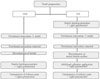
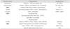
Eighteen extracted permanent molars without caries were stored in distilled water. The teeth were ground flat to expose occlusal dentin surface. The flat dentin surface was polished with wet 600-grit silicon carbide paper. The exposed dentin surface was evaluated for the presence of remaining enamel under operating microscope (OPMI pico; Carl zeiss, Obercohen, Germany), which was removed by additional trimming. The teeth were then randomly assigned to two groups of 9 teeth each, according to application of the dentin adhesive such as IDS and DDS (Figure 1).
2. Experimental group
Delayed dentin sealing (DDS)
The teeth for DDS group were restored with a provisional restoration material (Clip; VOCO, Cuxhaven, Germany), and immersed in saline solution for 1 week. Following that delay, the provisional restoration was removed and dentin was cleaned by an intraoral air abrasion device (Danville Engineering Inc., Danville, CA, USA) filled with 50 µm aluminum oxide particles. And this group was divided into 3 subgroups according to dentin bonding adhesive. All dentin bonding adhesives (Table 1) were manipulated and applied to the dentin surfaces according to the manufacturers' instructions (Table 2). The specimen was light polymerized with LED curing unit (Bluephase; Ivoclar Vivadent, Shann, Liechtenstein) at 600 mW/cm2.
Immediate dentin sealing (IDS)
The teeth for IDS group were also divided into 3 subgroups. Like DDS group, all dentin bonding adhesives were manipulated and applied to the fresh dentin surfaces according to the manufacturers' instructions. The specimen was light polymerized with LED curing unit at 600 mW/cm2.
Polymerization of the adhesive was followed by the application of an air-blocking barrier (glycerin jelly) and 10 seconds of additional light exposure with the same light unit to polymerize the oxygen-inhibition layer as proposed by Magne4). The bonded surfaces were then isolated with petroleum gel not to attach between bonding surface and resin-based provisional restoration. Teeth were restored with a provisional restoration material, and immersed in saline solution for 1 week. Following that delay, the provisional restoration was removed and the sealed dentin was cleaned by an intraoral air abrasion device filled with 50 µm aluminum oxide particles. An adhesive resin was applied once and polymerized.
3. Cementation
Eighteen indirect composite discs of 7mm in height and 10mm in diameter (Sinfony; 3M ESPE, St. Paul, MN, USA) were prepared to simulate overlying laboratory-processed resin composite restorations. The surface of each composite disc was sandblasted with 50µm aluminum oxide particles for 10 seconds from a distance of approximately 5mm. The composite disc surface to be cemented was silanized with a Monobond-S (Ivoclar vivadent AG, Schaan, Liechtenstein) for one minute, and then air dried. Rely X ARC resin cement (3M ESPE, St. Paul, MN, USA) was mixed according to the manufacturers' instructions, and applied to the tooth specimen. A5 kg mass was applied to the composite disc during cementation. The excess resin cement was removed with a probe. The resin cement was photopolymerized for 40 seconds at 600 mW/cm2. The restored specimens were subsequently stored in distilled water at room temperature for 24 hours before testing.
4. Microtensile bond strength (µTBS) testing
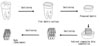
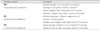
The teeth were then cut longitudinally into six or seven sections perpendicular to the tooth / adhesive interface, with each slab being 1mm thick and 10mm long using the hard tissue cutter (Accutom-50; Struers, Rфdovre, Denmark) under water cooling. The sections were left attached to the remainder of the tooth for further sectioning to obtain sticks approximately 1×1mm thick and 10mm long (Figure 2). Each group was consisted of 36 rods. The specimens were glued to the jig of microtensile testing machine (BISCO Inc, Schaumburg, IL, USA) using cyanoacrylate cement (Zapit; Dental Ventures of America, Corona, CA, USA). Tensile load was applied until specimen was failed. Failure load was recorded for each specimen and then the µTBS was calculated.
5. Fracture mode investigations
1) Optical microscopy observation
After testing, the failure mode of each beam was determined under operating microscope. Fractured test specimens were examined to record the type of bond failure (adhesive, cohesive, or mixed). Bond failure was characterized according to the area of resin remaining on the dentin surface. Adhesive failures were characterized as having less than 25% resin remaining at the interfacial bond area. Cohesive failures had greater than or equal to 75% resin remaining at the interfacial bond area, and mixed failures had 25% to 75% resin remaining at the interfacial bond area.
2) Scanning electron microscopy (SEM) observation
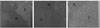
The dentin sides of 6 fractured beams (mixed failure or adhesive failure) from each group were air dried, sputter coated with gold / palladium (E1010 Ion Sputter, Hitachi Co., Mito City, Japan), and examined using SEM (S-3500N SEM, Hitachi Co., Mito City, Japan).
6. Statistical analysis
In each dentin bonding adhesive, the difference of µTBS between IDS and DDS were analyzed statistically by Student t-test. In IDS and DDS group, one-way ANOVA and Tukey's test were used to determined statistical difference of µTBS between the dentin bonding adhesives using SPSS 12.0 software (SPSS, Chicago, IL, USA). The level of significance was set at p < 0.05.
III. RESULTS
1. Microtensile bond strength (µTBS)
1) Comparison between IDS and DDS in each dentin bonding adhesive
Table 3 lists the µTBS values of three dentin bonding adhesives to dentin in IDS and DDS group. The mean µTBS values from 12 to 25 MPa.
In 3 step total-etch SB and 2 step self-etch SE subgroup, IDS group showed higher bond strength than DDS group (p < 0.05). However, in 1 step self-etch XE subgroup, there was no significant difference between IDS and DDS group (p > 0.05).
2) Comparison with each dentin bonding adhesive in IDS and DDS group
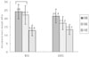
The µTBS values of three dentin bonding adhesives in IDS and DDS group were showed in Figure 3.
In IDS group, 3 step total-etch SB subgroup showed the highest µTBS, followed by 2 step self-etch SE, and 1 step self-etch XE subgroup. But, there was no significant difference between 3 step total-etch SB and 2 step self-etch SE subgroup (p > 0.05). 1 step self-etch XE subgroup showed the lowest µTBS value and was significantly different from the other subgroups (p < 0.05).
In DDS group, 3 step total-etch SB subgroup also exhibited the highest bond strength, followed by 2 step self-etch SE, and 1 step self-etch XE subgroup. And, there was significant difference among all groups (p < 0.05).
2. Fracture mode
1) Optical microscopy observation
The fracture mode was different among the dentin bonding adhesives. These results were summarized in Table 4. In IDS and DDS group, the fractured beams for 3 step total-etch SB and 2 step self-etch SE subgroup demonstrated cohesive or mixed failure, and there was no specimen with adhesive failure. However, 1 step self-etch XE subgroup showed adhesive failure both IDS and DDS group (25% and 22% respectively).
IV. DISCUSSION
This study compared the µTBS of three dentin bonding adhesives with IDS and DDS. The results of this study demonstrated that the IDS method used when cementing indirect resin composite restoration may affect the µTBS of indirect restoration, depending on the dentin bonding adhesive used.
For 3 step total-etch and 2 step self-etch subgroup, IDS group had a statistically higher mean value of bond strength than DDS group. Dietschi et al.15) suggested that using IDS and indirect bonded restorations, because of the delayed placement of the restoration and postponed occlusal loading, the dentin bond can increase over time and residual stress can dissipate, resulting in significantly improved restoration adaptation. And several studies5,6) incorporating various dentin bonding adhesives and application methods have shown that the IDS method increased bond strength values compared to the DDS method. This was related to formation of longer resin tags and a thicker hybrid zone. Moreover, IDS method can protect the tooth from the consequences of microleakage by sealing the dentin tubules that are vulnerable to bacteria invasion, immediately after completion of the preparation16). Sealing of the dentin tubules also reduces sensitivity by preventing hydraulic fluid flow within the dentin tubules, which is associated with postoperative sensitivity17).
In this study, 3 step total-etch subgroup in IDS group exhibited the highest bond strength. According to recent study18), total-etch systems have shown high bond strength to dentin. Bouillaguet et al.19) demonstrated 3 step total-etch adhesive exhibited significantly higher bond strength values than some self-etch systems. In total-etch system, an acid etching of dentin is necessary to efficiently dissolve the smear layer and the smear plugs and to promote a strong and impervious bond between dentin and adhesive mediated by a hybrid layer20). This system could be considered as more reliable in spite of their more time-consuming procedures and technique sensitivity.
The 2 step self-etch adhesive in this study presented good bond strength to dentin. The advantage of this adhesive is that it combines conditioning and priming into one step, avoiding a gap between inorganic component demineralization and primer infiltration. Tanumiharja et al.21) reported that 2 step self-etch adhesive provides the simplest bonding technique and exhibited the highest bond strength to dentin. Laboratory studies22,23) have demonstrated the capability of self-etch systems to bond equally as well as phosphoric acid-etch based systems which demineralize the tooth surface and require a wash and dry step prior to dentin application. In this study, SE Bond was used as 2 step self-etch adhesive, and then showed as high as µTBS of 3 step total-etch adhesive in IDS group. This is probably due to SE Bond's mild acidic monomer content and high filler particle content9). Another reason to show high µTBS of 2 step self-etch adhesive in IDS group described twice application of adhesive layer. First adhesive layer to the freshly cut dentin was done before impression taking. Second adhesive layer was applied before cementation. Several studies24,25) have reported that bond strength to dentin may be improved by application of a second adhesive layer. And due to the second adhesive layer, thicker adhesive layer contribute to greater reduction in polymerization shrinkage stress and the extent of microleakage in cavities24). IDS method is somewhat similar to a twice applications of adhesive on dentin bonding. Another advantage of 2 step self-etch adhesive has been associated with less postoperative sensitivity than 3 step total-etch adhesive26). It seems that 2 step self-etch adhesive in IDS showed as an alternative 3 step total-etch adhesive in IDS. But, the results of this study show that the bond strength of 2 step self-etch subgroup resulted in lower bond strength values than 3 step total-etch subgroup in DDS group.
In this study, XE subgroup bond strength is the lowest value in IDS and DDS group. 1 step self-etch adhesives have been shown to contain a higher concentration of acid derivatives, methacrylated phosphoric acid esters, water, and organic solvents than conventional bonding agents to simultaneously etch and infiltrate the dentin surface in 1 step. The low pH (1.5-2.5) of these 1 step self-etch adhesives makes them hydrolytically unstable as a result of the methacrylate-based components27). Meerbeek et al.8) described that µTBS of 1 step self-etch adhesive was significantly least favorable. Low bond strengths recorded with 1 step self-etch adhesive may indicate the single step material cannot yet fulfill all requirements for the production of effective adhesive layers. As reported in several studies28,29), most simplified 1 step self-etch adhesives are the least durable, while 2 step self-etch adhesives continue to show the best performance in terms of bond strength, aging, and stability of the bonded interface for IDS. Inoue et al.30) found that 1 step self-etch adhesive tended to have lower bond strengths than 2 step self-etch adhesive. In this study, in both IDS and DDS group, µTBS value of 1 step self-etch subgroup was lower than those of 2 step self-etch subgroup.
In this study, failure modes of tested dentin boning adhesive were mostly mixed or cohesive failure. Cohesive failures represent integrity in the adhesive layer, protecting the dentin. In contrast, adhesive failures denote a rupture at the dentin / resin interface, characterized by open dentinal tubule and intertubular dentin. In this study, 1 step self-etch subgroup showed adhesive failure. This result is somewhat supported by the SEM microscopes showing poorly infiltrated smear layer. The low µTBS and the relatively high number of adhesive failures of the 1 step self-etch adhesive strongly suggest that the incorporation of the smear layer might have decreased the adhesive properties or that they failed to optimally hybridize the smear layer covered dentin31).
For many years, some authors7,8) claimed the IDS method needed 3 step total-etch adhesive. But, the problem of 3 step total-etch adhesive showed technique sensitivity and postoperative sensitivity. To overcome this problem, 2 step self-etch adhesives were produced. In this study, the µTBS of 2 step self-etch subgroup was as high as that of 3 step total-etch subgroup and failure mode of 2 step self-etch subgroup are similar to that of 3 step total-etch subgroup. As the result of this study, when clinician use the IDS method, 2 step self-etch adhesive may be advocated as alternatives of 3 step total-etch adhesive. But, 1 step self-etch adhesive is not recommended.
V. Conclusion
With the limitations of the study, the following conclusions were drawn,
The IDS group showed significantly higher µTBS than DDS group in 3 step total-etch and 2 step self-etch adhesive (p < 0.05).
In IDS and DDS group, 3 step total-etch adhesive showed the highest µTBS value, followed by 2 step self-etch, and 1 step self-etch adhesive. In IDS group, the µTBS value for 1 step self-etch adhesive was significantly different from those of the other subgroups (p < 0.05), and in DDS group, there were statistical differences in all subgroups (p < 0.05).
Failure modes of tested dentin bonding adhesives were mostly mixed failure and only 1 step self-etch adhesive showed adhesive failure.


 XML Download