

 PDF
PDF ePub
ePub Citation
 Print
Print


I. INTRODUCTION
Recently tooth colored composite restorative materials are substituting enamel and dentin not only for the esthetic aspects but also mechanical properties, however, long-term serviceability is not yet guaranteed1-3). Current dentin adhesives favored user-friendliness and have been simplified into reduced-step systems. The inclusion of relatively high concentrations of acidic monomers and water, to permit ionization of those monomers and solution of calcium and phosphate, makes these polymers very hydrophilic. This hydrophilic nature of the adhesives rendered the resultant layer absorbing water over time, thereby decreasing stiffness4). During the service in the oral environment, water sorption within the adhesive interface may adversely affect stress distribution across the bonded interface and provide a weak link across the bonded complex4-6). A recent study correlated water sorption and the elastic modulus of dentin adhesives, using disc-shaped cured specimens and showed that water stored adhesives exhibited time-dependent decreases in modulus4). It was anticipated that lowered stiffness of adhesive layer coupling composite to dentin would result in poor load transfer across the interface, leading to joint failure.
Dentin has a localized structural divergence in location-specific tubular orientation, and there are difficulties in performing mechanical tests on small dentin specimens7). Recently, advances in nanoindentation technique made it possible to sample material properties with small applied loads and fine spatial resolutions8). It has been shown the accurate positioning capability of nanoindenter over the hybrid layer to yield hardness and elastic modulus from computerized measurements9,10). This study compared the modulus of elasticity of the bonded interfaces produced with three commercial dentin adhesives (three-step etch and rinse, two-step self etch, and one-step self etch system) using a nanoindentation technique. It also aimed to determine whether any change in that property would be induced by thermo-cycling, and the change would be different among three commercial dentin adhesives.
II. MATERIALS AND METHODS
Specimen Preparation
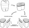
Twenty one freshly extracted human molars (extracted before less than 2 wks) stored in 5% chloramines-T solution were used. Flat dentin surfaces were produced using a low speed diamond saw (Isomet, Buehler Ltd., Lake Bluff, IL. USA). The exposed dentin was abraded with #500 silicon carbide papers to create a clinically relevant smear layer. The twenty one dentin substrates were randomly divided into three adhesive groups; OptiBond FL [OP], Clearfil SE Bond [CL], and Xeno III [XE] (Table 1) and bonded with one of the adhesives following the manufacturer's directions. A commercial composite (Premisa, Kerr, USA) was applied to form a core 1-mm in thickness and light-cured in the same condition (Figure 1). After storage in distilled water at 4℃ for 6 days, each tooth was sectioned into halves occluso-gingivally perpendicular to the adhesive interface along its midpoint with a diamond saw. One half of a tooth was additionally sectioned from the midline to make a slab with 2-mm thickness. The slab was polished using silicon carbide paper with grit size up to 1200. Final polishing was performed with 6 µm to 0.25 µm diamond paste. The polished specimens were stored in distilled water at 4℃ until nanoindentation testing. The other half of the tooth was thermocycled (5 - 55℃; dwell time 30 s; transfer time 5 s) for 10,000 cycles. After thermo-cycling, each tooth was sectioned and polished in the same way to yield a similar 2-mm slab to determine the effect from the thermocycling process.
Nanoindentation test
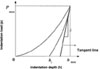
The indentation experiments were performed using a nano-based indentation system (Nano indenter XP, MTS Nano instruments, Oak Ridge, TN, USA). A load up to 20 mN was applied using a Berkovich diamond indenter with a calibrated area function at a rate of 0.5 mN/s. Indentations were made at 6 points in the middle of the adhesive and hybrid layer, respectively. Modified atomic force microscope (AFM) was used for an adequate location of the indenter with the 10 µm interval to avoid corruption of the examination surface. Following the Oliver-Pharr methodology, indenter force load and the depth of penetration into the specimen were continuously monitored, resulting in load-displacement curves to measure the elastic modulus11-13). The following equation was used to calculate the effective indentation elastic modulus (Er).
Where H is the hardness, Pmax is the peak indentation load, and AC is the projected contact area. The reduced elastic modulus is derived as follows:
Where S, the contact stiffness is the initial slope of the unloading curve on the load-depth curve (Figure 2). For a non-rigid indenter that contacts the sample surface, the reduced elastic modulus can be described as:
Where E and ν are the elastic modulus and Poisson's ratio of the specimen (0.2), respectively, and where i, as in Ei and νi, refers to the indenter material.
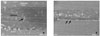
After nanoindentation test, specimens were dried in a desiccator for 5 days and subsequently gold-sputtered for microscopic observations. Field emission electron microscopy (FE-SEM) (S-4700, Hitachi, Pleasanton, CA, USA) was used to confirm the placement of the indentations.
Statistical analysis
An exploratory performance of a general linear model (GLM) revealed the significant inter-specimen differences (p = 0.0134). Mixed-level repeated analysis of variance (ANOVA) and Mixed-level repeated measures analysis of covariance (ANCOVA) were selected to compare the moduli of the three dentin adhesives at the immediate bonding and at the thermocycled stage, respectively, under consideration of six correlated measurements from the same specimen and differences among specimens. Modulus differences between the two conditions were separately compared using paired t-test. Mixed Procedure of statistical package SAS version 9.13 (SAS Institute, Cary, NC, USA) was used for analysis. A significance level of 0.05 was applied to identify statistically significant differences.
III. RESULTS
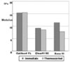
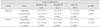
The mean values and standard deviations of elastic modulus obtained by nanoindentation are shown in Table 2. On the adhesive layer, OP showed the significantly higher modulus of elasticity than XE and CL at the immediate bonding, (p = 0.0094) and at the thermocycled stage (p = 0.0061). After thermocycling, the elastic modulus of OP rarely changed, while CL decreased slightly, though not in a statistically significant manner. On the other hand, XE significantly dropped by 3.6 GPa (p = 0.0246). For the hybrid layer, the elastic modulus of OP was the highest, followed by CL and XE. After thermocyling each group had the same tendency to a decrease, not in a statistically significant way. FE-SEM micrographs illustrating the indentation geometry and the accurate positioning capacity are presented in Figure 3. (Figure 4)
IV. DISCUSSION
There have been many attempts to verify the unstable bonded interface resulting from hydrolytic degradation. Water permeability in the hydrophilic adhesive interface has been well documented using various microscopic methodologies14-17). This hydrolysis susceptible area was clearly depicted in micrographs and weak junctions were often confirmed by bond strength tests, which determine fracture position1,17-20). Our study was designed to verify the mechanical properties of each component in the bonding interface by measuring the elastic modulus. Water sorption of polymers is known to make its structure more plasticized and loosely linked monomer particles can be dissolved in the hydrolytic condition. Adhesive materials incorporate hydrophilic primer within their systems and have demonstrated higher water sorption and solubility than the composite resin21,22). It was readily expected that simplified-step adhesive systems would allow higher water sorption and a higher degree of plasticization, resulting in greater deterioration of mechanical property. Previous in vitro experiments tended to standardize specimen fabrication using the polymerized sticks of the adhesive-primer mixture and measured their solubility and elastic modulus22-24). However, this method is far-removed from the clinical environment, where the bonding surface is rubbed with a primer dampened brush and dried with a light air stream, and additionally coated with adhesive. Moreover, it does not incorporate the hybrid layer, the resin impregnated organic substance, which is also subjected to the hydrolytic degradation. Our study tried to reproduce the actual clinical circumstance, following the manufacturer's direction in the bonding procedure. Another attempt given in the study is to minimize the divergence of dentin substrate by using the same tooth in comparison between before and after thermocycling. Two mirror-image surfaces were compared, having one surface represent the immediate bonding stage and the other the thermocycled one. A comparison of the mechanical property between these two adjacent interfaces was expected to be more relevant, because the two counterpart surfaces contain the similar histological features.
At the state of immediate bonding, the elastic modulus of the hybrid layer was higher than of the adhesive layer in all groups, as in previous studies25,26). This zone, in which resinous materials were penetrated into a partially decalcified dentin, still remained a rather stiff resin-dentin interdiffusion area, compared to the pure adhesive layer. After thermocycling, the differences between two layers existed (Table 2). OP which is relatively heavily loaded with fillers (52%) had a significantly higher elastic modulus in the adhesive layer than the other groups. With this traditional three-step etch and rinse system, the thick hydrophobic layer excluded the hydrophilic primed dentin and sustained thermocyling without signs of deterioration (Table 2). CL also adopted the separate bottle of hydrophobic adhesive. Its different filler system might contribute to the slight decrease in the modulus after thermocycling, but not significantly. XE showed a significantly great decrease after thermocycling. The hydrophilic interface produced by this all-in-one system might have been more prone to hydrolysis. After water entered the polymer matrix, the microstructure of adhesive layer was possibly changed due to progressive degradation through pore formation22). Residual monomers, oligomers and degradation products might have been released via these pores, impairing the structural rigidity. As for the hybrid layer, this tendency was similar to that shown in the overlying adhesive layer. OP showed the higher modulus, representing a stiffer backbone of polymer within the collagen network. However, even OP showed a decrease in modulus after thermocycling, although not significantly. There may be the assumption given that less complete polymerization within the collagen network accomplished and the unpolymerized monomers have been leached during thermodynamic process22). As water has been known to be one of the major causes for collagen degradation, disorganization of collagen fibrils may have played an additional role in the change in bonding stability1).
Nanoindentation technique allowed us to determine surface mechanical properties from load-displacement indentation data. In spite of the minute indent contact area, a filler embedded matrix structure might not provide the uniform surface for examination and this anisotropic nature could account for the large standard deviation observed in this study27). This also explained why other nanoindentation studies have allowed multiple measurements and put mean values into data processing26-29). Interspecimen differences were statistically significant (p < .00128), while differences were not significant among measurements within the same specimen. This suggested that consideration of the individual differences among specimens was inevitable and at the same time supported the protocol used here to minimize the structural divergence of dentin substrate. However, considering that the peripheral composite-enamel bond was shown to reduce the degradation rate in resin-dentin interface30,31), the results of our study may be exaggerated, because the bonding surfaces were directly exposed to hydrothermal environment. Another drawback in this study was that thremocycling process may not reflect degradation within the mouth, although 10,000 times of cycles was suggested to represent one service year32). Therefore, the limitations imposed on these in vitro experimental conditions needs to be considered.
V. CONCLUSION
In this study, three different versions (three-step, two-step, and all-in-one system) of commercial adhesive resins were compared to determine the effects of thermocycling on the elastic modulus of the adhesive or the hybrid layer. Only XE group, an all-in-one type adhesive had a significantly decreased modulus in the adhesive layer after 10,000 thermocycles. When adhesives contain more hydrophilic monomers, they may be more prone to hydrolytic degradation, resulting in a mechanically weakened bonding interface. Adhesive systems including separate hydrophobic adhesive with fillers may be more capable of sustaining the hydrous conditions of the oral cavity. Based on the result of this study, some all-in-one self-etch system may be more affected by the hydrolytic degradation over time. It seems that clinicians need to be considerate in selecting materials and procedures, which is predicative of a clinical longevity of bonded restorations.
 XML Download