

 PDF
PDF ePub
ePub Citation
 Print
Print


Abstract
Type II root canal was defined that two canals leave the chamber and merge to form a single canal at short of the apex. The aim of this study was to analyse the master apical file (MAF) size according to various instrumentation techniques in the type II root canal when each canal was enlarged to working length.
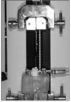
Eighty mesial roots of molar with ISO #15 initial apical file (IAF) size in type II root canals were randomly divided into four experimental groups with 20 teeth each. According to enlarging instruments, four groups are: K-FLEXOFILE® (KF), engine-driven Ni-Ti PROTAPER® (PT), HERO Shaper® (HS), K3 ™ (K3). All canals were enlarged to each working length with ISO #30 size: #30 in KF, F3 in PT, .04/30 in HS, and .06/30 in K3. The master apical file (MAF) size was confirmed by tactile sensation and universal testing machine (EZ test, Shimadzu Co., Kyoto, Japan). The mean MAF size was statistically compared using one-way ANOVA and Tukey HSD test at the 0.05 probability level.
These results show that the MAF size was appeared one or two sizes larger than the final enlarging instrument when all canal in type II configuration were enlarged to each working length. Therefore, the clinician have to confirm the apical stop once more after instrumentation of type II root canal.
Figures and Tables


References
1. Cohen S, Burns R. Pathways of the pulp. 1994. 6th ed. St. Louis: CV Mosby;128–178.
2. Lim SS. Clinical Endodontics. 1999. 2nd ed. Seoul: Uichihaksa;128–136.
3. Grossman L. Endodontic practice. 1985. 10th ed. Philadelphia: Lea & Febiger;207.
4. Weine FS. Endodontic therapy. 1996. 5th ed. St. Louis: Mosby Co.;256–340.
5. Ingle J, Bakland L. Endodontics. 1994. 4th ed. Philadelphia: Lea & Febiger;92–227.
6. Weine FS, Killy RF, Lio PJ. The effect of preparation procedures on original canal shape and on apical foramen shape. J Endod. 1975. 1(8):255–262.

7. Morgan LF, Montgomery S. An evaluation of the crown-down pressureless technique. J Endod. 1984. 10(10):491–498.
8. Roane J, Sabala C, Duncanson M. The balanced force concept for instrumentation of curved canals. J Endod. 1985. 11(5):203–211.
9. Walia H, Brantley WA, Gerstein H. An initial investigation of the bending and torsional properties of Nitinol root canal files. J Endod. 1988. 14(7):346–351.
10. Vertucci FJ. Root canal anatomy of the human permanent teeth. Oral Surg Oral Med Oral Pathol. 1984. 58(5):589–599.
11. Cohen S, Burns R. Pathways of the pulp. 2002. 8th ed. St. Louis: Mosby Inc.;710–713.
12. al-Omari MA, Dummer PMH, Newcombe RG. Comparison of six files to prepare simulated root canals. Part 1. Int Endod J. 1992. 25(2):57–66.
13. al-Omari MA, Dummer PMH, Newcombe RG. Comparison of six files to prepare simulated root canals. Part 2. Int Endod J. 1992. 25(2):67–81.
14. Tharuni SL, Parameswaran E, Spangberg LSW, Sukumaran VG. A comparison of canal preparation using the K-file and Lightspeed in resin blocks. J Endod. 1996. 22(9):474–476.
15. Oh HJ, Hong CU, Cho YB. The Effect of NITI Rotary Instrumentation on the Configuration of Apical Root Canal. J Korean Acad Conserv Dent. 1997. 22(1):244–253.
16. Leeb J. Canal orifice enlargement as related to biomechanical preparation. J Endod. 1983. 9(11):463–470.
 XML Download