

 PDF
PDF ePub
ePub Citation
 Print
Print


I. INTRODUCTION
The major goal of root canal treatment is to remove irritants from the root canal system, to obturate the cleaned and shaped root canals, and to prevent recontamination.
Ray and Trope1) proposed that coronal microleakage may be a major cause of endodontic failure. Many previous studies have examined that coronal leakage might occur, even in well obturated root canals2-4).
One of the most important concerns in root canal cleaning and shaping is how to remove the smear layer that is formed during root canal preparation.
The smear layer is created during instrumentation from the cutting action of the microinstruments and the compression of the resulting crumbs against the root canal walls. It is composed of inorganic particles of calcified tissue and organic elements such as pulp tissue debris, odontoblastic processes, microorganisms, and blood cells5,6). It delays the action of endodontic disinfectants7) and acts as a physical barrier which deteriorates adhesion and penetration of sealers into dentinal tubules8,9). Thus, it can decrease the sealing ability of root canal filling material. Some investigators suggested that the removal of smear layer might significantly improve the sealability especially in the coronal area10-12).
A new solution, MTAD (Biopure MTAD, Tulsa, OK, USA), was recently developed as root canal cleanser. MTAD contains a mixture of a tetracycline isomer, an acid, and a detergent. Recent studies on MTAD demonstrated that MTAD has antimicrobial effect and is able to effectively remove smear layer. Torabinejad et al. have shown that MTAD is able to safely remove the smear layer and is effective against Enterococcus faecalis, a microorganism resistant to the action of other antimicrobial medications such as calcium hydroxide13-19). Portenier et al. demonstrated that the full concentration (100%) MTAD and 0.2% chlorhexidine were both effectively killed Enterococcus faecalis20).
The smear layer removing ability of MTAD was also investigated by recent studies. Tay et al. demonstrated that root canal instrumentation produced 2-5 µm thick smear layer which could be effectively removed by EDTA and MTAD21). They reported that canal irrigation with EDTA and MTAD eroded dentin and created a zone of demineralized collagen matrices, which can form dentin hybridization by infiltration of hydrophilic adhesive and sealer. Ghoddusi et al. also reported that the use of EDTA or MTAD produced more resistance to bacterial leakage than sodium hypochlorite22) due to their superior smear layer removing ability. However Tay et al. pointed out the potential risk of nanoleakage between hydrophobic root canal sealers and collapsed collagen matrices, which was confirmed in another study of them23).
However, there have been very few studies comparing the leakage resistance of the root canals irrigated with MTAD (Biopure MTAD, Tulsa, OK, USA), EDTA and sodium hypochlorite. The purpose of this study was to evaluate the effect of a new root canal cleanser, MTAD, in comparison with EDTA and sodium hypochlorite, on the coronal leakage of obturated root canals using a bacterial leakage test model.
II. MATERIALS AND METHODS
Forty, extracted, noncarious, human, three-rooted maxillary molars with closed apices were used in this study. Other factors, such as age and sex of the patient at the time of extraction, were not considered. The teeth were immersed in 2.6 % sodium hypochlorite (NaOCl) for approximately 1 hour and the root surface was carefully debrided using curettes. Then, the teeth were stored in physiologic saline during the experiment. The teeth were randomly divided into 3 experimental groups of 10 teeth each and 2 control groups of 5 teeth each. Both buccal roots were removed at the furcation. The orifice of each canal was enlarged with a #330 bur (Komet Carbide FG 330, Lemgo, Germany) and was sealed with glass ionomer (GC Fuji II LC, Tokyo, Japan). Only the palatal root was used in this experiment.
After preparing a conventional access for each tooth, #10 K-type file (Maillefer, Zurch, Switzerland) was inserted to determine the working length by penetrating the apical foramen under the microscope (OPMI pico, Zeiss, Germany) and subtracting 1 mm. The root canal of each tooth was cleaned and shaped to a size 40 master apical file using 0.06 Taper ProFlie NiTi rotary systems (Maillefer Profile, Zurich, Switzerland) in a crown-down manner. The root canals in groups 1 to 4 were irrigated with 2 ml of 5.25 % sodium hypochlorite between instrumentations. The irrigant was delivered into each canal with a 28 gauge needle (Max-i-Probe irrigant system, KerrHawe, Switzland). The tip of the irrigating needle penetrated within 1 to 2 mm short of working length in each canal.
In group 1 (n = 5, positive control) and 2 (n = 5, negative control), the canals were irrigated with 2 ml of 5.25 % sodium hypochlorite between instrumentations.
In group 3 (n = 10), the canals were irrigated in the same manner as previous groups. After canal shaping, the canals were irrigated with 5 ml of 5.25 % sodium hypochlorite and left without a dry for 5 minutes.
In group 4 (n=10), the canals were irrigated in the same manner as previous groups. After canal shaping, the root canals were irrigated with 5 ml of 17 % EDTA (Roth International, Chicago, USA) and soaked for 5 minutes to remove the smear layer and rinsed with 5 ml of 5.25 % sodium hypochlorite, following the method used in previous study of Ghoddusi et al.22).
In group 5 (n = 10), the canals were irrigated using MTAD (Biopure MTAD, Tulsa, OK, USA) according to the manufacturer's instructions. The canals in this group were irrigated with 1.3 % NaOCl. After shaping, the root canals were soaked with 1 ml of MTAD for 5 min and rinsed with 4 ml MTAD. MTAD was freshly prepared before each usage according to Park's study24).
The canals were dried with paper points (Diadent, Seoul, Korea) and obturated as follows: all of the root canals were obturated using continuous wave of condensation technique. In group 1 (positive control group), canals were obturated with gutta-percha (Diadent, Seoul, Korea) without sealer. The canals in groups 2 to 5 were obturated with gutta-percha and AH 26 (Dentsply, York, PA, USA). All of the specimens were stored in 100 % humidity for 1 day. To standardize the length of obturation, the coronal filling was removed with a System B (Analytic Endodontics, Orange, CA, USA) and 10 mm of gutta percha filling was remained in the root canal. After confirming the length of obturation radiographically, all of the access openings were sealed temporarily with Caviton (GC, Tokyo, Japan) and the specimens were stored in an incubator with 100 % humidity at 37℃ for 30 days. Then, all of the specimens were sterilized with ethylene oxide (EO) gas. The external root surface of each tooth in group 1, 3, 4, and 5 was coated with triple layers of nail varnish excluding the apical foramen. The access opening of each tooth in negative controls (group 2) was filled with sticky wax, and all of the external surfaces were completely sealed with triple layers of nail varnish.
A dual chamber anaerobic bacteria model was assembled using a 5 ml irrigation syringe as the upper chamber and 20 ml scintillation vial (Iwaki, Tokyo, Japan) as the lower chamber. The syringe was secured via a hole drilled through the cap of the 20 ml scintillation vial. The syringe tip was placed into the chamber. The tooth was attached with sticky wax to the tip of the syringe to complete the upper chamber and the joint sealed with three coats of nail varnish. A cap to cover the tube opening of the upper chamber was made with putty. The assemblies of leakage models were placed in envelopes and sterilized in EO gas.
The vials were placed in an anaerobic chamber (Sheldon Manufacturing, Inc., Cornelius, OR) containing 85 % N2, 5 % CO2, and 10 % H2 for 48 hours. This was done to eliminate any oxygen in the system, reduce the media before inoculation, and check for sterility of the system. Brain Heart Infusion (BHI) broth supplemented with yeast extract (5 g/L), hemin (5 mg/L), and menadione (10 mg/L) was aseptically placed into the lower chamber until exposed root apex immersed completely in the BHI. Two millimeters of BHI broth turbid with Enterococcus faecalis (ATCC 29212) was pipetted into the upper chamber syringe reservoirs. The tube cap was replaced to prevent evaporation. Fresh BHI inoculated with Enterococcus faecalis was added to the upper chamber every 3 to 4 days. The vials were observed everyday for turbidity of the broth in the lower chamber, indicating bacterial growth from penetration of the bacteria past the root apex. The day of turbidity was recorded. The experiment was conducted for six months. Statistical analysis was performed using Fisher's Exact Test. (SAS 9.1, SAS Institute Inc., Cary, NC, USA)
III. RESULTS
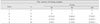
The positive controls showed turbidity at 1 or 2 days after inoculation. The negative controls did not leak for the entire experimental period. 1 and 2 samples were lost in group 3 and 4, respectively due to contamination during experiment. The number of leaking samples per group was described in Table 1.
On the 50th day of experiment, 3, 1, and 0 samples leaked in Group 3, 4, and 5, respectively. On the 80th day of experiment, 5, 3, and 1 samples leaked in group 3, 4, and 5 respectively. On the 180th day of experiment, 7, 4, and 5 samples leaked in group 3, 4, and 5, respectively. On the 180th day of experiment, there was no statistical difference among three experimental groups (p > 0.05).
IV. DISCUSSION
So far, to our knowledge, there have been very few studies regarding the effect of final rinsing with MTAD on bacterial leakage. Ghoddusi et al.22) compared the bacterial leakage of root canals which were rinsed with either MTAD or EDTA. There was no significant difference between MTAD and EDTA treated groups in bacterial leakage. However, in their study, they used streptococcus mutans, which could be easily destroyed by root canal irrigation with sodium hypochlorite or calcium hydroxide. In this experiment, we used Enterococcus faecalis which are known to be resistant to sodium hypochlorite and calcium hydroxide. This rendered our study to simulate the clinical situations more closely than previous studies.
The observation period of our study was 180 days, which is relatively longer that those of other leakage studies that used Enterococcus faecalis. Fathi et al.25) and Yucel et al.26) observed bacterial (Enterococcus faecalis) penetration for 60 days in their studies. In their studies, all the samples leaked after 60 days. But in our study, there were samples which didn't leak until 180 days, which indicated the superior ability of MTAD and EDTA to NaOCl as a smear layer removing agent.
Sundqvist reported that Enterococcus faecalis is important microorganism in endodontic failure27). In the study of Fabricius et al., Enterococci have been shown to have an ability to survive in root canals as single organisms without the support of other bacteria28). In most of recent bacterial leakage test, single bacterial inoculation with Enterococcus faecalis was used25,26,29). That is the reason why we used Enterococcus faecalis for this study.
In group 5, MTAD was used with 1.3% sodium hypochlorite because it is known to be more effective than MTAD only. In a previous study16), sodium hypochlorite was used as an irrigant to assist MTAD to remove the smear layer and there were no significant differences among 1.3 %, 2.6 %, and 5.25 % sodium hypochlorite in the ability as root canal irrigants when MTAD was used as a final rinse to remove the smear layer. Therefore, in our study, 1.3 % sodium hypochlorite was used because this is less toxic16). However, there still might be a possibility that the difference in the concentrations of sodium hypochlorite had an effect on the results of this study.
The leakage test model used in this study was modified from that of Ghoddusi et al.22). There are two major differences between the method of us and Ghoddusi et al. Firstly, they used streptococcus mutans and we used Enterococcus faecalis. Secondly, they didn't use NaOCl as final irrigant as we did in experimental group 3.
In this study, the resistances to bacterial leakage were not significantly different in group 3, 4 and 5. These results are different from the observations of Ghoddusi et al.22). They proposed that use of MTAD or EDTA as root canal irrigant had reduced bacterial leakage compared with sodium hypochlorite. The difference of the results of this study and those of Ghoddusi et al. might be explained from the experimental design. In this study, in group 3, the root canals were soaked with sodium hypochlorite for 5 minutes. This rendered the same experimental conditions as group 4 and 5, in which the root canals were soaked with MTAD and EDTA for 5 minutes. On the contrary, Ghoddusi et al. used the sodium hypochlorite as irrigant only between instrumentation and the root canals were not soaked with sodium hypochlorite. The 5 minutes soaking with sodium hypochlorite might have resolved possible tissue remnant, which might have caused a better seal.
On the other hand, the results of this experiment showed that the effect of MTAD and EDTA as root canal irrigant had a similar effect on bacterial leakage. These results are consistent with the findings of Park et al., who reported the similar bacterial leakage resistance between root canals irrigated with MTAD and EDTA24).
Doxycycline is a major component of MTAD. It is strongly adsorbed to tooth surfaces which makes tooth retain its antimicrobial activity30,31). The results of previous study showed that a short-term exposure of dental hard tissues to tetracycline may result in a long-lasting antibacterial capacity.
However, tetracycline is well-known cause of tooth discoloration when prescribed during tooth development32). Moreover, in recent studies33,34), tooth discoloration was observed when it was used as intracanal medicament. In this study, tooth discoloration was observed during observation period. This phenomenon was due to the photo-oxidation reaction. This photo-oxidation process was probably triggered by the use of sodium hypochlorite as an oxidizing agent34).
In contemporary endodontics, using both EDTA and NaOCl is an effective method for removal of smear layer. However, 17% EDTA solution may have an erosive effect on dentin structure. Thus, it should not be remained in the root canal more than 1 min during endodontic treatment35). But, MTAD does not have an erosive effect and is less destructive to the root dentin structure compared with EDTA when used as a root canal cleanser16,17).
The observation periods of this study was longer than any other leakage studies that compared efficiency of MTAD, EDTA and sodium hypochlorite. Moreover, Enterococcus faecalis was used in this experiment to test the leakage of bacterial species that are most resistant to endodontic disinfection. This study demonstrated that MTAD and EDTA showed similar effect in reducing bacterial leakage when used as root canal irrigants.
Considering the results of this experiment, use of MTAD as a final irrgant might be as effective as EDTA. And, the use of MTAD might be more effective than NaOCl in resistance to bacterial leakage, although there was no statistical difference.
 XML Download