

 PDF
PDF ePub
ePub Citation
 Print
Print


Abstract
This study was done to evaluate the reliability of the digital color analysis system (ShadeScan, CYNOVAD, Montreal, Canada) for dentistry.
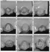
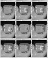
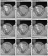
Sixteen tooth models were made by injecting the A2 shade chemical cured resin for temporary crown into the impression acquired from 16 adults. Surfaces of the model teeth were polished with resin polishing cloth. The window of the ShadeScan handpiece was placed on the labial surface of tooth and tooth images were captured, and each tooth shade was analyzed with the ShadeScan software. Captured images were selected in groups, and compared one another.
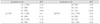
Two models were selected to evaluate repeatability of ShadeScan, and shade analysis was performed 10 times for each tooth.
And, to ascertain the color difference of same shade code analyzed by ShadeScan, CIE L*a*b*values of shade guide of Gradia Direct (GC, Tokyo, Japan) were measured on the white and black background using the Spectrolino (GretagMacbeth, USA), and Shade map of each shade guide was captured using the ShadeScan.
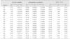
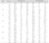
There were no teeth that were analyzed as A2 shade and unique shade. And shade mapping analyses of the same tooth revealed similar shade and distribution except incisal third.
Color difference (ΔE*) among the Shade map which analyzed as same shade by ShadeScan were above 3.
Within the limits of this study, digital color analysis instrument for dentistry has relatively high repeatability, but has controversial in accuracy.
Figures and Tables
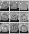
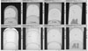 | Figure 8Shade Map of A1 shade guide on the black background (upper) and white background (below) according to thickness.
|
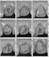
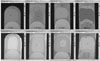 | Figure 9Shade Map of A3 shade guide on the black background (upper) and white background (below) according to thickness.
|

References
1. Sproull RC. Color matching in dentistry. Part II: Practical applications of the organization of color. J Prosthet Dent. 1973. 29:556–566.

2. Paravina RD, Power JM, Fay RM. Color comparison of two shade guides. Int J Prosthodont. 2002. 15:73–78.
3. Hwang IN, Oh WM. Colorimetric Analysis of Extracted Human Teeth and Five Shade Guides. J Korean Acad Conserv Dent. 1997. 22:769–781.
4. Preston JD. Current status of shade selection and color matching. Quintessence Int. 1985. 16:47–58.
5. Grajower R, Revah A, Sorin S. Reflectance spectra of natural and acrylic resin teeth. J Prosthet Dent. 1976. 36:570–579.
6. Bell AM, Kurzeja R, Gemberg MG. Ceramometal crowns and bridges: focus on failures. Dent Clin North Am. 1985. 29:763–778.
7. O'Brien WJ, Boenke KM, Groh CL. Coverage errors of two shade guides. Int J Prosthodont. 1991. 4:45–50.
8. Cernavin I. Effect of chlorine-containing disinfecting compounds on shade guides made of acrylic resin. J Prosthet Dent. 1996. 75:574.
9. Sykora O. Fabrication of a posterior shade guide for removable partial dentures. J Prosthet Dent. 1983. 50:287–288.
10. Lemire PA, Burk B. Color in dentistry. 1975. Hartford, CT: J.M Ney Co..
11. Grajower R, Revah A, Sorin S. Reflectance spectra of natural and acrylic teeth. J Prosthet Dent. 1976. 36:570–579.
12. Macentee M, Lakowski R. Instrumental color measurement of vital and extracted teeth. J Oral Rehabil. 1981. 8:203–208.
13. Goodkind RJ, Schwabacher WB. Use of a fiber-optic colorimeter for in vivo color measurements of 2830 anterior teeth. J Prosthet Dent. 1987. 58:535–542.
14. Park HK, Jeong JH. A Study on the Color of Korean Natural Teeth. J Korean Acad Prosthodont. 1988. 26:185–195.
15. Cho KM, Shin DH. Color Analysis of the Natural Teeth with a Modified Intraoral Spectrophotometer. J Korean Acad Conserv Dent. 1998. 23:223–235.
16. Freedman G. Communicating color. Dent Today. 2001. 20:78–80.
17. Chu SJ, Tarnow DP. Digital shade analysis and verification: A case report and discussion. Pract Proced Aesthet Dent. 2001. 13:129–136.
18. Chu SJ. Precision shade technology: Contemporary strategies in shade selection. Pract Proced Aesthet Dent. 2002. 14:79–83.
19. Paravina RD, Powers JM. Esthetic color training in dentistry. 2004. St. Louis, Missouri: Mosby Inc.;165–174.
20. Clark EB. An analysis of tooth color. J Am Dent Assoc. 1931. 18:2093–2103.
21. Clark EB. Tooth color selection. J Am Dent Assoc. 1933. 20:1065–1073.
22. Miller LL. Shade matching. J Esthet Dent. 1993. 5:143–153.
23. Miller LL. Organizing color in dentistry. J Am Dent Assoc. 1987. 12. Spec No:26E–40E.
24. Hayashi T. Medical color standard. V. Tooth crown. 1967. Tokyo: Japan Color Research Institute.
25. Goodkind RJ, Loupe MJ. Teaching of color in predoctoral and postdoctoral dental education in 1988. J Prosthet Dent. 1992. 67:713–717.
26. O'Brien WJ, Groh CL, Boenke KM. A one-dimensional color order system for dental shade guides. Dent Mater. 1989. 5:371–374.
27. Seghi RR, Hewlett ER, Kim J. Visual and instrumental colorimetric assessments of small color differences on translucent dental porcelain. J Dent Res. 1989. 68:1760–1764.
28. Gross MD, Moser JB. A colorimetric study of coffee and tee staining of four composite resins. J Oral Rehabil. 1977. 4:311–322.
29. Hwang IN, Lee KW. Translucency of Light Cured Composite Resins Depends on Thickness & its Influence on Color of Restorations. J Korean Acad Conserv Dent. 1999. 24:604–613.
30. Johnston WM, Kao EC. Assessment of appearance match by visual observation and clinical colorimeter. J Dent Res. 1989. 68:819–822.
31. Goodkind RJ, Keenan KM, Schwabacher WB. A comparison of Chromascan and spectrophotometric color measurement of 100 natural teeth. J Prosthet Dent. 1985. 53:105–109.
32. Schwabacher WB, Goodkind RJ. Three-dimensional color coordinates of natural teeth compared with three shade guide. J Prosthet Dent. 1990. 64:425–431.
33. Yeh CL, Powers JM, Miyagawa Y. Color of selected shades of composites by reflection spectrophotometry. J Dent Res. 1982. 61:1176–1179.
34. Robertson AR. Colorimetry. Rep Prog Phys. 1978. 41:471–510.
35. Thornton WA. How strong metamerism disturbs color spaces. Color Res Appl. 1998. 23:402–407.
36. Park EJ. The basis of color molding. 1996. 2nd ed. Seoul: Mijinsa;56–194.
37. Park DY. Practical Chromatics. 1992. Revised ed. Seoul: Bando publisher;99–120.
38. The Korean Industrial Standard. KS A 0065.
 XML Download