

 PDF
PDF ePub
ePub Citation
 Print
Print


Abstract
The purpose of this study was to measure the polymerization shrinkage and hygroscopic expansion of resin-based temporary filling materials and to evaluate microleakage at the interface between the materials and cavity wall.
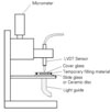
Five resin-based temporary filing materials were investigated: Fermit (Vivadent), Quicks (Dentkist), Provifil (Promedica), Spacer (Vericom), Clip (Voco). Caviton (GC) was also included for comparison. Polymerization shrinkage of five resin-based temporary filling materials was measured using the bonded disc method. For the measurement of hygroscopic expansion, the discs of six cured temporary filling materials were immersed in saline and a LVDT displacement sensor was used to measure the expansion for 7 days. For estimating of microleakage, Class I cavities were prepared on 120 extracted human molars and randomly assigned to 6 groups of 20 each. The cavities in each group were filled with six temporary filling materials. All specimens were submitted to 1000 thermo-cycles, with temperature varying from 5℃/55℃. Microleakage was determined using a dye penetration test.
The results were as follows:
Fermit had significantly less polymerization shrinkage than the other resin-based temporary filling materials. Fermit (0.22 %) < Spacer (0.38 %) < Quicks (0.64 %), Provifil (0.67 %), Clip (0.67 %)
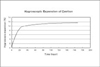
Resin-based temporary filling materials showed 0.43 - 1.1 % expansion in 7 days.
Fermit showed the greatest leakage, while Quicks exhibited the least leakage.
There are no correlation between polymerization shrinkage or hygroscopic expansion and microleakage of resin-based temporary filling materials.
Figures and Tables
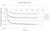 | Figure 2Representative curves of axial polymerization shrinkage of temporary filling materials as a function of time.
|
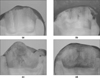 | Figure 4The representative pictures for different microleakage patterns.
(a) Score 0: no leakage
(b) Score 1: leakage up to half of the cavity depth
(c) Score 2: leakage in the entire cavity depth without reaching the pulpal wall
(d) Score 3: leakage in the entire cavity depth even reaching the pulpal wall
|
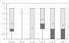 | Figure 5Boxplot display of mean and median leakage score for each group.
The plot illustrates a summary of the microleakage scores based on the median, quartiles, and extreme values. The box represents the interquartile range which contains the 50% of values, the whiskers represent the highest and lowest microleakage values and the bold black line across the box indicates the median microleakage scores. The mark ◇ represents the mean microleakge scores.
|
References
1. Paul SJ, Scharer P. Effect of provisional cements on the bond strength of various adhesive bonding systems on dentine. J Oral Rehabil. 1997. 24:8–14.

2. Garber DA, Goldstein RE. Porcelain and composite inlays & onlays. 1994. Quintessence Publishing Co, Inc;57–61.
3. Tulunoglu O, Uctasli MB, Ozdemir S. Coronal microleakage of temporary restorations in previously restored teeth with amalgam and composite. Oper Dent. 2005. 30(3):331–337.
4. Deveaux E, Hildelbert P, Neut C, Romond C. Bacterial microleakage of Cavit, IRM, TERM, and Fermit : a 21-day in vitro study. J Endod. 1999. 25(10):653–659.
5. Mayer T, Eickholz P. Microleakage of temporary restorations after thermocycling and mechanical loading. J Endod. 1997. 23(5):320–322.
6. Anderson RW, Powell BJ, Pashley DH. Microleakage of three temporary endodontic restorations. J Endod. 1988. 14(10):497–501.
7. Bobotis HG, Anderson RW, Pashley DH, Pantera EA. A microleakage study of temporary restorative materials used in endodontics. J Endod. 1989. 15(12):569–572.
8. Uctasli MB, Tinaz AC. Microleakage of different types of termporary restorative materials used in endodontics. J Oral Sci. 2000. 42(2):63–67.
9. Hosoya N, Cox CF, Arai T, Nakamura J. The walking bleach procedure: an in vitro study to measure microleakage of five temporary sealing agents. J Endod. 2000. 26(12):716–718.
10. Noguera AP, McDonald NJ. A comparative in vitro coronal microleakage study of new endodontic restorative materials. J Endod. 1990. 6(11):523–527.
11. Watts DC, Cash AJ. Determination of polymerization shrinkage kinetics in visible-light-cured materials: methods development. Dent Mater. 1991. 281–287.
12. Lee IB, Cho BH, Son HH, Um CM. The effect of consistency, specimen geometry and adhesion on the linear polymerization shrinkage measurement of light cured composites. Dent Mater. 2006. 2(11):1071–1077.
13. Campos PEGA, Sampaio Filho HR, Barceleir MO. Occlusal loading evaluation in the cervical intergrity of class II cavities filled with composite. Oper Dent. 2005. (6):727–732.
14. de Gee AJ, Feilzer AJ, Davidson CL. True linear polymerization shrinkage of unfilled resins and composite determined with a linometer. Dent Mater. 1995. 9:11–14.
15. Davidson CL, Feilzer AJ. Polymerization shrinkage and polymerization shrinkage stress in polymer-based restoratives. J Dent. 1997. 25:435–440.
16. Scotti R, Ciocca L, Baldissara P. Microleakage of temporary endodontic restorations in overdenture tooth abutments. Int J Prosthodont. 2002. 15:479–482.
17. Jacquot BM, Panighi MM, Steinmetz P, G'sell C. Evaluation of temporary restorations' microleakage by means of electrochemical impedance measurements. J Endod. 1996. 22(11):586–589.
18. Teplitsky PE, Meimaris IT. Sealing ability of Cavit and TERM as intermediate restorative materials. J Endod. 1988. 14(6):278–282.
19. Lee YC, Yang SF, Hwang YF, Chueh LH, Chung KH. Microleakage of endodontic temporary restorative materials. J Endod. 1993. 19:516–520.
20. Widerman FH, Eames WB, Serene TP. The physical and biological properties of Cavit. JADA. 1971. 82:378–382.
21. Webber RT, del Rio CE, Brady JM, Segall RO. Sealing quality of a temporary filling material. Oral Surg. 1978. 46:123–130.
22. Uranga A, Blum JY, Esber S, Parahy E, Prado C. A comparative study of four coronal obturation materials in endodontic treatment. J Endod. 1999. 25(3):178–180.
23. Qvist V. The effect of mastication on marginal adaptation of composite restorations in vivo. J Dent Res. 1983. 62:904–906.
 XML Download