

 PDF
PDF ePub
ePub Citation
 Print
Print


Abstract
The purpose of this study was to evaluate the effect of a new resin monomer, filler size and polishing technique on the surface roughness of composite resin restorations using confocal laser scanning microscopy. By adding new methoxylated Bis-GMA (Bis-M-GMA, 2,2-bis[4-(2-methoxy-3-methacryloyloxy propoxy) phenyl] propane) having low viscosity, the content of TEGDMA might be decreased. Three experimental composite resins were made: EX1 (Bis-M-GMA/TEGDMA = 95/5 wt%, 40 mm nanofillers); EX2 (Bis-M-GMA/TEGDMA = 95/5 wt%, 20 mm nanofillers); EX3 (Bis-GMA/TEGDMA = 70/30 wt%, 40 mm nanofillers). Filtek Z250 was used as a reference.
Nine specimens (6 mm in diameter and 2 mm in thickness) for each experimental composite resin and Filtek Z250 were fabricated in a teflon mold and assigned to three groups. In Mylar strip group, specimens were left undisturbed. In Sof-lex group, specimens were ground with #1000 SiC paper and polished with Sof-lex discs. In DiaPolisher group, specimens were ground with #1000 SiC paper and polished with DiaPolisher polishing points. The Ra (Average roughness), Rq (Root mean square roughness), Rv (Valley roughness), Rp (Peak roughness), Rc (2D roughness) and Sc (3D roughness) values were determined using confocal laser scanning microscopy. The data were statistically analyzed by Two-way ANOVA and Tukey multiple comparisons test (p = 0.05).
The type of composite resin and polishing technique significantly affected the surface roughness of the composite resin restorations (p < 0.001). EX3 showed the smoothest surface compared to the other composite resins (p < 0.05). Mylar strip resulted in smoother surface than other polishing techniques (p < 0.05).
Bis-M-GMA, a new resin monomer having low viscosity, might reduce the amount of diluent, but showed adverse effect on the surface roughness of composite resin restorations.
Figures and Tables


Table 3
The Ra, Rq, Rv, Rp, Rc, and Sc values of experimental composite resins according to the polishing techniques
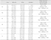
† The numbers in parentheses are the standard deviations. (n = 21)
¶ Means within same columns with different superscript letters in each parameter are significantly different (p < 0.05), and means within same rows with different superscript numbers in each parameter are significantly different (p < 0.05).
§ The values of Filtek Z250 were also measured as a reference.
![]()
References
1. Lu H, Roeder LB, Powers JM. Effect of polishing systems on the surface roughness of microhybrid composites. J Esthet Restor Dent. 2003. 15:297–304.

2. van Noort R, Davis LG. The surface finish of composite resin restorative materials. Br Dent J. 1984. 157:360–364.
3. Toledano M, De La Torre FJ, Osorio R. Evaluation of two polishing methods for resin composites. Am J Dent. 1994. 7:328–330.
4. Chan KC, Fuller JL, Hormati AA. The ability of foods to stain two composite resins. J Prosthet Dent. 1980. 43:542–545.
5. Bouvier D, Duprez JP, Lissac M. Comparative evaluation of polishing systems on the surface of three aesthetic materials. J Oral Rehabil. 1997. 24:888–894.
6. Kaplan BA, Goldstein GR, Vijayaraghvan TV, Nelson IK. The effect of three polishing systems on the surface roughness of four hybrid composites; a profilometric and scanning electron microscopy study. J Prosthet Dent. 1996. 76:34–38.
7. Turssi CP, Saad JRC, Duarte SLL, Rodirigues AL. Composite surfaces after finishing and polishing techniques. Am J Dent. 2000. 13:136–138.
8. Lee JY, Shin DH. Surface roughness of universal composites after polishing procedures. J Korean Acad Conserv Dent. 2003. 28:369–377.
9. Fruits TJ, Miranda FJ, Coury TL. Effects of equivalent abrasive grit sizes utilizing differing polishing motions on selected restorative materials. Quintessence Int. 1996. 27:279–285.
10. Roeder LB, Tate WH, Powers JM. Effect of finishing and polishing procedures on the surface roughness of packable composites. Oper Dent. 2000. 25:534–543.
11. Marigo L, Rizzi M, La Torre G, Rumi G. 3-D surface profile analysis: different finishing methods for resin composites. Oper Dent. 2001. 26:562–568.
12. Terry DA. Direct applications of a nanocomposite resin system; part 1- the evolution of contemporary composite materials. Pract Proced Aesthet Dent. 2004. 16:417–422.
13. Kalachandra S, Sankarapandian M, Shobha HK, Taylor DF, McGrath JE. Influence of hydrogen bonding on properties of Bis-GMA analogs. J Mater Sci Mater Med. 1997. 8:283–286.
14. Anseth KS, Newman SM, Bowman CN. Polymeric dental composites: properties and reaction behavior of multimethacrylate dental restorations. Adv Polym Sci. 1995. 122:177–217.
15. Braden M. The formulation of composite filling materials. Oper Dent. 1978. 3:97–102.
16. Kim JW, Kim LU, Kim CK, Cho BH, Kim OY. Characteristics of novel dental composites containing 2,2-bis[4-(2-methoxy-3-methacryloxy-propoxy) phenyl] propane as a base resin. Biomacromolecules. 2006. 7:154–160.
17. Bae JH, Kim YK, Yoon PY, Lee MA, Cho BH. Effect of a new resin monomer on the microleakage of composite resin restorations. J Korean Acad Conserv Dent. 2007. 32:469–475.
18. González-Cabezas C, Fontana M, Dunipace AJ, Li Y, Fischer GM, Proskin HM, Stookey GK. Measurement of enamel remineralization using microradiography and confocal microscopy. A correlational study. Caries Res. 1998. 32:385–392.
19. Al-Nawas B, Grotz KA, Götz H, Heinrich G, Rippin TG, Stender TE, Duschner H, Wagner W. Validation of three-dimensional surface characterising methods: scanning electron microscopy and confocal laser scanning microscopy. Scanning. 2001. 23:227–231.
20. Al-Shammery HA, Bubb NL, Youngson CC, Fasbinder DJ, Wood DJ. The use of confocal microscopy to assess surface roughness of two milled CAD-CAM ceramics following two polishing techniques. Dent Mater. 2007. 23:736–741.
21. Ross M, Romrell LJ, Kaye GI. Histology. A test and atlas. 1995. 3rd Ed. Williams & Wikins;12–13.
22. Mohan B, Kandaswamy D. A confocal microscopic evaluation of resin-dentin interface using adhesive systems with three different solvents bonded to dry and moist dentin in vitro study. Quintessence Int. 2005. 36:511–521.
23. Banerjee A, Boyde A. Autofluorescence and mineral content of carious dentine: scanning optical and backscattered electron microscopic studies. Caries Res. 1998. 32:219–226.
24. Hachiya Y, Iwaku M, Hosoda H, Fusayama T. Relation of finish to discoloration of composite resins. J Prosthet Dent. 1984. 52:811–814.
25. Okazaki M, Douglas WH. Comparison of surface layer properties of composite resin by ESCA, SEM and X-ray diffractiometry. Biomaterials. 1984. 5:284–290.
26. Lee IB, Lee JH, Cho BH, Son HH, Lee ST, Um CM. Rheological properties of resin composites according to the change of monomer and filler compositions. J Korean Acad Conserv Dent. 2004. 29:520–531.
27. Rong MZ, Zhang MQ, Pan SL, Friedrich K. Interfacial effects in polypropylene-silica nanocomposites. J Appl Polym Sci. 2004. 92:1771–1781.
 XML Download